The MicroRNA-217 Functions as a Potential
Tumor Suppressor in Gastric Cancer by
Targeting GPC5
Hui Wang1‡, Xiaolin Dong2‡, Xin Gu3, Rong Qin1, Hongping Jia1, Jianpeng Gao1*
1Department of Gastroenterology, The Affiliated YanAn Hospital of Kunming Medical University, Kunming, 650051, Yunnan, China,2Department of neurology, The Affiliated YanAn Hospital of Kunming Medical University, Kunming, 650051, Yunnan, China,3Department of General Surgery, The Affiliated YanAn Hospital of Kunming Medical University, Kunming, 650051, Yunnan, China
‡HW and XD are co-first authors on this work.
*ggq4fv@163.com
Abstract
Gastric cancer (GC) is one of the most common malignancies worldwide. Emerging evi-dence has shown that aberrant expression of microRNAs (miRNAs) plays important roles in cancer progression. However, little is known about the potential role of miR-217 in GC. In this study, we investigated the role of miR-217 on GC cell proliferation and invasion. The expression of miR-217 was down-regulated in GC cells and human GC tissues. Enforced expression of miR-217 inhibited GC cells proliferation and invasion. Moreover, Glypican-5 (GPC5), a new ocncogene, was identified as the potential target of miR-217. In addition, overexpression of miR-217 impaired GPC5-induced promotion of proliferation and invasion in GC cells. In conclusion, these findings revealed that miR-217 functioned as a tumor sup-pressor and inhibited the proliferation and invasion of GC cells by targeting GPC5, which might consequently serve as a therapeutic target for GC patients.
Introduction
Gastric cancer (GC) is the fourth most common human malignancies and the second leading cause of cancer-related deaths worldwide, with estimated one million new cases per year[1–4]. Despite recent advances in diagnostic method, surgical technique and new chemotherapy
regi-mens, the long-term survival rate for GC is still quite low[5,6]. In many patients, GC is
diag-nosed at advanced stage with extensive invasion and lymphatic metastasis. Successful
therapeutic strategies are limited and the mortality is high[7]. Therefore, it is urgent to investi-gate the fundamental molecular mechanisms underlying the drug resistance, histological het-erogeneity, and development of metastasis to identify novel markers for the diagnosis and treatment for GC.
MicroRNAs (miRNAs) are small, approximately 22-nucleotide, non-coding RNAs that
function as negative regulators of protein coding genes at the posttranscriptional level[8,9]. By
binding to the complementary sequences in the 3’-untranslated regions (3’-UTR) of their target a11111
OPEN ACCESS
Citation:Wang H, Dong X, Gu X, Qin R, Jia H, Gao J (2015) The MicroRNA-217 Functions as a Potential Tumor Suppressor in Gastric Cancer by Targeting GPC5. PLoS ONE 10(6): e0125474. doi:10.1371/ journal.pone.0125474
Editor:Jun Li, Sun Yat-sen University Medical School, CHINA
Received:October 10, 2014
Accepted:March 24, 2015
Published:June 22, 2015
Copyright:© 2015 Wang et al. This is an open access article distributed under the terms of the
Creative Commons Attribution License, which permits unrestricted use, distribution, and reproduction in any medium, provided the original author and source are credited.
Data Availability Statement:All relevant data are within the paper and its Supporting Information files.
Funding:The authors have no support or funding to report.
mRNAs, miRNAs can induce direct mRNA degradation or translational inhibition[10–13]. Increasing evidence has indicated that miRNAs are involved in many important biological pro-cesses, including cell proliferation, differentiation, apoptosis, angiogenesis and immune response. Deregulation of miRNAs may lead to aberrant gene expression in various diseases including gastric cancer[14–16]. However, the understanding of the role and function of miR-NAs in the GC is still in the early stage. Likewise, the roles of many other aberrantly expressed miRNAs in GC development are still unknown.
Downregulation of miR-217 is a frequent event in various cancers, suggesting its important role in tumorigenesis[17–19]. However, little is known about the potential role of miR-217 in GC. In this study, the expression of miR-217 was decreased in GC cell lines and tissues. In addition, lower expression of miR-217was associated with pTNM stage. Overexpression of miR-217 suppressed GC cell invasion and proliferation. Furthermore, luciferase reporter assay and western blot confirmed that miR-217might function as a tumor suppressor in GC by targe-tingGlypican-5(GPC5).
Materials and Methods
Ethics Statement
All patients agreed to participate in the study and gave written informed consent. This study and consent was approved by the ethical board of the institute of The Affiliated YanAn Hospi-tal of Kunming Medical University and complied with Declaration of Helsinki.
Tissue samples
Samples of human GC tissues and paired-adjacent non-tumor gastric tissues that were farther than 5 cm from the tumors were obtained from 50 patients who underwent surgery resection at the The Affiliated YanAn Hospital of Kunming Medical University. Fresh samples were snap frozen inliquid nitrogen immediately after resection and stored at -80°C. All samples
were obtained with patients’informed consent and were histological confirmed by staining
with hematoxylin–eosin. The histological grade of cancers was assessed according to criteria set by the World Health Organization. None of the patients received radiotherapy or
chemo-therapy before surgery. The characteristics of patients are described inS1 Table.
Cell lines and cell culture
Human gastric cancer cell lines SGC-7901, HGC-27, MGC-803, MKN-45 and one normal gas-tric epithelial cell line GES-1 (as control) were purchased from the Institute of Biochemistry and Cell Biology at the Chinese Academy of Sciences(Shanghai, China). SGC-7901, HGC-27, MGC-803, MKN-45 were propagated in RPMI 1640 medium (Invitrogen, Carlsbad, CA, USA) and GES-1 was propagated in Dulbecco’s modified Eagle’s medium (Invitrogen). All the media were supplemented with10% fetal bovine serum. Cell lines were cultured at 37°C in a humidi-fied incubator of 5% CO2.
RNA extraction and qRT-PCR
Real-Time PCR System (Applied Biosystems, Foster City, CA) in duplicates. The expression of miR-217 was defined based on the threshold cycle (Ct), and relative expression levels were
calcu-lated as22-[(Ct of miR-217)-(Ct of U6)]after normalization with reference to expression of U6 small
nuclear RNA[20,21] (S2 Table).
Western blot
Total protein was extracted from frozen specimens (or the cells) using a Total Protein Extrac-tion Kit (KeyGen, Nanjing, China) according to the manufacturer’s instrucExtrac-tions. Measurement of protein concentration was done using a BCA Protein Assay Kit (KeyGen). Western blot analysis was performed as previously described[22]. The primary antibodies used for western blot were rabbit polyclonal anti-GPC5(Santa Cruz Biotechnology, Santa Cruz, CA, USA), mouse monoclonal anti-GAPDH(Cell Signaling Technology, Beverly, MA, USA).
Cell transfection
The miR-217 mimic, mimic control (scramble), miR-217 inhibitor, inhibitor control,
pEZ-GPC5 and control vector were purchased from RiboBio (Guangzhou, China). The HGC-27 cells were seeded in six-well plates at 30% confluence one day prior to transfection. Lipofec-tamine2000 (Invitrogen) was used for transfection of plasmid alone or together with RNA oligonucleotides.
Luciferase assays
cells of 8×10³were plated into 96-well plates. After 24-hincubation, a mixture of 100-ng
pLUC-3’-UTR, 5-pmol negative control, miR-217mimic was cotransfected with 20 ng Renilla into cells using Lipofectamine 2000. Twenty four hoursafter transfection, firefly and Renilla luciferase activities were measured with a Dual-Luciferase Reporter System (Promega, Madison, WI, USA). The transfection efficiency was normalized by cotransfection with a Renilla reporter vector.
Cell proliferation
Cells (3,000/well) were collected and seeded in 96-wellplates and incubated at 37°C after trans-fection. After incubation for 1 to 5 days, to the media of each well was added 10% Cell Count-ing Kit-8 (CCK-8; Dojindo, Kumamoto,Japan), and the plates were further incubated for another 3 hours at 37°C. Then, the media was replaced with 150 mL dimethyl sulfoxide (DMSO; Sigma-Aldrich), and the absorbance was measured at 450 nm using a microplate reader (Sunrise). The assay was repeated 3 times with six replicates.
Northern blot
Total RNA was isolated from each tissue using TRIzol reagent (Invitrogen Life Technologies) and Northern blotting was conducted as previous described[23]. Following Perfect Hyb Plus hybridization at 68°C, membranes were developed and analyzed. Northern blots hybridized with an 18S ribosomal RNA (rRNA) cDNA were used as controls.
Cell invasion
Invasion assays were performed in triplicate using Transwell invasion chambers coated with Matrigel (BD, USA) as described in the manufacturer’s protocol. Cells were transfected with-miR-217 mimics, inhibitor or negative control oligonucleotide, cultured for 48 h, and
per Transwell). DMEM containing 10% fetal calf serum was added to the lower chambers. After incubation for 24 h, cells that remained on the top of the filter were scrubbed off and cells that migrated to the lower surface were fixed in90% alcohol and followed by crystal violet stain.
Histology
Histological diagnosis was according to the triple-site gastric biopsy method. Tissues were
fixed overnight in buffered formalin, embedded in paraffin, cut to 3-μm thickness, and stained
with hematoxylin-eosin (H&E) staining.
Statistical analysis
Statistical analyses were performed using the SPSS 17.0 statistical software package. Experi-ments were repeated independently at least three times, and data presented as means ± SD.
The association between miR-217 and GPC5 was analyzed using Spearman’s correlation test.
Comparisons between groups for statistical significance were conducted with Student’s paired
two tailed t-test or One-way ANOVA. P<0.05 was considered as statistically significant.
Result
The expression of miR-217 is downregulated in GC cell lines
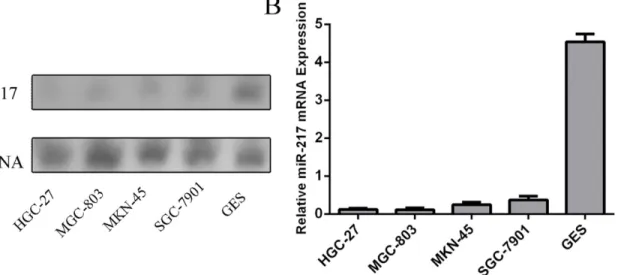
We firstly quantified the expression level of miR-217 in four human GC cell lines (SGC-7901, HGC-27, MGC-803, MKN-45) and GES-1 using northern blot (Fig 1A). The expression level of miR-217 wasdecreased in GC cell lines compared with GES-1. QuantitativeRT-PCR also showed that the expression of miR-217 was decreased in GC cell lines (SGC-7901, HGC-27, MGC-803, MKN-45) compared with GES-1, a normal gastric epithelial cell line(Fig 1B).
MiR-217 is downregulated in GC tissues
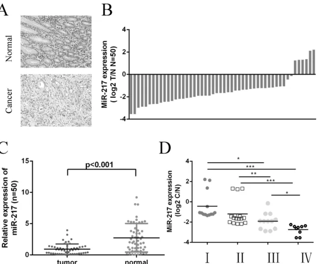
Quantitative RT-PCR was used to examine miR-217 expression in 50 GC tissues and their paired adjacent noncancerous tissues. The representative histological characteristics of GC and
its paired adjacent noncancerous tissues were shown inFig 2A. Among 50 tumor tissues, 44
Fig 1. The expression of miR-217 is downregulated in GC cell lines.(A) The expression level of miR-217 in four human GC cell lines (SGC-7901, HGC-27, MGC-803, MKN-45) and GES-1 was quantified using Northern blot.(B) The expression of miR-217 was assessing in GC cell lines (SGC-7901, HGC-HGC-27, MGC-803, MKN-45)andGES-1using Quantitative RT–PCR.
cases exhibited decreased miR-217 expression compared with the adjacent normal tissues
(88%, 44 of 50,Fig 2B). The expression of miR-217 was lower in GC tissues than in adjacent
noncancerous tissues (Fig 2C). Moreover, lower levels of miR-217 expression associated with the pTNM stage of GC patients (Fig 2D).
miR-217 inhibits GC cell proliferation and invasion
The expression of miR-217was increased in transfected HGC-27 cells using miR-217 mimics (Fig 3A) and decreased using miR-217 inhibitor (Fig 3B). The proliferation was reduced in the HGC-27 cells transfected withmiR-217 mimics compared with cells transfected with scramble or untreated (Fig 3C). Meanwhile, the proliferation was increased in HGC-27 cells transfected withmiR-217inhibitor compared with cells transfected with control or untreated (Fig 3D). The invasiveness of cells transfected with miR-217 mimics was decreased compared with the scram-ble group or control group cells and miR-217 inhibitor increased cell invasion compared with the scramble group or control group cells (Fig 3E).
Fig 2. MiR-217 is downregulated in GC tissues.(A) The tissues were histological confirmed using H&E staining. (Original magnification, ×100). (B) miR-217 was detected in 50 GC patients by real-time PCR. Data are presented as log 2 T/N of fold change of GC tissues relative to non-tumor adjacent tissues. (C) The expression of miR-217 in GC tissues compared with normal tissues. The expression of miR-217 was normalized to U6 snRNA. (D) The statistical analysis of the association between miRNA level and pTNM stage (I, II, III and IV).*p<0.05, and**p<0.01,***p<0.001. One-way ANOVA was performed for analysis.
Fig 3. miR-217 inhibits GC cell proliferation and invasion.(A) Real-time RT-PCR analysis of miR-217 in HGC-27 cells upon transfection ofmiR-217 mimic. The expression of miR-217 in HGC-27 cells transfected with 217 mimics was up-regulated.U6 snRNA was used as internal control. (B) The expression of miR-217 in HGC-27 cells transfected with miR-miR-217inhibitor was down-regulated.U6 snRNA was used as internal control. (C) Ectopic miR‑217 expression significantly inhibited cell proliferation, as demonstrated by CCK8 assay. (D) Inhibition of miR-217 expression significantly promoted cell proliferation, as demonstrated by CCK8 assay. (E) Invasion analysis of HGC-27cells after treatment withmiR-217 mimics, inhibitors or scramble or control; the relative ratio of invasive cells per field is shown below,*p<0.05,**p<0.01, and
***p<0.001.
miR-217 posttranscriptional reduces GPC5 expression by directly
targeting its 3
’
UTR
Analysis using available algorithms indicated that GPC5 was a theoretical target gene of miR-217 (Fig 4A) [24]. To prove that miR-miR-217 directly targeted the GPC53’UTR, we performed luciferase reporter gene assays. Ectopic of miR-217 reduced luciferase activity in the GPC5 wild-type reporter gene but not the mutant GPC5 3’UTR, indicating that miR-217 directly tar-geted the GPC5 3’UTR (Fig 4B). The mRNA level of GPC5 was decreased after transfection with miR-217 mimics and increased after transfection with miR-217 inhibitor using qRT-PCR (Fig 4C). Consistent with this result, the ability of miR-217 to regulate the expression of the GPC5 protein was verified by western blotting (Fig 4D).
Restoration of miR-217 inhibits GPC5-mediated GC cell proliferation
and invasion
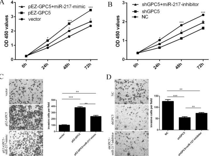
Overexpression of GPC5 promoted the GC cell proliferation and invasion. When miR-217 mimic and pEZ-GPC5 were cotransfected into HGC-27 cells, miR-217 expression reduced the
GPC5-induced GC cell proliferation and invasion (Fig5Aand5C). Inhibition of GPC5
reduced the GC cell proliferation and invasion. When miR-217 inhibitor and shGPC5 were cotransfected into HGC-27 cells, miR-217 inhibitor promoted the shGPC5-inhibited cell
pro-liferation and invasion (Fig5Band5D).
Fig 4. miR-217 posttranscriptional reduces GPC5 expression by directly targeting its 3’UTR.(A) The 3'-UTR of GPC5 mRNA contains the binding sequences of miR-217. (B) Relative luciferase activity of the indicated GPC5reporter construct in HGC-27cells is shown. Firefly luciferase values were normalized to Renilla luciferase activity and plotted as relative luciferase activity. (C) miR-217 overexpression significantly reduced the GPC5 mRNA levels in the HGC-27 cells and miR-217 inhibitor significantly enhanced the GPC5 mRNA levels in the HGC-27 cells. (D) Western blotting was performed to examine the effects of miR-217 on the expression of GPC5. GAPDH was also detected as a loading control.
GPC5 is upregulated in GC cell lines and specimens
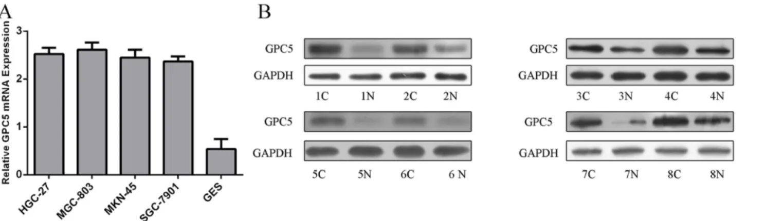
The expression of GPC5 was higher in GC cell lines (SGC-7901, HGC-27, MGC-803, MKN-45) compared with GES-1, a normal gastric epithelial cell line (Fig 6A). The protein levels of GPC5 were also higher in GC tissues than that in adjacent noncancerous tissues using western blot (Fig 6B).
Discussion
GC causes nearly one million deaths worldwide per year[25]. Recently, accumulating evidence has suggested that miRNAs play a crucial role in the pathogenesis of GC through regulating cell proliferation, apoptosis, migration, ads invasion[26–28]. In this study, miR-217 was fre-quently downregulated in human GC cell lines and tissues and the lower level of miR-217 was associated with pTNM stage of GC. Further experiments indicated that overexpression of miR-217 can suppress GC cell migration and invasion. GPC5 was identified as a direct and
Fig 5. Restoration of miR-217 inhibits GPC5-mediated GC cell proliferation and invasion.(A) The cell growth in HGC-27co-transfected with either miR-217 mimic or 2.0μg pEZ-GPC5 or pCDNA empty vector using CCK-8 proliferation assay.(B) The cell growth in HGC-27co-transfected with either miR-217 inhibitor or 2.0μg shGPC5 (knocks down GPC5) or pCDNA empty vector using CCK-8 proliferation assay.(C) The cell invasive in HGC-27co-transfected with either miR-217 mimic or 2.0μg pEZ-GPC5 or pCDNA empty vector using invasion assay.(D) The cell invasive in HGC-27co-transfected with either miR-217inhibitor or 2.0μg shGPC5 or pCDNA empty vector using invasion assay.*p<0.05,**p<0.01, and***p<0.001.
functional target of miR-217. We conclude that miR-217 appears to be a novel tumor suppres-sor in GC and that downregulated miR-217 may contribute to tumor development and pro-gression in GC patients. Downregulation of miR-217 is a frequent event in various cancers, suggesting its important role in tumorigenesis[17–19]. Li and his colleagues showed that miR-217 was down-regulated in clear cell renal cell carcinoma (ccRCC) compared to paired normal tissue[29]. Lower miR-217 expression level was associated with higher tumor grade and stage. In this study, miR-217 was frequently downregulated in GC. Intriguingly, patients with lower expression of miR-217 tended to have more advanced TNM stage, suggesting that low expres-sion ofmiR-217was associated with GC progresexpres-sion. Further studies demonstrated that overex-pression of miR-217 suppressed GC cell invasion and proliferation. Together with previous results, these data suggest that miR-217might play a crucial role in GC tissue homeostasis and deregulatedmiR-217 might contribute to the development of a stomach neoplasia.
To investigate the molecular mechanism of the tumor suppressor role of miR-217 in GC, we used luciferase reporter assay and western blot to confirm that GPC5 was a target of miR-217
in GC cells. To confirm the direct regulation of GPC5 by miR-217, we used GPC 3’UTR
reporter vector bearing the potential miR-217 binding site in the fluorescent reporter. Further-more, qRT-PCR and western blot assay showed that overexpression of miR-217 inhibited GPC expression. Glypicans are a family of proteoglycans that are linked to the exocytoplasmic
sur-face of the plasma membrane via a glycosyl phosphatidylinositol anchor[30,31]. Six glypicans
have been identified in mammals (GPC1 to GPC6)[32,33]. GPC5 is mainly expressed in
devel-oping central nervous system, limbs, kidney, lung and liver[34,35]. Recent studies have
indi-cated that some GPCs, especially GPC3 andGPC5, might play an important role in regulating
cancer progression[35,36]. For example,Zhanget al. showed that GPC5 was highly expressed
in SACC-M(high lung-metastatic cell line) and in clinical samples of salivary adenoid cystic carcinoma (SACC) cases with lung metastasis[36]. The overall expression level of GPC5 in clinical cases of SACC with lung metastasis was higher. Williamson et al. showed that the gene encoding GPC5was amplified in 20% of patients with alveolar rhabdomyosarcoma (RMS) and that this glypican was overexpressed in RMS patients. Moreover, down-regulation of GPC5 expression by siRNA inhibited the proliferation rate of RMS cells. Another study found that high levels of GPC5 expression predicted poor postsurgical survival times for curatively respected NSCLC patients, suggesting the value of GPC5 as a molecular prognostic indicator [35,37]. In our study, the expression of GPC5 was higher in GC cell lines and the protein levels Fig 6. GPC5 is upregulated in GC cell lines and specimens.(A) The mRNA expression of CPG5 was assessing in GC cell lines (SGC-7901, HGC-27, MGC-803, MKN-45) and GES-1 using Quantitative RT–PCR. (B) The protein level of CPG5 in eight human GC tissues and its adjacent normal controls was quantified using western blot.
of GPC5 were also higher in GC tissues than in adjacent noncancerous tissues. Inhibition of GPC5 reduced the GC cell proliferation and invasion. Restoration of miR-217 inhibited GPC5-mediated GC cell proliferation and invasion. These results demonstrated that miR-217might act as a tumor suppressor in gastric cancer by targeting GPC5.
In conclusion, the present study demonstrated that miR‑217was downregulated in GC tis-sues and cell lines. Low levels of miR-217 expression were associated with pTNM stage of patients. Ectopic miR‑217 expression resulted in inhibition of GC cell proliferation and inva-sion. Further investigation revealed that GPC5 was a potential target of miR‑217. miR‑217 may serve as a predictor for prognosis and a therapeutic target for GC patients.
Supporting Information
S1 Table. Summary of clinicopathological parameters of patients with gastric cancer. (DOCX)
S2 Table. Primer sequence. (DOCX)
Author Contributions
Conceived and designed the experiments: HW XD XG RQ HJ JG. Performed the experiments: HW XD XG RQ HJ JG. Analyzed the data: HW XD XG RQ HJ JG. Contributed reagents/mate-rials/analysis tools: HW XD XG RQ HJ JG. Wrote the paper: HW XD XG RQ HJ JG.
References
1. Zhang WH, Gui JH, Wang CZ, Chang Q, Xu SP, Cai CH, et al. The identification of miR-375 as a poten-tial biomarker in distal gastric adenocarcinoma. Oncology research. 2012; 20(4):139–47. PMID: 23461060
2. Fei B, Wu H. MiR-378 Inhibits Progression of Human Gastric Cancer MGC-803 Cells by Targeting MAPK1 In Vitro. Oncology research. 2013; 20(12):557–64.
3. Li Z, Lei H, Luo M, Wang Y, Dong L, Ma Y, et al. DNA methylation downregulated mir-10b acts as a tumor suppressor in gastric cancer. Gastric cancer: official journal of the International Gastric Cancer Association and the Japanese Gastric Cancer Association. 2014.
4. Yang L. Incidence and mortality of gastric cancer in China. World journal of gastroenterology: WJG. 2006; 12(1):17–20. PMID:16440411
5. Katada T, Ishiguro H, Kuwabara Y, Kimura M, Mitui A, Mori Y, et al. microRNA expression profile in undifferentiated gastric cancer. International journal of oncology. 2009; 34(2):537–42. PMID:19148490
6. Thiel A, Ristimaki A. Gastric cancer: basic aspects. Helicobacter. 2012; 17 Suppl 1:26–9. doi:10.1111/ j.1523-5378.2012.00979.xPMID:22958152
7. Yasui W, Sentani K, Sakamoto N, Anami K, Naito Y, Oue N. Molecular pathology of gastric cancer: research and practice. Pathology, research and practice. 2011; 207(10):608–12. doi:10.1016/j.prp. 2011.09.006PMID:22005013
8. Yu X, Li Z, Shen J, Wu WK, Liang J, Weng X, et al. MicroRNA-10b Promotes Nucleus Pulposus Cell Proliferation through RhoC-Akt Pathway by Targeting HOXD10 in Intervetebral Disc Degeneration. PloS one. 2013; 8(12):e83080. doi:10.1371/journal.pone.0083080PMID:24376640
9. Han G, Wang Y, Bi W. C-Myc overexpression promotes osteosarcoma cell invasion via activation of MEK-ERK pathway. Oncology research. 2012; 20(4):149–56. PMID:23461061
10. Lee H, Jee Y, Hong K, Hwang GS, Chun KH. MicroRNA-494, upregulated by tumor necrosis factor-alpha, desensitizes insulin effect in C2C12 muscle cells. PloS one. 2013; 8(12):e83471. doi:10.1371/ journal.pone.0083471PMID:24349514
12. Xiao X, Tang C, Xiao S, Fu C, Yu P. Enhancement of proliferation and invasion by MicroRNA-590-5p via targeting PBRM1 in clear cell renal carcinoma cells. Oncology research. 2013; 20(11):537–44. doi: 10.3727/096504013X13775486749335PMID:24063284
13. Wang Z, Yin B, Wang B, Ma Z, Liu W, Lv G. MicroRNA-210 promotes proliferation and invasion of peripheral nerve sheath tumor cells targeting EFNA3. Oncology research. 2014; 21(3):145–54.
14. Zhu ED, Li N, Li BS, Li W, Zhang WJ, Mao XH, et al. miR-30b, Down-Regulated in Gastric Cancer, Pro-motes Apoptosis and Suppresses Tumor Growth by Targeting Plasminogen Activator Inhibitor-1. PloS one. 2014; 9(8):e106049. doi:10.1371/journal.pone.0106049PMID:25170877
15. Yamashita S, Yamamoto H, Mimori K, Nishida N, Takahashi H, Haraguchi N, et al. MicroRNA-372 is associated with poor prognosis in colorectal cancer. Oncology. 2012; 82(4):205–12. doi:10.1159/ 000336809PMID:22456107
16. Ye Z, Jingzhong L, Yangbo L, Lei C, Jiandong Y. Propofol inhibits proliferation and invasion of osteo-sarcoma cells by regulation of microRNA-143 expression. Oncology research. 2014; 21(4):201–7.
17. Williams KJ, Telfer BA, Shannon AM, Babur M, Stratford IJ, Wedge SR. Inhibition of vascular endothe-lial growth factor signalling using cediranib (RECENTIN; AZD2171) enhances radiation response and causes substantial physiological changes in lung tumour xenografts. Br J Radiol. 2008; 81 Spec No 1: S21–7. doi:10.1259/bjr/59853976PMID:18819995
18. Yin H, Hu M, Zhang R, Shen Z, Flatow L, You M. MicroRNA-217 promotes ethanol-induced fat accumu-lation in hepatocytes by down-regulating SIRT1. The Journal of biological chemistry. 2012; 287(13): 9817–26. doi:10.1074/jbc.M111.333534PMID:22308024
19. Zhao WG, Yu SN, Lu ZH, Ma YH, Gu YM, Chen J. The miR-217 microRNA functions as a potential tumor suppressor in pancreatic ductal adenocarcinoma by targeting KRAS. Carcinogenesis. 2010; 31(10):1726–33. doi:10.1093/carcin/bgq160PMID:20675343
20. Song L, Lin C, Wu Z, Gong H, Zeng Y, Wu J, et al. miR-18a impairs DNA damage response through downregulation of ataxia telangiectasia mutated (ATM) kinase. PloS one. 2011; 6(9):e25454. doi:10. 1371/journal.pone.0025454PMID:21980462
21. Jiang L, Mao P, Song L, Wu J, Huang J, Lin C, et al. miR-182 as a prognostic marker for glioma progres-sion and patient survival. The American journal of pathology. 2010; 177(1):29–38. doi:10.2353/ajpath. 2010.090812PMID:20472885
22. Li Z, Yu X, Liang J, Wu WK, Yu J, Shen J. Leptin downregulates aggrecan through the p38-ADAMST pathway in human nucleus pulposus cells. PloS one. 2014; 9(10):e109595. doi:10.1371/journal.pone. 0109595PMID:25299465
23. Yang J, Zhang HM, Liu XY, Li J, Lv MF, Li PP, et al. Identification of 23 novel conserved microRNAs in three rice cultivars. Gene. 2014; 548(2):285–93. doi:10.1016/j.gene.2014.07.048PMID:25038275
24. Shen J, Niu W, Zhou M, Zhang H, Ma J, Wang L. MicroRNA-410 suppresses migration and invasion by targeting MDM2 in gastric cancer. PloS one. 2014; 9(8):e104510. doi:10.1371/journal.pone.0104510 PMID:25136862
25. Zhang X, Nie Y, Du Y, Cao J, Shen B, Li Y. MicroRNA-181a promotes gastric cancer by negatively reg-ulating tumor suppressor KLF6. Tumour biology: the journal of the International Society for Oncodeve-lopmental Biology and Medicine. 2012; 33(5):1589–97. doi:10.1007/s13277-012-0414-3PMID: 22581522
26. Zhang H, Cai X, Wang Y, Tang H, Tong D, Ji F. microRNA-143, down-regulated in osteosarcoma, pro-motes apoptosis and suppresses tumorigenicity by targeting Bcl-2. Oncology reports. 2010; 24(5): 1363–9. PMID:20878132
27. Yan K, Gao J, Yang T, Ma Q, Qiu X, Fan Q, et al. MicroRNA-34a inhibits the proliferation and metasta-sis of osteosarcoma cells both in vitro and in vivo. PloS one. 2012; 7(3):e33778. doi:10.1371/journal. pone.0033778PMID:22457788
28. Zhao G, Cai C, Yang T, Qiu X, Liao B, Li W, et al. MicroRNA-221 induces cell survival and cisplatin resistance through PI3K/Akt pathway in human osteosarcoma. PloS one. 2013; 8(1):e53906. doi:10. 1371/journal.pone.0053906PMID:23372675
29. Li H, Zhao J, Zhang JW, Huang QY, Huang JZ, Chi LS, et al. MicroRNA-217, down-regulated in clear cell renal cell carcinoma and associated with lower survival, suppresses cell proliferation and migration. Neoplasma. 2013; 60(5):511–5. doi:10.4149/neo_2013_066PMID:23790169
30. Zhang C, Zhang S, Zhang D, Zhang Z, Xu Y, Liu S. A lung cancer gene GPC5 could also be crucial in breast cancer. Molecular genetics and metabolism. 2011; 103(1):104–5. doi:10.1016/j.ymgme.2011. 02.005PMID:21396872
32. Li Y, Yang P. GPC5 gene and its related pathways in lung cancer. Journal of thoracic oncology: official publication of the International Association for the Study of Lung Cancer. 2011; 6(1):2–5.
33. Yu W, Inoue J, Imoto I, Matsuo Y, Karpas A, Inazawa J. GPC5 is a possible target for the 13q31-q32 amplification detected in lymphoma cell lines. Journal of human genetics. 2003; 48(6):331–5. PMID: 12721791
34. Joslyn G, Wolf FW, Brush G, Wu L, Schuckit M, White RL. Glypican Gene GPC5 Participates in the Behavioral Response to Ethanol: Evidence from Humans, Mice, and Fruit Flies. G3 (Bethesda). 2011; 1(7):627–35.
35. Yang X, Zhang Z, Qiu M, Hu J, Fan X, Wang J, et al. Glypican-5 is a novel metastasis suppressor gene in non-small cell lung cancer. Cancer letters. 2013; 341(2):265–73. doi:10.1016/j.canlet.2013.08.020 PMID:23962560
36. Zhang Y, Wang J, Dong F, Li H, Hou Y. The role of GPC5 in lung metastasis of salivary adenoid cystic carcinoma. Archives of oral biology. 2014; 59(11):1172–82. doi:10.1016/j.archoralbio.2014.07.009 PMID:25093697