Volume 2013, Article ID 721383,9pages http://dx.doi.org/10.1155/2013/721383
Research Article
BCG and BCG/DNAhsp65 Vaccinations Promote Protective
Effects without Deleterious Consequences for Experimental
Autoimmune Encephalomyelitis
Sofia Fernanda Gonçalves Zorzella-Pezavento,
1Clara Pires Fujiara Guerino,
1Fernanda Chiuso-Minicucci,
1Thais Graziela Donegá França,
1Larissa Lumi Watanabe Ishikawa,
1Ana Paula Masson,
2Célio Lopes Silva,
2and Alexandrina Sartori
11Departamento de Microbiologia e Imunologia, Instituto de Biociˆencias, Universidade Estadual Paulista (UNESP),
18618-970 Botucatu, SP, Brazil
2Departamento de Bioqu´ımica e Imunologia, Universidade de S˜ao Paulo (USP), 14049-900 Ribeir˜ao Preto, SP, Brazil
Correspondence should be addressed to Alexandrina Sartori; [email protected]
Received 22 April 2013; Accepted 16 September 2013
Academic Editor: Carlos Barcia
Copyright © 2013 Soia Fernanda Gonc¸alves Zorzella-Pezavento et al. his is an open access article distributed under the Creative Commons Attribution License, which permits unrestricted use, distribution, and reproduction in any medium, provided the original work is properly cited.
A prime-boost strategy conserving BCG is considered the most promising vaccine to control tuberculosis. A boost with a DNA vaccine containing the mycobacterial gene of a heat shock protein (pVAXhsp65) ater BCG priming protected mice against experimental tuberculosis. However, anti-hsp65 immunity could worsen an autoimmune disease due to molecular mimicry. In this investigation, we evaluated the efect of a previous BCG or BCG/pVAXhsp65 immunization on experimental autoimmune encephalomyelitis (EAE) development. Female Lewis rats were immunized with BCG or BCG followed by pVAXhsp65 boosters. he animals underwent EAE induction and were daily evaluated for weight loss and clinical score. hey were euthanized during recovery phase to assess immune response and inlammatory iniltration at the central nervous system. Previous immunization did not aggravate or accelerate clinical score or weight loss. In addition, this procedure clearly decreased inlammation in the brain.
BCG immunization modulated the host immune response by triggering a signiicant reduction in IL-10 and IFN-�levels induced
by myelin basic protein. hese data indicated that vaccination protocols with BCG or BCG followed by boosters with pVAXhsp65 did not trigger a deleterious efect on EAE evolution.
1. Introduction
Tuberculosis (TB) is an infection caused byMycobacterium tuberculosisand this disease remains one of the most impor-tant causes of death worldwide [1,2]. Factors as coinfection with human immunodeiciency virus and emergence of drug resistance inM. tuberculosisstrains have hampered TB control [3,4].
he only available vaccine against TB is the attenuatedM. bovisBacillus Calmette-Gu´erin (BCG) that is recommended by the World Health Organization for all infants under 1 year of age. Around 100 million newborn children receive this vaccine and the global vaccine coverage is estimated to be
80% [5, 6]. In spite of this extensive use, numerous well-documented trials showed signiicant variation, from 0 to 80%, in BCG protective eicacy [7]. his has been attributed to variability in BCG vaccine strains and environmental fac-tors as well as host genetic background [8,9].
ability to induce a strong h1 type of response, could con-tribute to TB control. DNA constructs encoding mycobacte-rial antigens as 65 kDa heat shock protein (hsp65), Ag85A, Ag85B, and PstS3 induced signiicant protective immunity [15–17]. We previously demonstrated that a DNA plasmid encoding theMycobacterium leprae65 kDa heat shock pro-tein exhibited prophylactic [18] and therapeutic activity in a TB murine model [19, 20]. In spite of these successful results with homologous vaccination protocols, heterologous prime-boost regimens, capable to increase BCG or rBCG eiciency, are considered more promising for future TB control [11]. In this context, we observed that pVAXhsp65 and BCG similarly primed neonate mice for a strong immune response to pVAXhsp65 boosters administered later, at the adult stage [21]. Prime-boost strategies combining these two vaccines were also able to protect mice and guinea pigs against experimental TB [22,23].
One of the arguments against the potential use of pVAXhsp65 alone or combined with BCG is the fact that hsp65 fromM. leprae, whose gene is inserted in this DNA vaccine, presents a high degree of homology with its equiv-alent mammalian protein [24]. heoretically, an anti-hsp65 immunity started with BCG and boosted by pVAXhsp65 could provoke or worsen an autoimmune disease. In support of this argument, many studies revealed immune response against bacterial hsp65 in diabetes [25], atherosclerosis [26], arthritis [27], and multiple sclerosis [28]. In addition, CpG motifs that are frequently present in bacterial plasmid vectors could trigger or exacerbate an autoimmune response [29,30]. Even though BCG has been described as a safe vaccine, a few publications suggested its implication as a possible trigger of autoimmunity [31,32]. In this context, the present study was designed to investigate if a vaccination protocol against TB, using BCG alone or a priming with BCG followed by boosters with pVAXhsp65, could aggravate or accelerate experimental autoimmune encephalomyelitis.
2. Material and Methods
2.1. Experimental Design. Female Lewis rats were immunized with BCG or with BCG plus pVAXhsp65. he animals underwent EAE induction by immunization with myelin basic protein (MBP). he efect of BCG or BCG/pVAXhsp65 on EAE was evaluated by clinical follow-up (weight variation and clinical score), histopathological analysis of the brain and lumbar spinal cord, and also by cytokine production. Nonimmunized and pVAX (empty vector) injected animals were included as control groups.
2.2. Animals. Female Lewis rats (4–6 weeks old) were pur-chased from CEMIB (UNICAMP, S˜ao Paulo, SP, Brazil). he animals were fed with sterilized food and waterad libitumand were manipulated in accordance with the ethical guidelines adopted by the Brazilian College of Animal Experimentation. All experimental protocols were approved by the local Ethics Committee (Ethics Committee for Animal Experimentation, Medical School, Universidade Estadual Paulista).
2.3. Genetic Vaccine Construction and Puriication. he vac-cine pVAXhsp65 was derived from the pVAX vector (Invit-rogen, Carlsbad, CA, USA), previously digested with BamHI and NotI (Gibco BRL, Gaithersburg, MD, USA) by inserting a 3.3 kb fragment corresponding to theM. lepraehsp65 gene and the CMV intron A. he empty pVAX vector was used as a control. DH5�E. colicells transformed with plasmid pVAX or the plasmid carrying the hsp65 gene (pVAXhsp65) were cultured in LB liquid medium (Gibco BRL, Gaithersburg, MD, USA) containing kanamycin (100�g/mL). he plasmids were puriied using the Concert High Purity Maxiprep System (Gibco BRL, Gaithersburg, MD, USA). Plasmid con-centrations were determined by spectrophotometry at �= 260 and 280 nm by using the Gene Quant II apparatus (Phar-macia Biotech, Buckinghamshire, UK).
2.4. Immunization with BCG and pVAXhsp65. Lewis rats were immunized with BCG or with BCG followed by pVAXhsp65 boosters. he M. bovis BCG Moreau-Rio de Janeiro (2 to 10 × 105UFC) was inoculated a single time by subcutaneous route at the base of the tail. pVAXhsp65 was injected twice (300�g each) by intramuscular route (quadriceps muscle), being the irst dose administered 15 days ater BCG and the second one 15 days later. Control groups received the same volume of saline or the same concentration of pVAX (empty vector).
2.5. EAE Induction and Evaluation. EAE was induced as previously described [33]. Briely, 15 days ater the last DNA immunization, EAE was induced by inoculation of 25�g of MBP (Sigma Aldrich, St. Louis, MO, USA) emulsiied with complete Freund’s adjuvant (CFA) containing 5 mg/mL of
Mycobacterium butyricum, in the hind let footpad. Animals were daily evaluated for weight loss and clinical score. Signs of disease were graded as 0 (zero): no disease; 1: loss of tonicity in the distal portion of the tail; 2: total loss of tail tonicity; 3: hind limb weakness (partial paralysis); 4: complete hind limb paralysis and urinary incontinence; and 5: moribund.
10
5 0 −5 −10
−20 Control Control BCG BCG pVAX pVAXhsp65
EAE
W
eig
h
t va
ri
at
io
n (%)
−15
BCG
(a)
4 3
2 1
0
1 4 5 6 7 8 9 10 11 12 13 14 15 16 17 18 19 20
Clinical s
co
re
Days after EAE induction
Control EAE BCG/EAE
BCG pVAX/EAE BCG pVAXhsp65/EAE
(b)
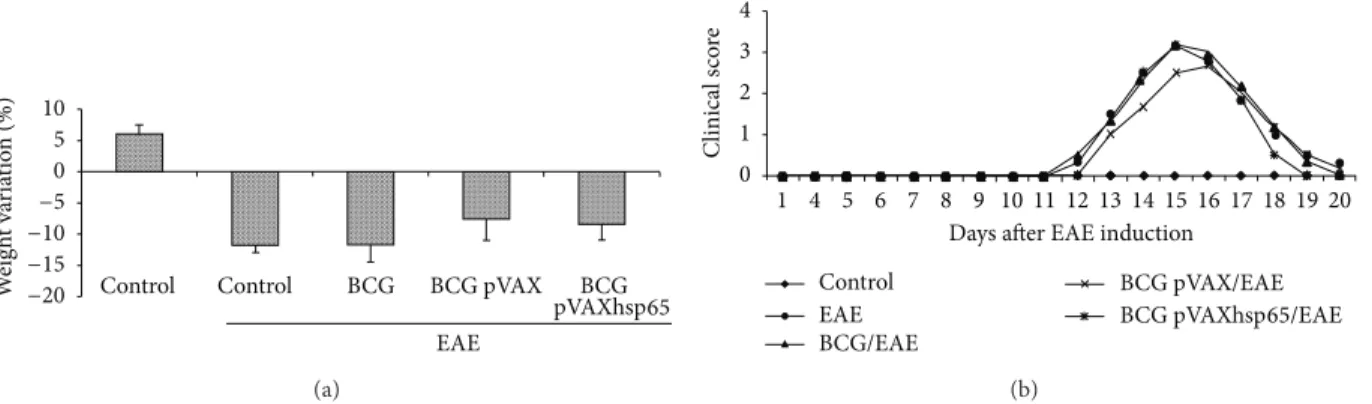
Figure 1: Efect of previous immunization with BCG and BCG/pVAXhsp65 in clinical EAE development. Female Lewis rats were immunized with BCG alone or with BCG followed by pVAXhsp65 boosters and then underwent EAE induction by inoculation of MBP emulsiied with
CFA. Animals were daily evaluated for weight variation (a) and clinical score (b). Data are presented by mean±SEM for 4–6 rats.
described elsewhere [34]. he values were expressed as�m2 of cellular iniltrate per mm2of organ section (�m2/mm2).
2.7. Cell Culture Conditions and IFN-�and IL-10 Production.
Control and immunized rats (BCG or BCG/DNA) were euthanized eight weeks ater initial BCG immunization. Lymph node (popliteal + inguinal) and spleen cells were collected and adjusted to 2.5 × 10⁶/mL and 5 × 10⁶/mL, respectively. Cells were cultured in complete RPMI medium (RPMI supplemented with 5% FCS, 20 mM glutamine, and 40 IU/mL of gentamicin), in the presence of 10�g/mL of rhsp65, 10�g/mL of MBP, or 5�g/mL of Concanavalin A (Sigma Aldrich). IFN-�levels were assayed in lymph node cell cultures whereas IL-10 production was evaluated in spleen cell cultures. Cytokine levels in culture supernatants were evaluated 48 h later by ELISA according to manufacturer’s instructions (R&D Systems). Briely, ninety-six well plates (NUNC) were coated with capture antibodies for IFN-� (DY 585) or IL-10 (DY 522) diluted in PBS at 2�g/mL and 4�g/mL, respectively. Plates were incubated overnight and then blocked during 2 h with 1% albumin in PBS. Standard rat cytokines and culture supernatants were added and the plates were incubated during 2 h. Biotinylated anti-IFN-� and anti-IL-10 were added (150 and 100 ng/mL, resp.) and plates were incubated for additional 2 h at room temperature. Ater incubation at room temperature for 30 minutes with streptavidin, the plates were revealed by adding H2O2+ OPD (Sigma Aldrich, St. Louis, MO, USA). Color development was stopped with H2SO4 and optical density was measured at 492 nm.
2.8. Statistical Analysis. Statistical analysis was performed using SigmaStat statistical sotware (Jandel Corporation, San Rafael, CA, USA). Cytokine data was expressed as mean± standard error of the mean (SEM) and tested for statistical signiicance by Kruskal-Wallis nonparametric test or one-way ANOVA followed by Tukey’s test. Morphometric analysis of the brain and lumbar spinal cord was tested by one-way ANOVA followed by Holm-Sidak method. A�value of less than 0.05 was considered statistically signiicant.
3. Results
3.1. EAE Evolution is Not Aggravated by Previous Immu-nization with BCG or BCG/pVAXhsp65. EAE development caused, as expected, a weight loss that varied from 12 to 15% of the original weight. Previous vaccination with BCG alone, associated with pVAXhsp65 or with the empty vector, did not afect weight loss (Figure 1(a)). In the control EAE group, that is, in the sick group that was not previously immunized against TB, clinical symptoms appeared 11 or 12 days ater MBP inoculation and clinical scores reached 2.5 to 3.0 (Figure 1(b)). A very similar clinical evolution was observed in the groups that were previously immunized with BCG, BCG/pVAXhsp65, or BCG/pVAX.
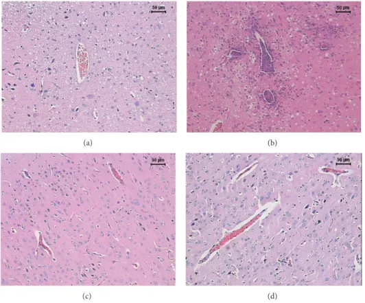
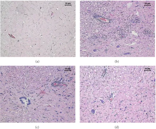
3.2. Both Vaccination Strategies Decreased Inlammation in the Brain. he severity of the inlammatory reaction in the CNS from the EAE group (positive control) clearly correlated with the observed clinical symptoms. Sections from the brain and lumbar spinal cord showed typical inlammatory foci, dominated by mononuclear cells that were localized around small vessels, as can be observed in Figures 2(b) and3(b), respectively. As expected, control animals without EAE did not present any inlammatory foci in the brain (Figure 2(a)) and lumbar spinal cord (Figure 3(a)). However, a previous vaccination with BCG alone (Figure 2(c)) or combined with pVAXhsp65 (Figure 2(d)) or pVAX, the empty vector (not shown), clearly decreased inlammation at the brain in comparison to the EAE group (positive control). hese results were further ascertained by a morphometric analysis, which indicated the presence of signiicantly lower inlammatory iniltrates in animals immunized with BCG alone or combined with pVAXhsp65, in comparison to the EAE group (positive control) (Table 1). he previous immunization with BCG, associated or not with pVAXhsp65, did not afect the intensity of inlammation in the lumbar spinal cord as demonstrated in Figures3(c)and3(d)and in Table 1.
(a) (b)
(c) (d)
Figure 2: Efect of tuberculosis vaccines in brain inlammation associated with EAE. Female Lewis rats were immunized with BCG alone or with BCG followed by pVAXhsp65 boosters and then underwent EAE induction by inoculation of MBP emulsiied with CFA. Animals were euthanized during the recovery phase (20th day ater MBP injection) and the brains were removed and further stained by haematoxylin and eosin. Normal control (a), EAE control (b), rats immunized with BCG before EAE (c), and rats immunized with BCG/pVAXhsp65 before EAE induction (d).
Table 1: Morphometric analysis of perivascular inlammatory inil-trate in the brain and lumbar spinal cord samples from rats immu-nized with BCG alone or associated with pVAXhsp65 before EAE induction.
Brain
� = 4
Lumbar spinal cord
� = 4
(�m2of mononuclear iniltrate/mm2of organ section)†
Control
EAE 3.55 ± 0.72 6.56 ± 2.72
BCG EAE 1.17 ± 0.38∗ 7.38 ± 2.53
BCG pVAXhsp65 EAE
1.50 ± 0.71∗ 4.46 ± 0.79
∗� < 0.05versus control EAE.
†Morphometric analysis was done in 5-micron thick sections ater
haema-toxylin and eosin stain using a Nikon Microphot-FXA optical microscope connected to a computer and employing the KS300 sotware.
immunized group. In all experimental groups with EAE, independently of a preceding immunization, IFN-� levels were very low, that is, similar to the normal control group (Figure 4(a)). As can be observed inFigure 4(d), IL-10 levels in rhsp65 activated cultures presented a very distinct pattern: the highest levels were found in EAE and BCG/pVAXhsp65
EAE groups. BCG group presented detectable but very low IL-10 levels.
he production of these two cytokines, in cultures stim-ulated with MBP, followed the same pattern (Figures 4(b) and4(e)). In this case, the highest levels of both cytokines were found in the EAE group. Previously immunized groups, that were later submitted to encephalomyelitis induction presented lower IFN-�(Figure 4(b)) and IL-10 (Figure 4(e)) levels in comparison to the control EAE group. IFN-�and IL-10 were not detected in the BCG group.
IFN-�levels were similarly elevated in all groups stim-ulated with ConA (Figure 4(c)). On the other hand, even though there was some IL-10 production by cultures from the BCG group, the levels of this cytokine were signiicantly high in all groups with encephalomyelitis, independently of a previous immunization (Figure 4(f)).
4. Discussion
(a) (b)
(c) (d)
Figure 3: Efect of tuberculosis vaccines in spinal cord inlammation associated with EAE. Female Lewis rats were immunized with BCG alone or with BCG followed by pVAXhsp65 boosters and then underwent EAE induction by inoculation of MBP emulsiied with CFA. Animals were euthanized during the recovery phase (20th day ater MBP injection) and the lumbar spinal cord was removed and further stained by haematoxylin and eosin. Normal control (a), EAE control (b), rats immunized with BCG before EAE (c), and rats immunized with BCG/pVAXhsp65 before EAE induction (d).
oligo and polyarticular arthritis were detected in approx-imately 3% of patients with bladder carcinoma that were treated with intravesicular BCG [35]. It is also being described that mycobacteria precipitates an SLE-like syndrome in NOD mice [36].
Otherwise, the maintenance of BCG in a new prophylaxis against TB is highly expected because this vaccine is widely accepted in the developing countries, it has protective efect against the most severe forms of TB in children and it is also endowed with immunomodulatory properties [37–39]. In this scenario, safety aspects need to be experimentally validated.
In the last years, our group analyzed a DNA vaccine containing the hsp65 gene from M. leprae (pVAXhsp65). he results showed that this vaccine, by itself or associated with BCG, is able to confer protection in diferent TB experimental models [22]. his genetic construction includes the mycobacterial hsp65 gene that presents a high homology degree with the corresponding hsp60 mammalian gene. A speciic immune response against this bacterial protein could cross-react with the corresponding mammalian protein and, therefore, trigger an autoimmune pathology.
In this context, the most relevant contribution of this work was the demonstration that the previous contact with BCG or BCG followed by pVAXhsp65 boosters did not dele-teriously afect the clinical EAE development in Lewis rats.
his was initially demonstrated by the very similar clinical evolution of the disease in vaccinated and nonvaccinated animals; they equally lost weight, the average clinical score was the same, and acute and remission phases occurred at comparable time periods. In addition of this absence of a detrimental efect, the previous immunization with BCG or BCG/pVAXhsp65 also determined a clear anti-inlammatory reaction in the brain. his anti-inlammatory activity did not reach the spinal cord; this could explain the absence of a beneicial clinical efect over EAE clinical score. he mechanism of this diferential protective efect over the brain and the spinal cord was not investigated. However, considering that interaction of circulating leukocytes with the endothelium of blood-brain and blood-spinal cord barriers is fundamental in inlammatory pathologies of the CNS and that they difer in many aspects, we could hypothesize that diferences in adhesion molecules, chemokines, or their receptors could explain this inding [40].
600 500 400 300 200 100 0 MBP ∗ IFN-𝛾 (pg/mL) 200 180 160 140 120 100 80 60 40 20 0 ∗
Control BCG Control BCG BCG
pVAXhsp65 EAE rh sp65 BCG pVAX IFN-𝛾 (pg/mL) Spleen ∗
Control BCG Control BCG BCG
pVAX BCG pVAXhsp65 EAE
Control BCG Control BCG BCG
pVAX BCG pVAXhsp65 EAE 12000 10000 8000 6000 4000 2000 0 Co n A IFN-𝛾 (pg/mL)
Control BCG Control BCG BCG
pVAXhsp65
EAE BCG pVAX
Control Control BCG BCG
pVAXhsp65 EAE
BCG pVAX
Control Control BCG BCG
pVAXhsp65 EAE BCG pVAX BCG BCG Lymph node IL -10 (pg/mL) 100 90 80 70 60 50 40 30 20 10 0 180 160 140 120 100 80 60 40 20 0 6000 5000 4000 3000 2000 1000 0 IL -10 (pg/mL) IL -10 (pg/mL) MBP Co n A ∗ ∗ ∗ ∗ ∗ (a) (d) (b) (e) (c) (f) rh sp65
Figure 4: Efect of tuberculosis vaccines in IFN-�and IL-10 production. Female Lewis rats were immunized with BCG alone or with BCG
followed by pVAXhsp65 boosters and then underwent EAE induction by inoculation of MBP emulsiied with CFA. Animals were euthanized during the recovery phase (20th day ater MBP inoculation) and spleen and lymph node cell cultures were stimulated with rhsp65 ((a) and
(d)); MBP ((b) and (e)); and ConA ((c) and (f)). IFN-�levels were evaluated in spleen cell cultures ((a), (b), and (c)) and IL-10 levels were
evaluated in lymph node cell cultures ((d), (e), and (f)). Data are presented by mean±SEM for 4–6 animals.∗Represents the diference
between immunized and control groups.� < 0.05.
previous immunization with live BCG clearly reduced clinical severity in murine EAE.
he protective ability of hsp65 is even more widely accepted and investigated. Heat shock proteins, especially hsp65, are understood as targets for regulatory T cells due to their enhanced expression in inlamed tissues. here are also very strong evidence that they are able to induce anti-inlammatory T cell responses [45,46]. Our previous experi-ence indicates that DNAhsp65 had a similar protective efect
over arthritis, diabetes, and encephalomyelitis. However, this protective efect was clearly more accentuated in arthritis and diabetes. In these two conditions, its immunomodulatory efect was strong enough to determine clinical improvement [47, 48]. In the case of EAE, DNAhsp65 determined less inlammation but no improvement was detected in clinical scores [49].
in the prime-boost, in comparison to the BCG immunized group. However, these two groups presented similar clinical evolution. his inding is in contrast to our recent experience with a similar prime-boost in NOD mice. In this model, priming with BCG followed by two boosters with pVAXhsp65 prevented pancreas inlammation and clinical diabetes devel-opment [50].
he analysis of IFN-� and IL-10 levels, produced by peripheral lymphoid organs, answered some mechanistic questions. We initially asked how BCG decreased inlamma-tion in the brain. In this sense, the most interesting inding was the accentuated drop of IFN-�and IL-10 levels induced by MBP, in EAE experimental groups previously vaccinated, in comparison to the EAE control group. A possible expla-nation for this inding could be the trapping of nervous tissue-speciic T cells in peripheral BCG inlammatory sites as elegantly demonstrated by Sewell et al. in 2003 [44]. Alter-natively, these autoreactive T cells could be in lower numbers due to an apoptotic process occurring in the periphery. his phenomenon was clearly demonstrated by O’Connor et al. in 2005 [51]. hese authors detected high levels of apop-tosis among activated CD4+ T cells in BCG experimental infection. Interestingly and concerning to the model used by us, these authors also described that the high apoptotic degree occurred simultaneously with a milder experimental encephalomyelitis course. Even though we did not observed improvement in clinical parameters, a signiicant drop in brain inlammation was detected. his lower IFN-� and IL-10 production could also be linked to the migration of myelin-speciic T clones to the brain, where they could exert the detected anti-inlammatory activity. IFN-� is described as able to shape the immune iniltration of the CNS by controlling chemokine expression. In addition, this cytokine accentuates apoptosis of iniltrating encephalitogenic T cell clones [52,53]. Additionally, the production of IL-10 by Tr1 regulatory cells has been widely accepted as one of the mech-anisms responsible for MS and EAE downregulation [54].
5. Conclusion
hese results indicate that immunization procedures with BCG or BCG/pVAXhsp65, as used in this investigation, did not deleteriously afect EAE development.
Acknowledgments
he authors are grateful to S˜ao Paulo Research Foundation (FAPESP) Grant no. 2007/05353-8 and Conselho Nacional de Desenvolvimento Cient´ıico e Tecnol´ogico (CNPq) Grant no. 473351/2004-8 that supported this study.
References
[1] D. B. Young and K. Duncan, “Prospects for new interventions in
the treatment and prevention of mycobacterial disease,”Annual
Review of Microbiology, vol. 49, pp. 641–673, 1995.
[2] J. L. Flynn and J. Chan, “Tuberculosis: latency and reactivation,”
Infection and Immunity, vol. 69, no. 7, pp. 4195–4201, 2001.
[3] K. L. Dierberg and R. E. Chaisson, “Human immunodeiciency virus-associated tuberculosis: update on prevention and
treat-ment,”Clinics in Chest Medicine, vol. 34, no. 2, pp. 217–228, 2013.
[4] E. Pontali, A. Matteelli, and G. B. Migliori, “Drug-resistant
tuberculosis,”Current Opinion in Pulmary Medicine, vol. 19, no.
3, pp. 266–272, 2013.
[5] P. Andersen and T. M. Doherty, “he success and failure of
BCG—implications for a novel tuberculosis vaccine,” Nature
Reviews Microbiology, vol. 3, no. 8, pp. 656–662, 2005. [6] Centers for Disease Control and Prevention, “Global routine
vaccination coverage, 2011,”MMWR Morbidity and Mortality
Weekly Report, vol. 61, no. 43, pp. 883–885, 2012.
[7] G. A. Colditz, T. F. Brewer, C. S. Berkey et al., “Eicacy of BCG vaccine in the prevention of tuberculosis: meta-analysis
of the published literature,”Journal of the American Medical
Association, vol. 271, no. 9, pp. 698–702, 1994.
[8] V. H. Springett and I. Sutherland, “A re-examination of the vari-ations in the eicacy of BCG vaccination against tuberculosis in
clinical trials,”Tubercle and Lung Disease, vol. 75, no. 3, pp. 227–
133, 1994.
[9] M. A. Behr, M. A. Wilson, W. P. Gill et al., “Comparative genomics of BCG vaccines by whole-genome DNA microarray,”
Science, vol. 284, no. 5419, pp. 1520–1523, 1999.
[10] P. E. M. Fine, “Variation in protection by BCG: implications of
and for heterologous immunity,”he Lancet, vol. 346, no. 8986,
pp. 1339–1345, 1995.
[11] Y. A. W. Skeiky and J. C. Sadof, “Advances in tuberculosis
vaccine strategies,”Nature Reviews Microbiology, vol. 4, no. 6,
pp. 469–476, 2006.
[12] I. M. Orme, “Beyond BCG: the potential for a more efective TB
vaccine,”Molecular Medicine Today, vol. 5, no. 11, pp. 487–492,
1999.
[13] D. N. McMurray, “A coordinated strategy for evaluating new
vaccines for human and animal tuberculosis,”Tuberculosis, vol.
81, no. 1-2, pp. 141–146, 2001.
[14] P. Andersen, “TB vaccines: progress and problems,”Trends in
Immunology, vol. 22, no. 3, pp. 160–168, 2001.
[15] R. E. Tascon, M. J. Colston, S. Ragno, E. Stavropoulos, D. Gregory, and D. B. Lowrie, “Vaccination against tuberculosis
by DNA injection,”Nature Medicine, vol. 2, no. 8, pp. 888–892,
1996.
[16] D. B. Lowrie, C. L. Silva, M. J. Colston, S. Ragno, and R. E. Tascon, “Protection against tuberculosis by a plasmid DNA
vaccine,”Vaccine, vol. 15, no. 8, pp. 834–838, 1997.
[17] A. Tanghe, S. D’Souza, V. Rosseels et al., “Improved immuno-genicity and protective eicacy of a tuberculosis DNA vaccine
encoding Ag85 by protein boosting,”Infection and Immunity,
vol. 69, no. 5, pp. 3041–3047, 2001.
[18] V. L. D. Bonato, V. M. F. Lima, R. E. Tascon, D. B. Lowrie, and C. L. Silva, “Identiication and characterization of protective T cells
in hsp65 DNA-vaccinated and Mycobacterium tuberculosis
-infected mice,”Infection and Immunity, vol. 66, no. 1, pp. 169–
175, 1998.
[19] D. B. Lowrie, C. L. Silva, and R. E. Tascon, “DNA vaccines
against tuberculosis,”Immunology and Cell Biology, vol. 75, no.
6, pp. 591–594, 1997.
[20] V. L. D. Bonato, E. D. C. Gonc¸alves, E. G. Soares et al., “Immune regulatory efect of pHSP65 DNA therapy in pulmonary
tuber-culosis: activation of CD8+ cells, interferon-�recovery and
reduction of lung injury,”Immunology, vol. 113, no. 1, pp. 130–
[21] A. Pelizon, D. R. Martins, S. F. G. Zorzella et al., “Genetic vaccine for tuberculosis (pVAXhsp65) primes neonate mice for
a strong immune response at the adult stage,”Genetic Vaccines
and herapy, vol. 5, article 12, 2007.
[22] L. de Paula, C. L. Silva, D. Carlos et al., “Comparison of diferent delivery systems of DNA vaccination for the induction of protection against tuberculosis in mice and guinea pigs,”
Genetic Vaccines and herapy, vol. 5, article 2, 2007.
[23] E. D. C. Gonc¸alves, V. L. Bonato, D. M. da Fonseca et al., “Improve protective eicacy of a TB DNA-HSP65 vaccine by
BCG priming,”Genetic Vaccines and herapy, vol. 5, article 7,
2007.
[24] U. Feige and W. van Eden, “Infection, autoimmunity and
autoimmune disease,”EXS, vol. 77, pp. 359–373, 1996.
[25] L. Horv´ath, L. Cervenak, M. Oroszl´an et al., “Antibodies against diferent epitopes of heat-shock protein 60 in children with type
1 diabetes mellitus,”Immunology Letters, vol. 80, no. 3, pp. 155–
162, 2002.
[26] P. Keren, J. George, A. Shaish et al., “Efect of hyperglycemia and hyperlipidemia on atherosclerosis in LDL receptor-deicient mice: establishment of a combined model and association with
heat shock protein 65 immunity,”Diabetes, vol. 49, no. 6, pp.
1064–1069, 2000.
[27] I. R. Cohen, “Autoimmunity to chaperonins in the pathogenesis
of arthritis and diabetes,”Annual Review of Immunology, vol. 9,
pp. 567–589, 1991.
[28] G. Birnbaum, “Stress proteins: their role in the normal central nervous system and in disease states, especially multiple
scle-rosis,”Springer Seminars in Immunopathology, vol. 17, no. 1, pp.
107–118, 1995.
[29] I. Tsunoda, N. D. Tolley, D. J. heil, J. L. Whitton, H. Kobayashi, and R. S. Fujinami, “Exacerbation of viral and autoimmune
animal models for multiple sclerosis by bacterial DNA,”Brain
Pathology, vol. 9, no. 3, pp. 481–493, 1999.
[30] B. M. Segal, J. T. Chang, and E. M. Shevach, “CpG oligonu-cleotides are potent adjuvants for the activation of autoreactive
encephalitogenic T cells in vivo,”Journal of Immunology, vol.
164, no. 11, pp. 5683–5688, 2000.
[31] T. Nakamura, J.-I. Yamamura, H. Sato, H. Kakinuma, and H. Takahashi, “Vasculitis induced by immunization with Bacillus Calmette-Gu´erin followed by atypical mycobacterium antigen:
a new mouse model for Kawasaki disease,”FEMS Immunology
and Medical Microbiology, vol. 49, no. 3, pp. 391–397, 2007. [32] A. Dubaniewicz, “Mycobacterium tuberculosis heat shock
pro-teins and autoimmunity in sarcoidosis,”Autoimmunity Reviews,
vol. 9, no. 6, pp. 419–424, 2010.
[33] F. Chiuso-Minicucci, D. B. Van, S. F. G. Zorzella-Pezavento et al., “Experimental autoimmune encephalomyelitis evolution
was not modiied by multiple infections with Strongyloides
venezuelensis,”Parasite Immunology, vol. 33, no. 5, pp. 303–308, 2011.
[34] R. S. Peres, F. Chiuso-Minicucci, L. C. da Rosa et al., “Previous contact with Strongyloides venezuelensis contributed to
pre-vent insulitis in MLD-STZ diabetes,”Experimental Parasitology,
vol. 134, no. 2, pp. 183–189, 2013.
[35] M. Tischler and Y. Shoenfeld, “Tuberculose et immunite: ou en
sommes-nous?”Annales de L’Institut Pasteur/ActuaLit´es, vol. 7,
no. 2, pp. 133–136, 1996.
[36] A. G. Baxter, A. C. Horsfall, D. Healey et al., “Mycobacteria precipitate an SLE-like syndrome in diabetes-prone NOD mice,”
Immunology, vol. 83, no. 2, pp. 227–231, 1994.
[37] L. C. Rodrigues, V. K. Diwan, and J. G. Wheeler, “Protective efect of BCG against tuberculous meningitis and miliary
tuber-culosis: a meta-analysis,”International Journal of Epidemiology,
vol. 22, no. 6, pp. 1154–1158, 1993.
[38] G. A. Colditz, C. S. Berkey, F. Mosteller et al., “he eicacy of bacillus Calmette-Guerin vaccination of newborns and infants in the prevention of tuberculosis: meta-analyses of the
published literature,”Pediatrics, vol. 96, no. 1 I, pp. 29–35, 1995.
[39] J. A. C. Sterne, L. C. Rodrigues, and I. N. Guedes, “Does the eicacy of BCG decline with time since vaccination?”
International Journal of Tuberculosis and Lung Disease, vol. 2, no. 3, pp. 200–207, 1998.
[40] B. Engelhardt, “Molecular mechanisms involved in T cell
migration across the blood-brain barrier,” Journal of Neural
Transmission, vol. 113, no. 4, pp. 477–485, 2006.
[41] W. van Eden, J. E. R. holet, R. V. D. Zee et al., “Cloning of the mycobacterial epitope recognized by T lymphocytes in adjuvant
arthritis,”Nature, vol. 331, no. 6152, pp. 171–173, 1988.
[42] M. Harada, Y. Kishimoto, and S. Makino, “Prevention of overt diabetes and insulitis in NOD mice by a single BCG
vaccination,”Diabetes Research and Clinical Practice, vol. 8, no.
2, pp. 85–89, 1990.
[43] S. H. Baik, I. B. Park, K. M. Choi et al., “BCG vaccine prevents insulitis in low dose streptozotocin-induced diabetic mice,”
Diabetes Research and Clinical Practice, vol. 46, no. 2, pp. 91–97, 1999.
[44] D. L. Sewell, E. K. Reinke, D. O. Co et al., “Infection with Mycobacterium bovis BCG diverts traic of myelin oligoden-droglial glycoprotein autoantigen-speciic T cells away from the central nervous system and ameliorates experimental
autoim-mune encephalomyelitis,” Clinical and Diagnostic Laboratory
Immunology, vol. 10, no. 4, pp. 564–572, 2003.
[45] W. van Eden, R. van der Zee, and B. Prakken, “Heat-shock proteins induce T-cell regulation of chronic inlammation,”
Nature Reviews Immunology, vol. 5, no. 4, pp. 318–330, 2005. [46] W. van Eden, G. Wick, S. Albani, and I. Cohen, “Stress, heat
shock proteins, and autoimmunity: how immune responses to heat shock proteins are to be used for the control of chronic
inlammatory diseases,”Annals of the New York Academy of
Sciences, vol. 1113, pp. 217–237, 2007.
[47] R. R. Santos Jr., A. Sartori, M. De Franco et al., “Immunomodu-lation and protection induced by DNA-hsp65 vaccination in an
animal model of arthritis,”Human Gene herapy, vol. 16, no. 11,
pp. 1338–1345, 2005.
[48] R. R. Dos Santos Jr., A. Sartori, V. L. Deperon Bonato et al., “Immune modulation induced by tuberculosis DNA vaccine protects non-obese diabetic mice from diabetes progression,”
Clinical and Experimental Immunology, vol. 149, no. 3, pp. 570– 578, 2007.
[49] S. F. G. Zorzella-Pezavento, F. Chiuso-Minicucci, T. G. D. Franc¸a et al., “Immunization with pVAXhsp65 decreases inlam-mation and modulates immune response in experimental
encephalomyelitis,”NeuroImmunoModulation, vol. 17, no. 5, pp.
287–297, 2010.
[50] L. C. da Rosa, F. Chiuso-Minicucci, S. F. G. Zorzella-Pezavento et al., “BCG/DNAhsp65 prime-boost is protective against dia-betes in NOD mice but not in the STZ model of type 1 diadia-betes,”
Clinical and Experimental Immunology, vol. 173, no. 3, pp. 430– 437, 2013.
[51] R. A. O’Connor, S. Wittmer, and D. K. Dalton,
for suppression of autoimmunity during BCG infection,” Jour-nal of Autoimmunity, vol. 24, no. 2, pp. 93–100, 2005.
[52] E. H. Tran, E. N. Prince, and T. Owens, “IFN-� shapes
immune invasion of the central nervous system via regulation of
chemokines,”Journal of Immunology, vol. 164, no. 5, pp. 2759–
2768, 2000.
[53] R. Furlan, E. Brambilla, F. Ruini et al., “Intrathecal delivery of
IFN-�protects C57BL/6 mice from chronic-progressive
experi-mental autoimmune encephalomyelitis by increasing apoptosis
of central nervous system-iniltrating lymphocytes,”Journal of
Immunology, vol. 167, no. 3, pp. 1821–1829, 2001.
[54] F. Jadidi-Niaragh and A. Mirshaiey, “Regulatory T-cell as orchestra leader in immunosuppression process of multiple
sclerosis,” Immunopharmacology and Immunotoxicology, vol.
Submit your manuscripts at
http://www.hindawi.com
Stem Cells
International
Hindawi Publishing Corporation
http://www.hindawi.com Volume 2014
Hindawi Publishing Corporation
http://www.hindawi.com Volume 2014
Hindawi Publishing Corporation
http://www.hindawi.com Volume 2014
Behavioural
Neurology
Endocrinology
International Journal ofHindawi Publishing Corporation
http://www.hindawi.com Volume 2014
Hindawi Publishing Corporation
http://www.hindawi.com Volume 2014
Disease Markers
Hindawi Publishing Corporation
http://www.hindawi.com Volume 2014
BioMed
Research International
Oncology
Journal of Hindawi Publishing Corporationhttp://www.hindawi.com Volume 2014
Hindawi Publishing Corporation
http://www.hindawi.com Volume 2014
Oxidative Medicine and Cellular Longevity
Hindawi Publishing Corporation
http://www.hindawi.com Volume 2014
PPAR Research
The Scientiic
World Journal
Hindawi Publishing Corporation
http://www.hindawi.com Volume 2014
Immunology Research
Hindawi Publishing Corporation
http://www.hindawi.com Volume 2014 Journal of
Obesity
Journal ofHindawi Publishing Corporation
http://www.hindawi.com Volume 2014
Hindawi Publishing Corporation
http://www.hindawi.com Volume 2014 Computational and Mathematical Methods in Medicine
Ophthalmology
Journal ofHindawi Publishing Corporation
http://www.hindawi.com Volume 2014
Diabetes Research
Journal ofHindawi Publishing Corporation
http://www.hindawi.com Volume 2014
Hindawi Publishing Corporation
http://www.hindawi.com Volume 2014 Research and Treatment
AIDS
Hindawi Publishing Corporation
http://www.hindawi.com Volume 2014
Gastroenterology Research and Practice
Hindawi Publishing Corporation
http://www.hindawi.com Volume 2014
Parkinson’s
Disease
Evidence-Based Complementary and Alternative Medicine
Volume 2014