Pch2 Acts through Xrs2 and Tel1/ATM to Modulate
Interhomolog Bias and Checkpoint Function during
Meiosis
Hsuan-Chung Ho, Sean M. Burgess*
Department of Molecular and Cellular Biology, University of California Davis, Davis, California, United States of America
Abstract
Proper segregation of chromosomes during meiosis requires the formation and repair of double-strand breaks (DSBs) to form crossovers. Repair is biased toward using the homolog as a substrate rather than the sister chromatid. Pch2 is a conserved member of the AAA+-ATPase family of proteins and is implicated in a wide range of meiosis-specific processes
including the recombination checkpoint, maturation of the chromosome axis, crossover control, and synapsis. We demonstrate a role for Pch2 in promoting and regulating interhomolog bias and the meiotic recombination checkpoint in response to unprocessed DSBs through the activation of axial proteins Hop1 and Mek1 in budding yeast. We show that Pch2 physically interacts with the putative BRCT repeats in the N-terminal region of Xrs2, a member of the MRX complex that acts at sites of unprocessed DSBs. Pch2, Xrs2, and the ATM ortholog Tel1 function in the same pathway leading to the phosphorylation of Hop1, independent of Rad17 and the ATR ortholog Mec1, which respond to the presence of single-stranded DNA. An N-terminal deletion of Xrs2 recapitulates thepch2D phenotypes for signaling unresected breaks. We propose that interaction with Xrs2 may enable Pch2 to remodel chromosome structure adjacent to the site of a DSB and thereby promote accessibility of Hop1 to the Tel1 kinase. In addition, Xrs2, like Pch2, is required for checkpoint-mediated delay conferred by the failure to synapse chromosomes.
Citation:Ho H-C, Burgess SM (2011) Pch2 Acts through Xrs2 and Tel1/ATM to Modulate Interhomolog Bias and Checkpoint Function during Meiosis. PLoS Genet 7(11): e1002351. doi:10.1371/journal.pgen.1002351
Editor:Kim S. McKim, Waksman Institute, United States of America
ReceivedJune 14, 2011;AcceptedAugust 30, 2011;PublishedNovember 3, 2011
Copyright:ß2011 Ho, Burgess. This is an open-access article distributed under the terms of the Creative Commons Attribution License, which permits unrestricted use, distribution, and reproduction in any medium, provided the original author and source are credited.
Funding:This work was funded by NIH RO1 GM075119 awarded to SMB. The funders had no role in study design, data collection and analysis, decision to publish, or preparation of the manuscript.
Competing Interests:The authors have declared that no competing interests exist.
* E-mail: [email protected]
Introduction
Meiosis is a specialized cell division program to produce haploid gametes. To achieve faithful chromosome segregation during meiosis I (MI), cells utilize meiotic recombination to establish physical connections through the formation of chiasmata or crossing-over at the DNA level between homologous chromo-somes [1].
In budding yeast, meiotic recombination is initiated by programmed double-strand breaks (DSBs) catalyzed by a topo-isomerase II-like enzyme Spo11 [2]. The 59 ends of DSBs are resected to produce 39single-stranded DNA, at which Dmc1 and Rad51 load to mediate strand exchange with a homologous DNA sequence [3,4]. Unlike in vegetative cells where sister chromatids are preferred templates for DSB repair, most meiotic programmed DSBs are repaired using homologous non-sister chromatids [5,6,7]. A subset of DSBs is repaired to form crossovers (CO) through a double Holliday junction (dHJ) pathway [8,9,10]. CO formation and distribution is highly regulated during meiosis; each homolog must receive at least one CO to sustain reductional segregation in meiosis I [11].
Interhomolog bias is established and maintained by regulatory proteins associated with chromosome axis structures, including Hop1 and Mek1. In response to DSBs, the meiotic chromosome axis protein Hop1 is phosphorylated by Tel1/Mec1 (ATM/ATR
homologs) [12]. Phosphorylated Hop1 promotes dimerization and auto-activation of Mek1 kinase [13,14,15,16]. A Hop1 mutant that is refractory to Tel1/Mec1 phosphorylation fails to activate Hop1-dependent Mek1 phosphorylation and results in the loss of interhomolog bias [12]. Mek1 kinase plays dual roles by promoting interhomolog bias and checkpoint signaling in the presence of recombination intermediates [13].
The presence of unrepaired DSBs is monitored by DNA damage checkpoint proteins Mec1, Rad17, Rad24, Tel1, and the MRX (Mre11-Rad50-Xrs2) complex [17]. Mutants defective in the repair of meiosis-induced DSBs activate one or more pathways involving these proteins [17]. Different lesions appear to activate different checkpoint pathways. For example, unresected DSBs appear to activate a checkpoint requiring Tel1 (ATM homolog) while unrepaired resected breaks activate a Mec1 (ATR) pathway [18,19].
Pch2 is a member of the AAA+
defined [20]. In mouse the Pch2 homolog TRIP13 plays roles in axis morphogenesis and early steps of recombination [25,26,27]. InCaenorhabditis elegansand Drosophila melanogaster,PCH-2 plays a role in a checkpoint that monitors synapsis and/or axis formation [28,29,30]. Whether these seemingly disparate roles of Pch2 share mechanisms in common is an open question.
Pch2 was originally identified by mutation as a suppressor of the arrest/delay phenotype conferred by the deletion of ZIP1
[31], which encodes the transverse element of the synaptone-mal complex (SC) [32,33]. Suppression of the zip1D delay phenotype by pch2Dis enigmatic since the zip1Ddelay is also suppressed by deletion ofRAD17[20]. Multiple roles for Zip1 during meiosis are indicated by the pleiotropic phenotypes associated with the deletion mutation [1,34], therefore it is possible that Pch2 might signal more than one lesion during a challenged meiosis.
Our data support these key findings: 1. Pch2 and Rad17 contribute to suppression of intersister recombination through independent pathways with partially overlapping functions. 2. Pch2 and Tel1 function in the same epistasis pathway to regulate meiotic recombination checkpoint signaling, independent of Rad17 and Mec1. 3. Pch2 functions to signal the presence of unresected breaks leading to the phosphorylation of Hop1. 4. Pch2 physically interacts with the N-terminal region of Xrs2 containing putative BRCT repeats. Deletion of this non-essential region of Xrs2 leads to a defect in Pch2-dependent checkpoint signaling. 5. Xrs2 and Pch2 play a role in the synapsis checkpoint while Tel1 does not. These findings link multiple roles of Pch2 in budding yeast to the ATM homolog Tel1 and/or the MRX component Xrs2. We propose that phosphorylation of the meiotic chromosome axis protein Hop1 depends on two partially redundant pathways: one pathway involving Tel1, Pch2 and Xrs2 that responds to the presence of unprocessed DSBs and another pathway involving Mec1 and Rad17 that responds to the presence of resected DSB intermediates of homologous recom-bination.
Results
Pch2 and Rad17 prevent intersister repair during meiotic recombination
Deletion of both PCH2 and RAD17 causes a synergistic reduction in spore viability and accelerated meiotic progression compared to either single mutant or wild type. Spore inviability is suppressed in a spo13 mutant background suggesting that programmed DSBs are repaired, most likely using the sister chromatid as a template [20]. These combined phenotypes led us to suggest that Pch2 and Rad17 function in redundant pathways to suppress the use of sister chromatids to repair meiotic pro-grammed DSBs. To test this, we monitored the presence of intersister (IS) and interhomolog (IH) joint molecules that form as intermediates of meiotic DSB repair at theHIS4LEU2hot spot in
pch2D, rad17D and pch2D rad17D at various time points during meiotic progression in a synchronized cell culture using two-dimensional gel electrophoresis (Figure 1A, 1B). To detect maximal levels of these intermediates we used anndt80Dmutant background to block the resolution of dHJs to crossover products [8,35]. While thendt80Dpch2Dmutant gave,10% higher levels of IH-dHJ compared to thendt80D strain, the levels in ndt80D rad17D and ndt80D pch2D rad17D were reduced by ,60% and 67%, respectively. By contrast, while thendt80D pch2D mutant gave,9% lower levels of IS-dHJ compared to thendt80Dstrain, this species was increased in ndt80D rad17D and ndt80D pch2D rad17D mutants by ,13% and 52%, respectively (based on averages of measurements from two independent time course experiments). Together these results suggest that Pch2 and Rad17 have independent and partially overlapping functions in promot-ing interhomolog bias.
In an independent test, we measured DSB levels in thedmc1D mutant background where DSBs form and are resected but their repair is blocked [3]. If the process of upholding interhomolog bias is compromised then breaks can be repaired using sister chromatids [7]. We found that steady-state DSB levels were decreased over two-fold in dmc1D pch2D rad17D compared to dmc1Dpch2Dand dmc1D rad17D (5 hours after transfer to SPM; Figure 1C and 1D). The observed decrease in DSBs in dmc1D pch2D rad17D (compare t = 3 hours and t = 5 hours), but not in sae2Dpch2Drad17Dwhere DSBs are not processed [20], suggests that repair of DSBs occurs using a sister chromatid. These results suggest that Pch2 and Rad17 are required to uphold the barrier to sister chromatid recombination.
Pch2 and Rad17 promote Hop1 phosphorylation and Mek1 activation
From the findings above, we reasoned that Pch2 and Rad17 might independently promote phosphorylation of Hop1 in response to DSBs. In wild-type cells, Hop1 was transiently phosphorylated starting at about t = 3.5, as revealed by slow-migrating bands in a western blot using an a-Hop1 antibody (Figure 2A). Slow-moving Hop1 isoforms were abundant in both
pch2Dandrad17Dsingle mutants but dramatically reduced in the pch2Drad17Ddouble mutant. These results suggest that Pch2 and Rad17 function in different pathways leading to Hop1 phosphor-ylation.
We next examined the phosphorylation status of Mek1 using an
a-Akt-substrate antibody to the T327 residue in the activation loop [36]. While phosphorylation of the T327 residue was present in thepch2Dandrad17Dsingle mutants, it was completely abolished in thepch2D rad17D double mutant, similar to the results seen above for Hop1 (Figure 2B). The reduction in Mek1–3HA phosphorylation inrad17Dwas more dramatic than the reduction Author Summary
Sexually reproductive organisms utilize meiosis to produce gametes (e.g. egg and sperm). During meiosis, chromo-some numbers reduce to half (haploid) and fertilization restores their numbers to a diploid state so that ploidy can be maintained throughout generations. Meiosis involves two successive divisions (meiosis I and meiosis II) that follow a single round of DNA replication. In meiosis I homologous chromosomes segregate, whereas in meiosis II sister chromatids segregate. Failure to properly segre-gate chromosomes leads to the formation of aneuploid gametes, which are a leading cause of birth defects and pregnancy loss in humans. In most organisms, proper chromosome segregation in meiosis I requires meiotic recombination, where the repair of deliberately introduced double-strand breaks (DSBs) generates physical connec-tions between homologous chromosomes. Importantly, DSBs must be repaired in a timely fashion and coordinated with the meiotic cycle by the recombination checkpoint. Here we investigated the role of Pch2, an AAA+-ATPase
of Hop1 phosphorylation in the same background. One interpretation of this result is that Rad17 not only regulates signaling upstream of Hop1 but also impacts the Hop1-dependent autophosphorylation of Mek1. Consistent with this notion,rad17D shows aberrant SC formation [37] perhaps indicating aberrantly formed axial elements. Together, these results demonstrate that two independent pathways defined by Pch2 and Rad17, respectively, regulate the activation status of the meiotic chromosome axis proteins Hop1 and Mek1. The failure to phosphorylate Hop1 and Mek1 in the absence of both Pch2 and Rad17 may account for the loss of interhomolog bias in thepch2D rad17Ddouble mutant background.
Pch2 acts together with Tel1 to promote spore viability and normal MI division timing
A hallmark of mutants defective in interhomolog bias is the formation of largely inviable spore products due to reduced levels of interhomolog crossovers [1]. Consistent with this pattern, the
pch2D rad17D double mutant gives ,0.1% viable spores, while each single mutant gives higher levels (37.1% for rad17D and 92.2% forpch2D; Table 1) [20]. Like Rad17 and Pch2, the ATR/ ATM homologs Mec1 and Tel1 have also been shown to play partially redundant roles in meiotic interhomolog recombination by phosphorylating Hop1 [12]. SinceRAD17andMEC1are in the same epistasis group that mediates checkpoint signaling in the Figure 1. Pch2 and Rad17 prevent intersister repair.(A) Southern blot of 2D gel analysis of joint molecules in indicated strains 9 hr after transfer to SPM. (B) Quantitation of interhomolog double Holliday junctions (IH-dHJs) and intersister double Holliday junctions (IS-dHJs) as a percent of total DNA isolated from synchronized meiotic cultures at the indicated times after transfer to SPM (A). (C) Southern blot of 1D gel analysis of DSB turnover in indicated strains. The slow-migrating DSB species at late time points indmc1Drad17Danddmc1Dpch2Drad17Dare likely DNA hairpin
structures [35]. Rad17 may be involved in limiting formation of these structures. (D) Quantitation of DSBs (% total DNA) and percentage of cells that have completed at least the first meiotic division (post-MI) from the time course shown in (C).
doi:10.1371/journal.pgen.1002351.g001
presence of ssDNA [19,37], one possibility is that Pch2 functions with Tel1 in a separate pathway, perhaps in response to unresected DSBs [18]. To test this, we examined spore viability in mutants containing pair-wise combinations ofpch2D, rad17D, tel1Dand mec1Dmutations. In the cases where we predicted the two genes would act in the same pathway (e.g.pch2D tel1Dand rad17Dmec1D), there was no decrease in spore viability compared to the single mutants (Table 1). By contrast, in the cases where we predicted the two genes would function in different pathways we observed a synergistic decrease in spore viability in the double mutants (2.9% forpch2Dmec1Dand,0.1% forrad17Dtel1D).
In a similar line of reasoning, checkpoint activation leads to a delay in MI division and can be triggered by loss of either Pch2 or Rad17, but not both. We showed previously that MI division kinetics in thepch2Drad17Ddouble mutant is faster than in wild type, yet is delayed in the two single mutant strains [20]. From a first approximation, the epistasis pattern described above for spore inviability holds true: i) the MI delay conferred by pch2D was suppressed bymec1Dto give divisions even faster than WT; and ii) the delay phenotype conferred byrad17Dwas suppressed bytel1D (Figure 3C and 3D). Notably, the MI delay in the pch2D tel1D double mutant was more severe than either single mutant, suggesting that each protein may function in additional pathways that do not involve the other.
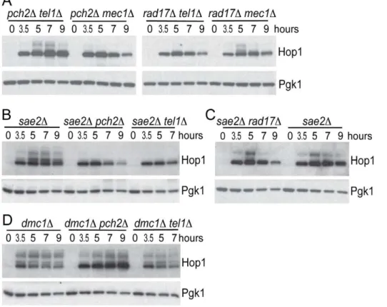
To further confirm the epistasis relationship observed above, we examined Hop1 phosphorylation in thepch2Dtel1D, rad17Dmec1D, pch2D mec1D and rad17D tel1D double mutant combinations. As expected, we observed abundant Hop1 phosphorylation inpch2Dtel1D andrad17Dmec1D, while only a low level of Hop1 phosphorylation was
seen inpch2Dmec1Dandrad17Dtel1Dwhich showed very low spore viability and fast meiotic progression (Figure 3E, 3F and Figure 4A). Together, these results suggest Pch2 acts together with Tel1 to promote an essential meiotic process, perhaps by ensuring interhomolog bias through Hop1 phosphorylation.
Pch2 is involved in signaling unprocessed DSBs
Tel1 is required to signal the presence of unprocessed DSBs during meiosis [17,18]. Specifically, deletion ofTEL1 eliminates the signaling of unresected DSBs to Hop1 [12]. To test if signaling of unprocessed DSBs also requires Pch2, we examined Hop1 phosphorylation in both pch2D and tel1D mutants in a sae2D mutant background where breaks are unprocessed to give blunt ends. Hop1 was phosphorylated in asae2Dmutant but not insae2D pch2Dorsae2Dtel1D(Figure 4B), as expected if Tel1 and Pch2 are specifically required for unprocessed DSBs signaling. By contrast, Rad17 was not required for Hop1 phosphorylation in thesae2D background (Figure 4C), which is also expected since Rad17 is involved in signaling resected DSBs. As a control, we measured Hop1 phosphorylation in the dmc1D mutant background where DSBs are resected to give ssDNA. Hop1 phosphorylation was not affected indmc1Dpch2Danddmc1Dtel1D(Figure 4D).
Figure 2. Pch2 and Rad17 promote Hop1 and Mek1 activation.
(A) Western blot analysis of WT,rad17D,pch2Dandpch2Drad17Dat
indicated time points after transfer to SPM usinga-Hop1 antibody. Pgk1
Western blot was used as the loading control. The phosphorylated isoforms of Hop1 are detectable as slow-moving species. (B) Mek1–3HA immunoprecipitates from WT, rad17D, pch2D and pch2D rad17D at
indicated time points were analyzed by Western blot usinga -phospho-Akt substrate (recognizing pT327 of Mek1) anda-HA antibodies. *IgG
heavy chain. Cell lysates were analyzed by Western blot usinga-HA and
a-Pgk1 antibodies.
doi:10.1371/journal.pgen.1002351.g002
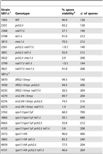
Table 1.Spore viability.
Strain
SBY#1 Genotype
% spore
viability3 #of spores
1903 WT 96.9 128
2351 pch2D 92.2 128
2368 rad17D 37.1 140
3798 tel1D 91.0 212
3815 mec1D 70.3 212
2361 pch2Drad17D ,0.1 140
3801 pch2Dtel1D 92.9 212
3822 pch2Dmec1D 2.9 208
3799 rad17Dtel1D ,0.1 144
3821 rad17Dmec1D 41.8 208
SBY#2
3870 XRS2-13myc 99.3 140
3930 XRS2-13myc pch2D 94.5 200
4235 XRS2-13myc rad17D 26.5 204
4270 xrs2DN-13myc 89.7 204
4276 xrs2DN-13myc pch2D 74.5 216
4273 xrs2DN-13myc rad17D 1.4 216
3763 spo11/spo11yf 68.3 700
3866 spo11/spo11yf tel1D 50.1 688
4063 spo11/spo11yf pch2D 35.8 212
4155 spo11/spo11yf pch2Dtel1D 3.8 208
3572 spo11-HA 96.0 400
3862 spo11-HA tel1D 85.3 340
4059 spo11-HA pch2D 77.5 204
4151 spo11-HA pch2Dtel1D 46.6 204
1MATa/MATaho::hisG/
0leu2::hisG/0ura3(DSma-Pst)/0 his4-X::LEU2-(NBam)-URA3/HIS4::LEU2-(NBam); all mec1Dstrains also contain sml1D.
2MATa/MATaho::hisG/0lys2/0leu2::hisG/0ura3D::hisG/0trp1::hisG/0GAL3/0;
spo11/spo11yf = spo11-HA/spo11Y135F-HA.
3Four-spore tetrads were analyzed.
We noticed that Hop1 protein levels were elevated in thedmc1D pch2D double mutant (Figure 4D) compared to the dmc1D single mutant. On the other hand,sae2Dpch2Dshowed no increase in Hop1 levels compared to thesae2Dsingle mutant (Figure 4B). We reasoned that this effect ofpch2Ddoes not relate to the role of Pch2 in promoting Hop1 phosphorylation per se since pch2D only affected Hop1 phosphorylation in thesae2Dbackground (where Hop1 levels were not altered) but not in thedmc1Dbackground (where Hop1 levels were increased). Thetel1Dstrain did not show such an effect either, again suggesting this aspect of Pch2 function is independent of its role in Tel1 signaling to Hop1. We speculate that the increase in Hop1 levels (or reduced Hop1 protein turnover) shown here by western blotting likely reflects altered Hop1 abundance/distribution shown previously by immunostaining [21] and is related to Pch29s role in axis organization and CO control. Interestingly, this effect is manifested at a ‘‘post resection’’ stage of DSB repair since increased Hop1 levels were observed in dmc1D but not insae2D. CO designation is also thought to occur around this stage of meiotic prophase [38,39].
Pch2 physically interacts with the region of Xrs2 containing putative BRCT repeats
The ATM homolog Tel1 physically interacts with Xrs2 and promotes the phosphorylation of Sae2 and Hop1 [12,40,41]. We
thus tested if Pch2 also interacts with components of the MRX complex using pair-wise bait-prey combinations of Pch2 with Mre11, Rad50 and Xrs2 for yeast two-hybrid analysis. In this trial, Pch2 interacted with Xrs2, but not Mre11 or Rad50 (Figure 5A). Mre11 and Rad50 two-hybrid constructs were functional since we detected interaction between Rad50-Mre11 (Figure 5B) and Mre11-Xrs2 (Figure 5C). We narrowed the Pch2-binding region of Xrs2 to a 187 amino acid region in the Xrs2(126–313)-Gal4AD construct (Figure 5D – 5F). This region contains two putative BRCT repeats, similar to the human ortholog Nbs1 [42]. Point mutations created to abolish FHA domain function present in Xrs2(1–313)-Gal4AD did not abolish interaction with LexA-Pch2 [43] (Figure 5F).
xrs2DNrecapitulates pch2Deffects on Hop1
phosphorylation and spore viability
The first 313 amino acids of Xrs2 are dispensable for the formation of normal levels of DSBs and crossover recombination products yet DSB turnover and MI division are delayed [44]. We created the allelexrs2DN-13mycthat deleted the first 313 amino acid coding region of XRS2 and found it delayed MI division (Figure 5G and 5H), presumably due to the slow turnover of DSBs as in thepch2Dmutant [20,21,45]. We wondered ifxrs2DN-13myc,
Figure 3. Meiotic progression analysis in single and double mutant strains.(A–F) Percentage of cells that have completed at least the first meiotic division (post-MI) in indicated strains. All data were from the same time course experiment.
doi:10.1371/journal.pgen.1002351.g003
likepch2D,would suppress the MI delay conferred byrad17D(and vice versa). We found this to be the case with MI division timing in
xrs2DN-13myc rad17D occurring earlier than either single mutant (Figure 5H). By contrast, MI division was delayed inxrs2DN-13myc pch2D(Figure 5G).
Spore viability of xrs2DN-13myc rad17D (1.4%; Table 1) was dramatically decreased compared to xrs2DN-13myc and XRS2-13myc rad17D (89.7% and 26.5%, respectively), while xrs2D N-13myc pch2D gave only a modest reduction of spore viability compared toXRS2-13myc pch2D(74.5% and 94.5%, respectively). To test ifxrs2DN-13mycaffects checkpoint signaling in a similar manner to pch2D, we examined the effect of this mutation on Hop1 phosphorylation inxrs2DN-13myc rad17Dandxrs2DN-13myc pch2Ddouble mutants as well as insae2Danddmc1Dbackgrounds. We found Hop1 phosphorylation was greatly reduced inxrs2D N-13myc rad17D but not in xrs2DN-13myc pch2D (Figure 6A). Furthermore, xrs2DN-13myc only abrogated Hop1 phosphoryla-tion insae2Dbut not indmc1Dbackgrounds (Figure 6B and 6C). The absence of Hop1 phosphorylation insae2Dxrs2DN-13mycwas not due to reduced DSB levels (Figure 6D). Notably, as withdmc1D pch2D, dmc1D xrs2DN-13myc accumulated more Hop1 protein (Figure 6C). Taken together, these results suggest that the interaction of Pch2 with the N-terminal region of Xrs2, and perhaps the putative BRCT repeats specifically, is required for Pch29s role(s) in the recombination checkpoint and axis organi-zation during meiosis.
xrs2DN but nottel1Drecapitulates pch2Deffects on the
zip1D-induced MI delay
Budding yeast pch2D was originally isolated in the BR background as a mutation that suppresses the meiotic arrest that
occurs in the absence of Zip1 [31] and Pch2 has been thought to be involved in a ‘‘synapsis checkpoint’’ [46]. In SK1,zip1Dcaused a meiosis I delay that is partially suppressed bypch2D[20]. We found that xrs2DN-13myc, but not tel1D, suppressed the zip1D -induced meiotic delay, suggesting that interaction with Xrs2 may also be required for Pch29s role in the synapsis checkpoint (Figure 6E). In fact MI delays conferred bytel1Dand zip1Dare independent since the double mutant exhibits a more severe delay. It is thus possible that Xrs2-Pch2 interaction is required for most, if not all, functions of Pch2; while Pch2 and Tel1 may perform independent functions besides their concerted role in the recombination checkpoint.
Pch2 and Tel1 regulate spore viability independently when DSBs are reduced
Deletion ofPCH2has been shown to sensitize strains carrying hypomorphic alleles ofspo11to give lower levels of spore viability [22,23]. Through our studies we found that the deletion ofTEL1
also gave a modest reduction of spore viability in a spo11-HA/ spo11Y135F-HAbackground (50.1% versus 68.3%; Table 1), but not to the extent ofpch2D(35.8%). When both Pch2 and Tel1 are absent, spore viability was dramatically reduced in this back-ground (3.8%). Similar effects were also observed in thespo11-HA
homozygous mutant background (Table 1). These data suggest that Pch2 and Tel1 independently influence an essential meiotic process that is sensitive to DSB levels. Identification of this process will require analysis of thepch2Dtel1Dspo11-HA/spo11Y135F-HA strain for defects in other meiotic chromosome events including DSB repair, crossover control, chromosome axis morphogenesis and/or synapsis.
Figure 4. Hop1 phosphorylation in various mutants.(A–D) Hop1 phosphorylation was analyzed in indicated strains usinga-Hop1 antibody similar to Figure 2A.
Discussion
This work presents evidence defining new functions for Pch2 and Xrs2 in promoting proper chromosome segregation during meiosis: First, Pch2 and Xrs2 function in the same epistasis pathway as Tel1 (ATM) to activate the recombination checkpoint in response to unprocessed DSBs. Second, Pch2 and Xrs2 function together in the synapsis checkpoint, independent of Tel1. Third, Pch2 interacts with the N-terminus of Xrs2 containing tandem BRCT repeats. The N-terminus of Xrs2 is required to activate both the recombination and synapsis checkpoints. Finally, a role for Pch2 in preventing intersister recombination events is revealed when one branch of the recombination checkpoint is abolished by
deletion ofRAD17. The separate roles for Pch2 and Rad17 in mediating the recombination checkpoint via sequential phosphor-ylation of Hop1 and Mek1 may account for their combined role in preventing intersister repair of DSBs. These findings may help to address the seemingly disparate roles Pch2 plays among synaptic organisms, including meiotic recombination, chromosome axis formation, checkpoint signaling and crossover control [21,47].
Recombination checkpoint
We propose that phosphorylation of the meiotic chromosome axis protein Hop1 is regulated by two partially redundant pathways: one pathway requires Tel1, Pch2 and Xrs2 and Figure 5. Pch2 interacts with putative BRCT repeats of Xrs2. (A–C) Two-hybrid spot assay of Pch2 and MRX complex components. Transformants carrying both LexA DNA binding domain (LexA)- and Gal4 activation domain (Gal4AD)-fusions were spotted on SC-Leu-Trp (left) and SC-Leu-Trp-His plus 1mM 3-AT (right) plates. (D–F) Mapping of the Pch2-interacting region of Xrs2. Xrs2(1–313)-fha: Two amino acids (S47 and T50) in the FHA domain were altered to alanine, shown as ‘‘AA’’ in (D). (G–H) Percentage of cells that have completed at least the first meiotic division (post-MI) in indicated strains.xrs2DN-13mycencodes 13myc-tagged Xrs2(314–854). Data were from the same time course experiment.
doi:10.1371/journal.pgen.1002351.g005
responds to the presence of unprocessed DSBs; a second pathway requires Mec1 and Rad17 and responds to the presence of resected DSB intermediates of homologous recombination (Figure 6F). This model is directly analogous to the different roles of Tel1/Xrs2 and Mec1/Rad17 in the DNA damage response during vegetative growth [18] with Pch2 providing a regulatory feature specific to meiotic chromosomes that coordinate the events of meiotic recombination with axis organization.
The physical association of Pch2 with Xrs2 suggests a mechanism to promote interhomolog bias near sites of DSBs by bringing the Tel1/ATM kinase near its substrate Hop1, a component of the chromosome axis. Pch2 might utilize the binding and/or hydrolysis of ATP to promote conformational changes in axis structure that enable the phosphorylation of Hop1 by Tel1. Alternatively or in addition, Pch2 interaction with Xrs2 might function to stabilize the association of the MRX complex at Figure 6.xrs2DN-13mycphenocopiespch2D.(A–C) Hop1 phosphorylation was analyzed in indicated strains usinga-Hop1 antibody similar to
Figure 2A. (D) Southern blot of 1D gel analysis of DSBs insae2Dandsae2Dxrs2DN-13myc. (E) Percentage of cells that have completed at least the first
the chromosome axis analogous to the interaction of Mdc1 protein (Mediator of DNA damage Checkpoint) with the BRCT repeats of the mammalian Xrs2 ortholog, Nbs1 [48]. In this case, Mdc1 stabilizes the association of Nbs1 at sites of DNA damage, thus creating a microenvironment to promote phosphorylation of H2AX by ATM [49].
It is not clear if Pch2 interacts with Xrs2 (Nbs1) in other organisms. The mouse ortholog of Pch2, TRIP13, is implicated in early recombination steps that follow DSB resection but precede Rad51 focus formation [25]. It is possible that TRIP13-NBS1 interaction could establish a precondition that facilitates a later step of recombination. Indeed in yeast, deletion ofPCH2results in the slow turnover of resected DSBs [20,21,45]. InDrosophila, NBS is required for DSB repair [50], but its role in meiotic recombination has not been explored to date. While Xrs2/Nbs1 proteins are conserved from vertebrates to fungi, there is no apparent ortholog inC. elegans. It remains possible that Pch2 plays a role in a recombination checkpoint inC. elegansthat has not yet been uncovered experimentally.
Synapsis checkpoint
Pch2 orthologs in worm and fly are implicated in a checkpoint activated by the failure to synapse chromosome and/or by disruptions in axis formation. The synapsis checkpoint functions in these instances even in absence of DSB formation [28,30]. Although synapsis is dependent on DSB formation in budding yeast, several examples implicate Pch2 in a synapsis checkpoint that responds to defects in synapsis and/or axis structure in situations where DSBs are efficiently repaired [13,51]. Strong evidence in support of a synapsis checkpoint comes from our previous observation that MEK1-GST, an artificially activated form ofMEK1, acts as a genetic enhancer ofzip1Dby causing MI arrest [13]. Since DSBs are efficiently repaired in this situation [13], this result suggests that synapsis and/or axis defects trigger the arrest, not the persistence of unrepaired DNA breaks. Deletion of PCH2, but not TEL1, can bypass this arrest, suggesting an independent role for Pch2 in a synapsis checkpoint (unpublished data). Similarly, deletion ofPCH2suppresses the meiosis I arrest phenotype that is activated by the presence of aberrantly synapsed chromosomes caused by the non-null allelezip1-4LA,which also repairs DNA breaks efficiently [51]. We found here thatxrs2D N-13myc, similar to pch2D, partially suppressed zip1D delay, suggesting that Xrs2, perhaps through association with Pch2, is required to execute the synapsis checkpoint. By contrast, Tel1 does not seem to be involved in this branch of Pch29s function. Borner and colleagues argued previously that Pch2 might mediate Mec1/ ATR activity with respect to sensing ‘‘structure-dependent interchromosome interactions’’ [21]. It is possible that a Pch2/ Xrs2/Mec1 pathway functions in this program.
Further understanding of the differential requirements for Xrs2 and Tel1 for Pch2 function in the recombination checkpoint versus the synapsis checkpoint (and possibly crossover control) may help to identify common mechanisms shared among synaptic organisms, where pairing and SC formation are not always coupled to recombination [52].
Materials and Methods
Strains
All strains are derivatives of SK1 except the strain used for yeast two hybrid spot assay is L40 (MATa trp1 leu2 his3 LYS2::lexA-HIS3 URA3::lexA-lacZ) [53]. Deletion mutants were generated by PCR-based gene disruption [54,55]. All themec1Dstrains also carried sml1Dto suppress inviability. MEK1-3HAand XRS2-13mycwere
made by using pFA6a-3HA-kanMX6 and pFA6a-13myc-kanMX6 modules, respectively [56].xrs2DN-13mycwas created by two-step allele replacement. Briefly, a PCR-amplifiedURA3 was used to replace the region encoding amino acid 1–313 of Xrs2-13myc. Then a PCR-generated fragment containing 385 bp upstream the first coding ATG fused to 375 bp downstream the ATG encoding amino acid 314 of Xrs2 was used to replace theURA3, resulting in
xrs2DN-13mycexpressing 13myc-tagged Xrs2(314–854) under the native promoter ofXRS2.spo11-HAandspo11Y135F-HAare a gift from Scott Keeney and crossed into our strain background.
SBY strain numbers are listed in Table 1. Additional strains used in this study are: strains isogenic to SBY1903 (MATa/MATaho::hisG/0 leu2::hisG/0 ura3(DSma-Pst)/0 his4-X::LEU2-(NBam)-URA3/HIS4:: LEU2-(NBam)) except the indicated mutations: SBY3055 (ntd80D); SBY3280 (ntd80D pch2D); SBY3277 (ntd80D rad17D); SBY3274 (ntd80Dpch2Drad17D); SBY2591 (dmc1D); SBY2597 (dmc1Dpch2D); SBY2594 (dmc1Drad17D); SBY2606 (dmc1Dpch2Drad17D); SBY3800 (dmc1Dtel1D); SBY2611 (sae2D); SBY2625 (sae2Dpch2D); SBY3843 (sae2D tel1D); SBY2616 (sae2D rad17D); SBY4684 (sae2D xrs2D N-13myc); SBY3589 (MEK1-3HA); SBY3595 (MEK1-3HA pch2D); SBY3592 (MEK1-3HA rad17D); SBY3598 (MEK1-3HA pch2D rad17D); strains isogenic to SBY4056 (MATa/MATaho::hisG/0lys2/
0leu2::hisG/0 ura3D::hisG/0trp1::hisG/0 GAL3/0) except the indicated mutations: SBY3560 (dmc1D), SBY4517 (dmc1D xrs2DN-13myc), SBY3644 (sae2D), SBY4514 (sae2Dxrs2DN-13myc), SBY4445 (zip1D), SBY4451 (zip1Dpch2D), SBY4448 (zip1D tel1D), SBY4404 (zip1D xrs2DN-13myc);tel1D, sml1D, andndt80Dare marked with hphMX; meclDis marked withnatMX; all other mutations are marked with kanMX.
Sporulation conditions
Time courses were conducted by the SPS method [20]. Briefly, cells were patched on YPG plates (3% glycerol, 2% bactopeptone, 1% yeast extract, 2% bactoagar, 0.01% adenine sulphate, 0.004% tryptophan) for ,14 hr and then stripped on YPD plates (2% glucose, 2% bactopeptone, 1% yeast extract, 2% bactoagar, 0.01% adenine sulphate, 0.004% tryptophan) and grown for 2 days. Single colonies were used to inoculate 5 ml YPD (plus 0.002% uracil ifura3strains were used) liquid cultures and grown for at least 24 hr before diluted into SPS (1% potassium acetate, 0.5% yeast extract, 1% bactopeptone, 0.17% yeast nitrogen base, 0.5% ammonium sulphate, 1.02% potassium biphthalate) (plus 0.002% uracil ifura3 strains were used) at O.D.600 = 0.16. SPS cultures were grown for,15.5 hr, washed with H2O, and then
resuspended into SPM (1% potassium acetate, 0.02% raffinose, 0.009% amino acid powder) at O.D.600 = 2–3. Spore viability data were obtained by sporulation on solid SPM media. All procedures were performed at 30uC.
DNA physical assay and meiotic progression analysis DNA extraction, gel electrophoresis and southern blot were performed as previously described [9]. Meiosis I division timing was determined by calculating the percentage of post-MI cells at indicated time points. Briefly, meiotic cultures were fixed in 50% ethanol and stained with DAPI. Cells with more than 2 DAPI-stained nucleus bodies were counted as post-MI cells. 200 cells were counted for each time points.
Protein extraction, Western blotting, and immunoprecipitation
Denaturing whole-cell extracts were prepared as previously described [57] with modifications. Briefly, 1 mL meiotic cultures at indicated time points were spun down and resuspended in 1 mL
ice-cold water with 1 mM PMSF, 10 mM sodium fluoride and 10 mM sodium diphosphate. 150mL ice-cold 2 N NaOH / 8% 2-ME was then added and mixtures were incubated on ice for 10 min. After added 160mL ice-cold 50% TCA and incubated on ice for 10 min, mixtures were spun for 8 min at 14000 rpm. Pellets were washed by 500mL ice-cold acetone and spun for
5 min at 14000 rpm. Washed pellets were dried by spinning in the vacufuge for 8 min and then resuspended in 1X SDS sample buffer with PMSF, sodium fluoride and sodium diphosphate. A bath sonicator was used to facilitate resuspension in acetone and sample buffer. Proteins from denaturing whole-cell extracts were detected by Western blotting using a-Hop1 (S. Roeder), a-HA (Santa Cruz, sc-7392), a-phospho-Akt substrate (Cell signaling,
#9614), and a-Pgk1 (Invitrogen, A-6457). Immunoprecipitation
was performed as previously described [13] excepta-HA antibody (Santa Cruz, sc-7392) was used.
Plasmids and yeast two hybrid analysis
Mre11, Rad50, and Xrs2 yeast two-hybrid plasmids are a gift from S. Keeney [58]. Xrs2 truncation plasmids were constructed
by cloning PCR-generating fragments into the same plasmid for full-length Xrs2 (pACT2-2). Xrs2(1–313)-S47A H50A plasmid was created by QuikChange (Stratagene). LexA-Pch2 plasmid was made by cloning PCR amplified intronlessPCH2 coding region into pCA1 plasmid. Y2H spot assay was performed by spotting 5mL O.D.600 = 1 cultures onto Trp plates and
SC-Leu-Trp-His+1mM 3AT plates and grown for 3–5 days.
Acknowledgments
We thank Shirleen Roeder for the Hop1 antibody, Scott Keeney for providing MRX two-hybrid constructs, JoAnne Engebrecht and Daniel Chu for reading the manuscript and for discussions, and Ting-Fang Wang and Valentine Borner for sharing their unpublished results.
Author Contributions
Conceived and designed the experiments: SMB H-CH. Performed the experiments: H-CH. Analyzed the data: SMB H-CH. Contributed reagents/materials/analysis tools: SMB H-CH. Wrote the paper: SMB H-CH.
References
1. Zickler D, Kleckner N (1999) Meiotic chromosomes: integrating structure and function. Annu Rev Genet 33: 603–754.
2. Keeney S, Giroux CN, Kleckner N (1997) Meiosis-specific DNA double-strand breaks are catalyzed by Spo11, a member of a widely conserved protein family. Cell 88: 375–384.
3. Bishop DK, Park D, Xu L, Kleckner N (1992) DMC1: a meiosis-specific yeast homolog of E. coli recA required for recombination, synaptonemal complex formation, and cell cycle progression. Cell 69: 439–456.
4. Shinohara A, Ogawa H, Ogawa T (1992) Rad51 protein involved in repair and recombination in S. cerevisiae is a RecA-like protein. Cell 69: 457–470. 5. Kadyk LC, Hartwell LH (1992) Sister chromatids are preferred over homologs
as substrates for recombinational repair in Saccharomyces cerevisiae. Genetics 132: 387–402.
6. Bzymek M, Thayer NH, Oh SD, Kleckner N, Hunter N (2011) Double Holliday junctions are intermediates of DNA break repair. Nature 464: 937–941. 7. Schwacha A, Kleckner N (1997) Interhomolog bias during meiotic
recombina-tion: meiotic functions promote a highly differentiated interhomolog-only pathway. Cell 90: 1123–1135.
8. Allers T, Lichten M (2001) Differential timing and control of noncrossover and crossover recombination during meiosis. Cell 106: 47–57.
9. Hunter N, Kleckner N (2001) The single-end invasion: an asymmetric intermediate at the double-strand break to double-holliday junction transition of meiotic recombination. Cell 106: 59–70.
10. Schwacha A, Kleckner N (1995) Identification of double Holliday junctions as intermediates in meiotic recombination. Cell 83: 783–791.
11. Youds JL, Boulton SJ (2011) The choice in meiosis - defining the factors that influence crossover or non-crossover formation. J Cell Sci 124: 501–513. 12. Carballo JA, Johnson AL, Sedgwick SG, Cha RS (2008) Phosphorylation of the
axial element protein Hop1 by Mec1/Tel1 ensures meiotic interhomolog recombination. Cell 132: 758–770.
13. Wu HY, Ho HC, Burgess SM (2010) Mek1 kinase governs outcomes of meiotic recombination and the checkpoint response. Curr Biol 20: 1707–1716. 14. Niu H, Wan L, Baumgartner B, Schaefer D, Loidl J, et al. (2005) Partner choice
during meiosis is regulated by Hop1-promoted dimerization of Mek1. Mol Biol Cell 16: 5804–5818.
15. Wan L, de los Santos T, Zhang C, Shokat K, Hollingsworth NM (2004) Mek1 kinase activity functions downstream of RED1 in the regulation of meiotic double strand break repair in budding yeast. Mol Biol Cell 15: 11–23. 16. Kim KP, Weiner BM, Zhang L, Jordan A, Dekker J, et al. (2010) Sister cohesion
and structural axis components mediate homolog bias of meiotic recombination. Cell 143: 924–937.
17. Hochwagen A, Amon A (2006) Checking your breaks: surveillance mechanisms of meiotic recombination. Curr Biol 16: R217–228.
18. Usui T, Ogawa H, Petrini JH (2001) A DNA damage response pathway controlled by Tel1 and the Mre11 complex. Mol Cell 7: 1255–1266. 19. Lydall D, Nikolsky Y, Bishop DK, Weinert T (1996) A meiotic recombination
checkpoint controlled by mitotic checkpoint genes. Nature 383: 840–843. 20. Wu HY, Burgess SM (2006) Two distinct surveillance mechanisms monitor
meiotic chromosome metabolism in budding yeast. Curr Biol 16: 2473–2479.
21. Borner GV, Barot A, Kleckner N (2008) Yeast Pch2 promotes domainal axis organization, timely recombination progression, and arrest of defective recombinosomes during meiosis. Proc Natl Acad Sci U S A 105: 3327–3332.
22. Joshi N, Barot A, Jamison C, Borner GV (2009) Pch2 links chromosome axis remodeling at future crossover sites and crossover distribution during yeast meiosis. PLoS Genet 5: e1000557. doi:10.1371/journal.pgen.1000557. 23. Zanders S, Alani E (2009) The pch2Delta mutation in baker’s yeast alters
meiotic crossover levels and confers a defect in crossover interference. PLoS Genet 5: e1000571. doi:10.1371/journal.pgen.1000571.
24. Zanders S, Sonntag Brown M, Chen C, Alani E (2011) Pch2 Modulates Chromatid Partner Choice During Meiotic Double-Strand Break Repair in Saccharomyces cerevisiae. Genetics.
25. Roig I, Dowdle JA, Toth A, de Rooij DG, Jasin M, et al. (2010) Mouse TRIP13/ PCH2 Is Required for Recombination and Normal Higher-Order Chromosome Structure during Meiosis. PLoS Genet 6: e1001062. doi:10.1371/journal. pgen.1001062.
26. Wojtasz L, Daniel K, Roig I, Bolcun-Filas E, Xu H, et al. (2009) Mouse HORMAD1 and HORMAD2, two conserved meiotic chromosomal proteins, are depleted from synapsed chromosome axes with the help of TRIP13 AAA-ATPase. PLoS Genet 5: e1000702. doi:10.1371/journal.pgen.1000702. 27. Li XC, Schimenti JC (2007) Mouse pachytene checkpoint 2 (trip13) is required
for completing meiotic recombination but not synapsis. PLoS Genet 3: e130. doi:10.1371/journal.pgen.0030130.
28. Bhalla N, Dernburg AF (2005) A conserved checkpoint monitors meiotic chromosome synapsis in Caenorhabditis elegans. Science 310: 1683–1686. 29. Joyce EF, McKim KS (2009) Drosophila PCH2 is required for a pachytene
checkpoint that monitors double-strand-break-independent events leading to meiotic crossover formation. Genetics 181: 39–51.
30. Joyce EF, McKim KS (2010) Chromosome Axis Defects Induce a Checkpoint-Mediated Delay and Interchromosomal Effect on Crossing Over during Drosophila Meiosis. PLoS Genet 6: e1001059. doi:10.1371/journal. pgen.1001059.
31. San-Segundo PA, Roeder GS (1999) Pch2 links chromatin silencing to meiotic checkpoint control. Cell 97: 313–324.
32. Dong H, Roeder GS (2000) Organization of the yeast Zip1 protein within the central region of the synaptonemal complex. J Cell Biol 148: 417–426. 33. Sym M, Engebrecht JA, Roeder GS (1993) ZIP1 is a synaptonemal complex
protein required for meiotic chromosome synapsis. Cell 72: 365–378. 34. Lynn A, Soucek R, Borner GV (2007) ZMM proteins during meiosis: crossover
artists at work. Chromosome Res 15: 591–605.
35. Lao JP, Oh SD, Shinohara M, Shinohara A, Hunter N (2008) Rad52 promotes postinvasion steps of meiotic double-strand-break repair. Mol Cell 29: 517–524. 36. Niu H, Li X, Job E, Park C, Moazed D, et al. (2007) Mek1 kinase is regulated to suppress double-strand break repair between sister chromatids during budding yeast meiosis. Mol Cell Biol 27: 5456–5467.
37. Grushcow JM, Holzen TM, Park KJ, Weinert T, Lichten M, et al. (1999) Saccharomyces cerevisiae checkpoint genes MEC1, RAD17 and RAD24 are required for normal meiotic recombination partner choice. Genetics 153: 607–620.
38. Borner GV, Kleckner N, Hunter N (2004) Crossover/noncrossover differenti-ation, synaptonemal complex formdifferenti-ation, and regulatory surveillance at the leptotene/zygotene transition of meiosis. Cell 117: 29–45.
39. Bishop DK, Zickler D (2004) Early decision; meiotic crossover interference prior to stable strand exchange and synapsis. Cell 117: 9–15.
41. Cartagena-Lirola H, Guerini I, Viscardi V, Lucchini G, Longhese MP (2006) Budding Yeast Sae2 is an In Vivo Target of the Mec1 and Tel1 Checkpoint Kinases During Meiosis. Cell Cycle 5: 1549–1559.
42. Becker E, Meyer V, Madaoui H, Guerois R (2006) Detection of a tandem BRCT in Nbs1 and Xrs2 with functional implications in the DNA damage response. Bioinformatics 22: 1289–1292.
43. Matsuzaki K, Shinohara A, Shinohara M (2008) Forkhead-associated domain of yeast Xrs2, a homolog of human Nbs1, promotes nonhomologous end joining through interaction with a ligase IV partner protein, Lif1. Genetics 179: 213–225.
44. Shima H, Suzuki M, Shinohara M (2005) Isolation and characterization of novel xrs2 mutations in Saccharomyces cerevisiae. Genetics 170: 71–85.
45. Hochwagen A, Tham WH, Brar GA, Amon A (2005) The FK506 binding protein Fpr3 counteracts protein phosphatase 1 to maintain meiotic recombi-nation checkpoint activity. Cell 122: 861–873.
46. Macqueen AJ, Hochwagen A (2011) Checkpoint mechanisms: the puppet masters of meiotic prophase. Trends Cell Biol.
47. Joyce EF, McKim KS (2011) Meiotic checkpoints and the interchromosomal effect on crossing over in Drosophila females. Fly (Austin) 5.
48. Williams RS, Dodson GE, Limbo O, Yamada Y, Williams JS, et al. (2009) Nbs1 flexibly tethers Ctp1 and Mre11-Rad50 to coordinate DNA double-strand break processing and repair. Cell 139: 87–99.
49. Lukas C, Melander F, Stucki M, Falck J, Bekker-Jensen S, et al. (2004) Mdc1 couples DNA double-strand break recognition by Nbs1 with its H2AX-dependent chromatin retention. EMBO J 23: 2674–2683.
50. Mukherjee S, LaFave MC, Sekelsky J (2009) DNA damage responses in Drosophila nbs mutants with reduced or altered NBS function. DNA Repair (Amst) 8: 803–812.
51. Mitra N, Roeder GS (2007) A novel nonnull ZIP1 allele triggers meiotic arrest with synapsed chromosomes in Saccharomyces cerevisiae. Genetics 176: 773–787.
52. Bhalla N, Dernburg AF (2008) Prelude to a division. Annu Rev Cell Dev Biol 24: 397–424.
53. Vojtek AB, Hollenberg SM, Cooper JA (1993) Mammalian Ras interacts directly with the serine/threonine kinase Raf. Cell 74: 205–214.
54. Goldstein AL, McCusker JH (1999) Three new dominant drug resistance cassettes for gene disruption in Saccharomyces cerevisiae. Yeast 15: 1541–1553. 55. Wach A, Brachat A, Pohlmann R, Philippsen P (1994) New heterologous modules for classical or PCR-based gene disruptions in Saccharomyces cerevisiae. Yeast 10: 1793–1808.
56. Longtine MS, McKenzie A, 3rd, Demarini DJ, Shah NG, Wach A, et al. (1998) Additional modules for versatile and economical PCR-based gene deletion and modification in Saccharomyces cerevisiae. Yeast 14: 953–961.
57. Yaffe MP, Schatz G (1984) Two nuclear mutations that block mitochondrial protein import in yeast. Proc Natl Acad Sci U S A 81: 4819–4823. 58. Arora C, Kee K, Maleki S, Keeney S (2004) Antiviral protein Ski8 is a direct
partner of Spo11 in meiotic DNA break formation, independent of its cytoplasmic role in RNA metabolism. Mol Cell 13: 549–559.