CASE REPORT
J of Evidence Based Med & Hlthcare, pISSN- 2349-2562, eISSN- 2349-2570/ Vol. 2/Issue 6/Feb 09, 2015 Page 746
PRIMARY MULTILOCULAR HYDATID CYST OF NECK: A RARE CASE
REPORT
Deepak Ramraj1, Swaroop S2, Jagadeesha B. V. C3, Mahesh K4
HOW TO CITE THIS ARTICLE:
Deepak Ramraj, Swaroop S, Jagadeesha B. V. C, Mahesh K. ”Primary Multilocular Hydatid Cyst of Neck: A Rare Case Report”. Journal of Evidence based Medicine and Healthcare; Volume 2, Issue 6, February 9, 2015; Page: 746-750.
ABSTRACT: Hydatid disease, also known as echinococcosis or hydatidosis, is an infectious disease caused by Echinococcus. Echinococcus granulosus is the most common Echinococcus species affecting human beings. It may affect any organ and tissue in the body, in particular the liver and lung. Musculoskeletal or soft tissue hydatidosis accounts for about 0.5% 5% of all echinococcal infections in endemic areas, and is almost always secondary to the hepatic or pulmonary disease. Even in regions where echinococcosis is endemic, hydatidosis of cervicofacial region is extremely rare. Herein, we present exceptionally rare case in a 55 year old female with an unusual localization of primary multilocular hydatid cyst in the right supraclavicular region of the neck. A high index of suspicion is required to diagnose hydatid cyst in rare locations like this. Hydatid cyst should be considered in differential diagnosis of benign swellings of head and neck region, so that it can be managed during surgery to prevent acute anaphylaxis.
KEYWORDS: Hydatid cyst, multilocular cyst, neck swellings, Echinococcus.
INTRODUCTION: Hydatid disease, also known as echinococcosis or hydatidosis, is a zoonotic infection caused by the larval forms (metacestode) of Echinococcus granulosus that lives in the small intestines of adult dogs.Man is an accidental host. Hydatid cyst, develops most frequently in the liver (65%), the lungs (25%), remaining 10% occurs in muscle, spleen, bones, kidneys, brain, eye, heart, and pancreas.[1-3] Involvement of hydatid cyst is extremely rare in head and neck region even in geographical areas where ecchinococcal infestation is frequent. Only a few cases of hydatid cyst located in neck have been reported in literature.[4,5] We report a case of multilocular hydatid cyst in right supraclavicular fossa in a 50yr old female, which was diagnosed accidentally on ultrasound.
CASE REPORT: A 50year old female presented to our hospital with six months history of painless lump in right side of lower part of neck. On physical examination there was a solitary swelling of size 4x3.5 cm in right supraclavicular region, just above medial one third of clavicle. The swelling had smooth surface, was firm in consistency, borders were well made out, with restricted mobility. The skin over the swelling was hyperpigmented with no ulcerations or sinuses. There were no other swellings in any other part of body. Respiratory, cardiovascular, abdomen and neurological examination revealed no abnormality.
CASE REPORT
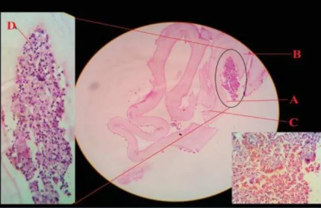
J of Evidence Based Med & Hlthcare, pISSN- 2349-2562, eISSN- 2349-2570/ Vol. 2/Issue 6/Feb 09, 2015 Page 747 disease of the right neck, as patient was from endemic area of hydatid disease. Complete surgical resection of the cystic mass from the surrounding tissue was performed without rupture. Histopathological examination of the specimen confirmed the diagnosis of Echinococcus cyst. The gross specimen comprised of multiloculated cyst filled with gelatinous membrane. (Figure 1, 3) Microscopic examination of the specimen revealed three layers of hydatid cyst-Inner germinal layer,Middle laminated layer, Outer adventitial layer with chronic inflammatory cells containing mainly histiocytes. (Figure 2) The patient was discharged from the hospital with advice of albendazole 400mg twice daily for six weeks.
DISCUSSION: Hydatid cyst is an infectious disease which is most commonly caused by the cestode, Echinococcus granulosus and less commonly by Echinococcus multilocularis. Dogs are the main host and animals like cattle, sheep, horse and pig are intermediate hosts in the disease. Parasite eggs that penetrate the organism hatch in the small intestine of the main host, pass into portal venous system or lymphatic system and reach the liver and lungs, and finally form hydatid cyst lesions. Moreover, they can cross the hepatic sinusoid or pulmonary capillary barriers, and embryos get into systemic circulation and can settle in all the organs and structures in the body.[6] Atypical localization of hydatid cyst may be a challenge in the diagnosis of hydatid disease.[7] Chevalier et al have reported the subcutaneous cyst hydatid incidence as 2%. However, this ratio also includes the secondary hydatid cyst cases.[8] Although the disease is generally asymptomatic,it may exhibit clinical symptoms depending on the size and location of the cyst and the pressure of the growing cyst. In our patient, there were no symptoms except a painless and mobile lump in the right supraclavicular area with no evidence of hydatid disease elsewhere in the body. Eroglu et al reported an unusual case of hydatid cyst found in the neck of a 66 year old woman and just like our case report, there was no pulmonary or hepatic involvement.[9]
Interesting aspect of solitary hydatid cysts in the absence of disease in lung and liver is to explain how these larvae produces solitary cysts after passing through the two filter sites. Although no route other than portal has been proven in humans, systemic dissemination through lymphatic route is a strong possibility in case of unusual presentation sites. Typically hydatid cyst consists of a single, unilocular cyst, however multiloculated cyst in the same or multiple organ are seen in 20 to 30% of the cases.[10]
CASE REPORT
J of Evidence Based Med & Hlthcare, pISSN- 2349-2562, eISSN- 2349-2570/ Vol. 2/Issue 6/Feb 09, 2015 Page 748 No serological tests and MRI was done in our case as patient was not affordable. For the evaluation of mass lesions in the cervical region, fine-needle aspiration cytology is beneficial however due to possibility of an anaphylactic reaction, dissemination of disease and recurrence as result of spillage of cyst contents, it is not recommended in the routine evaluation of suspected hydatid cysts.[13,14,15] So FNAC was not performed in our case and the case was subjected to surgical excision biopsy.
The diagnosis of hydatid cyst is confirmed by histology. Histopathological evaluation shows three layers of hydatid cyst. The inner most germinal layer which is thin and translucent. The embryonic tape worm, scolices, develops from an out pouching of the germinal layer and form hydatid sand. The middle laminated membrane is white 2mm thick and is easily ruptured. The outer layer or pericyst is a rigid protective layer representing response of the host to parasite.[15]
Surgical removal is the most effective treatment of hydatid cyst.[5,14] Surgeon must be careful to avoid spillage of cyst contents to avoid fatal anaphylaxis, recurrence and multiple hydatidosis.[3,15] If presurgical diagnosis is hydatid cyst, preliminary aspiration and instillation of hypertonic saline (20%), silver nitrate (0.5%), formalin, and other chemicals could be used to prevent seeding of the cyst contents and to inactivate the protoscolices.[5] Therapy with nontoxic scolocidal agents or combination chemotherapy with mebendazole is of therapeutic value in the treatment of patients with recurrence or a high risk of contamination.[13] There was complete cyst removal with no rupture and spillage of cyst contents in the present case. Albendazole is suggested to be given post operatively for 1–3 months. We treated the present case with albendazole for six weeks post operatively.
CONCLUSION: Hydatid disease is a widespread public health problem in developing countries. The possibility of hydatid disease, especially in endemic regions, should always be considered in the differential diagnosis of mesenchymal neoplasms or soft tissue masses in the neck or in the other parts of the body. Radiologic imaging modalities in such cases are mandatory for the diagnosis of unilocular or multilocular hydatid cyst with thin borders, thin walls, inner membranes, and a distinct appearance of characteristic cystic mass. The prognosis is excellent in hydatid cyst cases treated with total removal of the cyst without rupture.
REFERENCES:
1. Dagtekin A, Koseoglu A, Kara E, Karabag H, Acvi E, Torun F, Bagdatoglu C: Unusual location of hydatid cysts in pediatric patients. Pediatric Neurosurg 2009, 45: 379-383.
2. Engin G, Acunas B, Rozanes I, Acunas G: Hydatid disease with unusual localization. Eur Radiology 2000, 10: 1904-1912.
3. Kireşi DA, Karabacakoğlu A, Odev K, Karaköse S: Uncommon locations of hydatid cysts. Acta Radiol 2003, 44: 622-636.
4. Eroğlu A, Atabekoğlu S, Kocaoğlu H: Primary hydatid cyst of the neck. Eur Arch Otorhinolaryngol 1999, 256: 202-204.
CASE REPORT
J of Evidence Based Med & Hlthcare, pISSN- 2349-2562, eISSN- 2349-2570/ Vol. 2/Issue 6/Feb 09, 2015 Page 749 6. Celik A, Turanli M, Kutun S, Delibasi T, Mengi N, Comert E, Aslan S, et al. Unusual location
of hydatid cyst: soft tissue mass in the neck. Eur Arch Otorhinolaryngol 2006; 263 (12): 1147-1150.
7. Ahmad S, Jalil S, Saleem Y, Suleman BA, Chughtai N. Hydatid cysts at unusual sites: reports of two cases in the neck and breast. J Pak Med Assoc 2010; 60 (3): 232-234.
8. Dirican A, Unal B, Kayaalp C, Kirimlioglu V. Subcutaneous hydatid cysts occurring in the palm and the thigh: two case reports. J Med Case Reports 2008; 2: 273.
9. Eroglu A, Atabekoglu S, Kocaoglu H. Primary hydatid cyst of the neck. Eur Arch Otorhinolaryngol 1999; 256 (4): 202-204.
10.Rochidi Y, Raji A, Elhattab Y: A rare localization of hydatidosis: A cervical hydatid cyst. Fr ORL 2007, 92: 315-317.
11.Guney O, Ozturk K, Kocaogullar Y, Eser O, Acar O: Submandibullar and intracranial hydatid cyst in an adolescent. Laryngoscope 2002, 112: 1857-1860.
12.Polat P, Kantarci M, Alper F, Suma S, Koruyucu MB, Okur A: Hydatid disease from head to toe.RadioGraphics 2003, 23: 475-494.
13.Akal M, Kara M: Primary hydatid cyst of the posterior cervical triangle. J Laryngol Otol 2002, 116: 153-155.
14.Baglam T, Karatas E, Durucu C, Sirikci A, Kara F, Kanlikama M: Primary hydatid cyst of infratemporal fossa.J Craniofac Surg 2009, 20: 1200-1201.
15.Izci Y, Tüzün Y, Seçer HI, Gönül E: Cerebral hydatid cyst: Technique and pitfalls of surgical management. Neurosurg Focus 2008, 24: E15.
CASE REPORT
J of Evidence Based Med & Hlthcare, pISSN- 2349-2562, eISSN- 2349-2570/ Vol. 2/Issue 6/Feb 09, 2015 Page 750
AUTHORS:
1. Deepak Ramraj
2. Swaroop S.
3. Jagadeesha B. V. C.
4. Mahesh K.
PARTICULARS OF CONTRIBUTORS:
1. Post Gtaduate, Department of General Surgery, J. J. M. M. C, Davangere. 2. Post Graduate, Department of General
Surgery, J. J. M. M. C, Davangere. 3. Professor, Department of General
Surgery, J. J. M. M. C, Davangere.
4. Professor, Department of General Surgery, J. J. M. M. C, Davangere.
NAME ADDRESS EMAIL ID OF THE CORRESPONDING AUTHOR:
Dr. Deepak Ramraj,
# 12, J. J. M. M. C Boys Hostel, M. C. C. B Block, Davangere-577004. E-mail: drdeepakramraj@gmail.com
Date of Submission: 09/01/2015.
Date of Peer Review: 10/01/2015. Date of Acceptance: 16/01/2015. Date of Publishing: 06/02/2015.
Fig. 2: Microscopic appearance showing A-Outer adventitial layer, B-Middle laminated layer, C-Inner Germinal layer,
D-Chronic inflammatory cells with histiocytes