Fatal outcome in bronchus-associated lymphoid tissue lymphoma*
,**
Linfoma do tecido linfóide associado ao brônquio com evolução fatal
Romulo Loss Mattedi1, Fabiola del Carlo Bernardi2, Carlos Eduardo Bacchi3,
Sheila Aparecida Coelho Siqueira2, Thais Mauad2
Abstract
Primary pulmonary lymphoma is rare. The most common histological type is the bronchus-associated lymphoid tissue lymphoma. This type of lymphoma has an indolent course and excellent response to therapy. One-third of all cases are diagnosed incidentally. However, due to the rarity of this disease, little is known about its natural history in terms of dissemination and evolution. Herein, we report the unusual case of a 61-year-old man who refused treatment after being diagnosed with bronchus-associated lymphoid tissue lymphoma and died 2 years later from massive lung infiltration without dissemination to other organs.
Keywords: Lung; Autopsy; Lymphoma.
Resumo
Linfomas primários do pulmão são raros. O tipo histológico mais freqüente é o linfoma do tecido linfóide associado ao brônquio. Este tipo de linfoma tem curso indolente e excelente resposta à terapia. Um terço dos casos é descoberto incidentalmente. Devido à raridade desta doença, no entanto, pouco se conhece sobre sua história natural em termos de disseminação e evolução. Neste relato, descrevemos o caso incomum de um homem de 61 anos que recusou o tratamento após diagnóstico de linfoma do tecido linfóide associado ao brônquio e, 2 anos após o diagnóstico, morreu por infiltração pulmonar maciça sem disseminação para outros órgãos.
Descritores: Pulmão; Autópsia; Linfoma.
* Trabalho realizado no Departamento de Patologia da Faculdade de Medicina da Universidade de São Paulo – USP – São Paulo (SP) Brasil. 1. Pós-Graduando do Departamento de Patologia da Faculdade de Medicina da Universidade de São Paulo, São Paulo (SP) Brasil. 2. Doutor em Patologia pela Faculdade de Medicina da Universidade de São Paulo, São Paulo (SP) Brasil.
3. Doutor em Patologia pela Faculdade de Medicina da Universidade Estadual Paulista, Botucatu (SP) Brasil.
Endereço para correspondência: Thais Mauad. Departamento de Patologia da Faculdade de Medicina da USP, Av. Dr. Arnaldo, 455, Cerqueira César, CEP 01246-903, São Paulo, SP, Brasil.
Tel 55 11 3061-7173. Fax 55 11 3062-8098. E-mail: tmauad@usp.br Recebido para publicação em 19/7/06. Aprovado, após revisão, em 21/8/06.
** A versão completa em português deste artigo está disponível em www.jornaldepneumologia.com.br
Introduction
Primary pulmonary lymphoma is a rare entity, accounting for 0.5% of all lung tumors and less than 1% of all lymphomas.(1) The most common type of primary pulmo-nary lymphoma is the mucosa-associated lymphoid tissue (MALT) lymphoma, a mature B-cell lymphoma, which cells seem to be derived from the marginal zone of the lymphoid aggregates that compose the bronchus-associated lymphoid tissue (BALT) of the lungs. It affects both genders equally, typically appearing between the sixth and seventh decade of life.(2-4)
The disease frequently has an indolent course. In fact, before more accurate techniques to detect clonality at the tissue level became available, most cases were classified as pseudo-lymphomas.(2) It is believed that, in approximately one-third
to one-half of the patients, the disease is discovered inciden-tally on radiographs, the most common radiological findings being pulmonary infiltrates or one or more nodules.(4,5)
All case series of this disease report slow-growing tumors that have an excellent response to surgery, chemotherapy or combined treatment. Treatment provides excellent prog-nosis, with extremely low mortality rates, reaching 5-year survival rate of 100% in many studies.(3) Some authors have even suggested that, in asymptomatic patients with surgi-cally unresectable tumors, watchful waiting is an option.(2) However, due to the rarity of this entity, its natural history has yet to be fully understood.
to massive tumor growth within a two-year period. This shows that, despite the indolent course of this type of lymphoma, prompt treatment is necessary in order to limit disease progression.
Case report
A 61-year-old man was admitted to the hospital after experiencing progressive shortness of breath, productive cough and malaise for 3 months, without fever, chest pain, or hemoptysis. Upon arrival at the emergency room, the patient was in severe respi-ratory distress. He was severely tachypneic, and physical examination revealed peripheral cyanosis. Chest auscultation revealed decreased breath sounds and dullness to percussion at the mid-thorax level on the left side. An examination of the heart and abdomen revealed no abnormalities. The patient presented no fever, edema, joint abnormalities, rashes, jaundice, or lymphadenopathy.
The family of the patient reported that, for two years, he had experienced productive cough, occasional hemoptysis, hoarseness, and progres-sive weight loss (20 kg). One year earlier, he had undergone bronchoscopy and had been diagnosed with a lung tumor at another institution, but he had refused further treatment. No medical records were available. Computed tomography of the chest performed one month earlier had shown a large anterior mediastinal mass and atelectasis of the left upper lobe of the lung. The patient had stopped smoking 17 years earlier after having smoked for 30 years. No other diseases were reported.
At admission, the white blood cell count was 12,240/mm3, the hemoglobin level was 8.6 g/dL,
and oxygen saturation was 70% on room air. An orotracheal tube was inserted, and the patient was submitted to mechanical ventilation. Although empirical treatment with intravenous antibiotics was started, the patient became hypotensive and was unresponsive to vasopressors. Due to the clinical instability of the patient, no attempt was made to perform a biopsy. Despite the treatment, the patient died four days later. An autopsy was performed:
External examination revealed peripheral edema. Opening the chest wall revealed moderate serous pleural effusion and thin pleural adhesions.
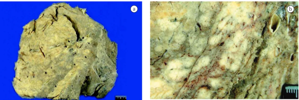
Both lungs presented increased volume; the left lung weighed 1650 g, and the right lung weighed 1050 g. The left lung had a fleshy consistency and was largely occupied by gray-whitish confluent nodules with no necrosis (Figures 1a and b). The lymph nodes in the pulmonary hilum had a similar aspect. The right lung was edematous, and a nodule with a diameter of 4 cm was observed in the right lower lobe. The peri-aortic lymph nodes were also altered. In the other organs there were no specific abnormalities other than generalized atherosclerosis.
We retrospectively consulted the files of the department of pathology where the transbron-chial biopsy had been analyzed 1.5 years prior to death. The pathology report confirmed a low-grade non-Hodgkin’s B-cell lymphoma with plasmacytic differentiation, suggestive of MALT lymphoma. In fact, the autopsy revealed extensive infiltra-tion of the left lung by a mature B-cell lymphoma. Large confluent masses of small to medium sized lymphocytes, some with a centrocytic aspect, formed
a
Figure 1 - Macroscopic aspects: a) View of the lung lymphoma; and b) Multiple, confluent, large whitish nodules occupy a large proportion of the lung parenchyma.
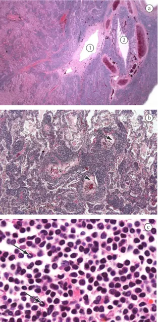
the bulk of the tumor. There was moderate plas-macytic differentiation, and numerous plasma cells were observed. At its border, the lymphoma dissemi-nation was of a predominantly bronchovascular type (Figures 2a, 2b and 2c), and the tumor extended to the pleura. Lymphoepithelial lesions were present. There was no necrosis. Although scattered immu-noblasts were observed, there were no areas of large-cell transformation. The non-neoplastic lung tissue presented post-obstructive pneumonia. Except for the hilar and peri-aortic lymph nodes, all other organs, including the bone marrow, were free of lymphoma. The immunohistochemical panel revealed that the lymphoma cells were positive for CD20 (Figure 3), CD79a, and Kappa, whereas they were negative for CD3, CD23, CD5, CD10, cyclin D1, CD43, and Lambda. The plasma cells were positive for CD 138. There was focal positivity for CD35. Fluorescent in situ hybridization using dual-color dual-fusion probes (Abbott Laboratories Inc., Abbott Park, IL, USA) confirmed the presence of the t(11;18)(q21;q21) translocation. This translocation was detected in 60% of the lymphoid cells using a morphometric image analysis system (MetaSystems, Altlussheim, Germany).
Discussion
Due to its rarity, relatively little is known about the natural history of BALT lymphomas in terms of local infiltration and dissemination. In a significant proportion of patients, the lymphoma is discovered incidentally.(5,6) In a study conducted in Japan,(6) 5 patients with BALT lymphoma were evaluated, and the authors reported that the mean interval between diagnostic suspicion (based on tomog-raphy findings) and a definitive diagnosis was 3.4 years, which reflects the indolent course of this disease. Our report shows that BALT lymphomas can occasionally progress more rapidly to massive lung infiltration, indicating that diagnostic efforts and treatment should not be delayed.
Patients with BALT lymphoma have an excel-lent prognosis. Interestingly, surgical treatment, radiotherapy, chemotherapy, or combinations of these strategies all seem to achieve good results.(7) One group of authors recently described the natural history of 22 patients with BALT lymphoma and showed that, regardless of the treatment received, the 5-year survival rate was 100%.(2) In a study
1
2
a
Figure 2 - Photomicrograph of the lung lymphoma: a) Massive invasion of the lung parenchyma by lymphoid infiltrates, with infiltration of large bronchovascular structures (H&E × 12.5). 1 = vessel and 2 = bronchus;
b) Note the vascular pattern of dissemination (arrows) (H&E ×50); and c) High magnification photomicrograph
of the lymphoma cells. Small- to medium-sized lymphocytes with plasmacytic differentiation (arrows) (H&E ×1000).
c b
lymphoma infiltration of the lungs.(8) The only case of primary pulmonary lymphoma found in that study was that of a primary T-cell lymphoma. In line with these findings, another study conducted in Japan showed that, among 24 cases of primary pulmonary lymphoma monitored for a mean period of 43 months, there were only two deaths, one in a patient with T-cell lymphoma and a patient with lymphomatoid granulomatosis(9); none of the patients with small B-cell lymphoma died.
It has yet to be established which factors are prog-nostic predictors in pulmonary MALT lymphomas. Disease progression has been associated with large-cell transformation of MALT lymphomas, which was not observed in our patient. One group of authors reported that 3 out of 13 patients with pulmonary MALT lymphoma died due to disease progression, large-cell transformation being observed in 2 of the deceased.(10) In another series of 13 patients with pulmonary MALT lymphoma,(5) death due to disease progression occurred in only 1 patient, in whom Sjögren’s syndrome was the underlying disease. However, another group of researchers,(11) studying a series of 50 MALT lung lymphomas, reported that factors such as age, gender, auto-immune disorders, monoclonal gammopathy, extent of disease within the lungs, pleural involvement, and positive lymph nodes (hilar or mediastinal) were not prognostic factors. The presence of the t(11;18)(q21;21) trans-location, which is particularly prevalent in patients with gastric (23.9%) and pulmonary (53.3%) MALT lymphomas,(12) does not seem to be related to disease recurrence.(13) Nor is there any evidence that this translocation is associated with chemotherapy resistance.
Although no detailed laboratorial screening had been performed, our patient had no history of chronic autoimmune diseases or HIV infec-tion, which is frequently found in patients with MALT lymphoma.(2,3) Since the patient remained in the hospital for only a short time, tests to iden-tify associated monoclonal gammopathy were not performed. Although smoking has been hypothe-sized as a possible risk factor for BALT lymphoma,(2) as a source of chronic antigen stimulation, it is unlikely that cigarette smoking acted as a trigger in our patient, since he had quit smoking 17 years before.
Interestingly, despite extensive involvement of the left lung, the disease was restricted to the thorax. This finding seems to be a characteristic of primary pulmonary MALT lymphomas, which remain restricted to the lungs for long periods of time before dissemination. It is possible that the specificity of lymphocyte homing patterns, the abscence of circulating marginal zone cells, and the antigen dependence of the lymphoma play a role in the dissemination pattern of this disease.(3)
Despite their indolent behavior and excellent prognosis, MALT lymphomas do tend to relapse frequently.(13) In a study of pulmonary MALT lymphoma patients in long-term follow-up treat-ment,(11) it was reported that the overall survival of such patients was significantly worse than that observed in age- and gender-matched controls. Our report suggests that the disease progression is not always indolent. Therefore, prompt and accurate diagnosis, as well as treatment and lifelong follow-up evaluation, are important in the management of this rare entity.
Acknowledgments
The authors thank Dr. Maísa Momesso de Quintal for retrieving the patient’s records at the Campinas University Hospital.
References
1. Cadranel J, Wislez M, Antoine M. Primary pulmonary lymphoma. Eur Respir J. 2002;20(3):750-62.
2. Ahmed S, Kussick SJ, Siddiqui AK, Bhuiya TA, Khan A, Sarewitz S, et al. Bronchial-associated lymphoid tissue lymphoma: a clinical study of a rare disease. Eur J Cancer. 2004;40(9):1320-6.
3. Zinzani PL, Tani M, Gabriele A, Poletti V, Stefoni V, Alinari L, et al. Extranodal marginal zone B-cell lymphoma of
type of the lung: single-center experience with 12 patients. Leuk Lymphoma. 2003;44(5):821-4.
4. Koss MN, Hochholzer L, Nichols PW, Wehunt WD, Lazarus AA. Primary non-Hodgkin’s lymphoma and pseudolymphoma of lung: a study of 161 patients. Hum Pathol. 1983;14(12):1024-38.
5. Wislez M, Cadranel J, Antoine M, Milleron B, Bazot M, Mayaud C, et al. Lymphoma of pulmonary mucosa-associated lymphoid tissue: CT scan findings and pathological correlations. Eur Respir J. 1999;14(2):423-9.
6. Takamori M, Noma S, Kobashi Y, Inoue T, Gohma I, Mino M, et al. CT findings of BALTOMA. Radiat Med. 1999;17(5):349-54.
7. Habermann TM, Ryu JH, Inward DJ, Kurtin PJ, Primary pulmonary lymphoma. Semin Oncol. 1999;26(3):307-15. 8. Costa MB, Siqueira SA, Saldiva PH, Rabe KF, Mauad
T. Histologic patterns of lung infiltration of B-cell, T-cell, and Hodgkin lymphomas. Am J Clin Pathol. 2004;121(5):718-26.
9. Tamura A, Komatsu H, Yanai N, Homma J, Nagase A, Nemoto E et al. Primary pulmonary lymphoma: relationship between
clinical features and pathologic findings in 24 cases. The Japan National Chest Hospital Study Group for Lung Cancer. Jpn J Clin Oncol. 1995;25(4):140-52.
10. O’Donnell PG, Jackson SA, Tung KT, Hassan B, Wilkins B, Mead GM. Radiological appearances of lymphomas arising from mucosa-associated lymphoid tissue (MALT) in the lung. Clin Radiol. 1998;53(4):258-63.
11. Kurtin PJ, Myers JL, Adlakha H, Strickler JG, Lohse C, Pankratz VS, et al. Pathologic and clinical features of primary pulmonary extranodal marginal zone B-cell lymphoma of MALT type. Am J Surg Pathol. 2001;25(8):997-1008. 12. Streubel B, Simonitsch-Klupp I, Müllauer L, Lamprecht A,
Huber D, Siebert R, et al. Variable frequencies of MALT lymphoma-associated genetic aberrations in MALT lymphomas of different sites. Leukemia. 2004;18(10):1722-6.