CARLOS ROBERTO EMERENCIANO BUENO
AVALIAÇÃO DA RESPOSTA TECIDUAL E
CAPACIDADE DE MINERALIZAÇÃO DE
CIMENTOS QUE CONTÉM COMPOSTOS
BIOCERÂMICOS, RESINOSOS E COM
HIDRÓXIDO DE CÁLCIO.
Dissertação apresentada à Faculdade de Odontologia de Araçatuba, Universidade Estadual Paulista “Júlio de Mesquita Filho” - UNESP como parte dos requisitos para obtenção do título de Mestre em Ciências Odontológicas.
Área de concentração: Endodontia.
Orientador: Prof. Adj. Eloi Dezan Junior
2
Dados Curriculares
CARLOS ROBERTO EMERENCIANO BUENO
Nascimento 01 de Junho de 1986 Campinas/SP
Filiação Carlos Roberto Bueno
Norma Pignataro Emerenciano Bueno
2004-2009 Curso de Graduação em Odontologia – Universidade Federal do Amazonas – UFAM
2010-2012
2011-2012
Curso de Especialização em Odontologia do Trabalho – Centro de Pesquisas Odontológicas São Leopoldo Mandic, Campinas - SP
Curso de Especialização em Endodontia –
Fundação Bauruense de Estudos Odontológicos – FunBeo/USP Bauru
2013-2015 Curso de Pós-Graduação em Endodontia, nível Mestrado – Faculdade de Odontologia de Araçatuba – Universidade Estadual Paulista “Júlio de Mesquita Filho” – UNESP
4
Aos meus pais Carlos e Norma
Pelo incentivo diário, pelas longas conversas e pela inspiração que vocês
são para mim. Meu pai sempre foi e sempre será meu ídolo. Minha mãe é
um poço sem fundo de carinho e amor com os filhos. Foram esse exemplos
que construíram meus princípios e me fizeram quem sou.
À minha noiva Vanessa (Tchula)
Por me levantar quando eu estava pra baixo e por comemorar comigo
quando eu estava por cima. Você é meu porto seguro, a quem sempre quero
voltar no final do dia.
Por trás de todo sucesso do homem há uma grande mulher. Este é um dos
meus grandes sucessos. Muito obrigado por você ser minha grande mulher.
Às minhas irmãs Mônica e Bruna
Por serem IRMÃS sempre que precisei! Conselhos e conversas a qualquer
hora do dia ou noite, por telefone ou Skype (junto com Cibele e Fabrício).
6
À Deus
Por todos os sonhos realizados.
Aos meus tio Marcos e tia Sandra
Por serem meus “segundos pais”. Pelas conversas e conselhos e por
fazerem eu me sentir em casa.
Aos meu primos Bruno e Paulo
Se meus tios são meus segundos pais, vocês são meus irmãos!
À minha família
Minha vovó Wanda, meu avô Antônio (in memoriam), Minha vovó Lúcia,
meu avô Hermínio (in memoriam). E a todos meus tios e tias de Natal e
Serra negra que torceram por mim!
Ao Prof. Eloi Dezan Junior
Pela chance, orientação e principalmente pela confiança.
Pelos conselhos e convivência dentro e fora da endodontia.
Ao amigo Diego Valentim
Por ser meu “segundo orientador”, quando precisei. Por ser meu irmão
mais velho na hora de puxar a orelha. Por ser meu irmão mais novo,
quando eu puxei a orelha. Mas principalmente por ser meu amigo.
Obrigado pela amizade.
Aos Dreibis
Jõao, Cláudia, Rodriguim e Rafa. Por toda a conversa, conselhos e
confiança que depositaram, sempre nos apoiando, desde pequenas decisões
ate grandes mudanças.
Aos docentes da disciplina de Endodontia
da FOA Prof. Luciano Tavares Ângelo Cintra, Prof. João Eduardo Gomes
Filho, Prof. Rogério Castilho, Prof. Gustavo Sivieri, pelas dúvidas tiradas
Aos meus amigos de Pós Graduação
da Endodontia da FOA/UNESP, Mariane Azuma (Tanomari), Gabi,
Loiane, Índia, Chris, Luciana, Vanessa e Renata, pela convivência,
amizade, apoio e ensinamentos! E estender um agradecimento especial à
Francine Benettinha, por ter me co-orientado nessa última semana.
Aos funcionários do departamento de endodontia da FOA
Peterson, Elaine e Nelci, fundamentais para o cotidiano harmônico do
departamento.
Ao professor
Emílio Carlos, por despertar meu interesse na endodontia e ao Professor
Marco Antônio Húngaro Duarte (Sal) por despertar meu interesse na pós
graduação.
Ao meu melhor amigo e anjo da guarda Adriano Brust
Por me guiar até aqui.
Juntos na faculdade, juntos pelas fronteiras do exército, juntos em Bauru.
Resolveu se eternizar mais cedo, mas...
... se passamos juntos por tudo isso... Com certeza estaremos juntos para
sempre!!
À Faculdade de Odontologia de Araçatuba
8
Bueno, CRE. Avaliação da Resposta Tecidual e Capacidade De Mineralização de Cimentos que Contém Compostos Biocerâmicos, Resinosos e Com Hidróxido De Cálcio, 2015. 56p. Dissertação (Mestrado em Endodontia) – Faculdade de Odontologia, Campus de Araçatuba, Universidade Estadual Paulista “Júlio de Mesquita Filho”.
Resumo
A obturação ideal é uma combinação de um cimento com um material sólido, geralmente guta percha, que corrobora com o escoamento do cimento fluido, espalhando-o e preenchendo possíveis espaços vazios. Em virtude da possibilidade de contato direto com os tecidos periapicais, estes cimentos devem ser biocompatíveis e, se possível, estimular a mineralização para proporcionar selamento apical. Com o objetivo de avaliar, in vivo, a resposta tecidual e a capacidade de mineralização dos cimentos endodônticos Smartpaste® Bio, Sealapex® e Acroseal®, foi realizado implante subcutâneo em 40 ratos Wistar e adotados os períodos experimentais de 7, 15, 30 e 60 dias (10 animais por período de tempo). Cada animal recebeu quatro implantes, três tubos de polietileno com os cimentos a serem testados e um tubo vazio como controle. Após cada período pós-operatório, os animais foram eutanasiados e os tubos de polietileno, juntamente com o tecido circunjacente foram removidos e fixados. Para a análise histológica da espessura da cápsula fibrosa, infiltrado inflamatório e mineralização as peças foram incluídas em historresina, e coradas em HE, Von Kossa ou permaneceram sem coloração para a luz polarizada. Os resultados foram submetidos ao teste de Kruskal Wallis e Dunn (p<0,05). Resultados: Todos os cimentos produziram reação inflamatória moderada nos períodos iniciais. O Smartpaste Bio®apresentou a menor reação inflamatória aos 15 dias (p<0,05). O Sealapex® induziu maior mineralização, seguido do Smartpaste Bio®. O Acroseal® não apresentou indução de mineralização. Conclusão: Ao final do experimento, todos os cimentos testados apresentaram compatibilidade tecidual. Com exceção do Acroseal, todos induziram mineralização.
10
Bueno, CRE. Biocompatibility and biomineralization assessment of bioceramic, epoxy- resin based and calcium hydroxide root canal sealers. Araçatuba, 2015. 56p. Dissertation (Master’s Degree in Endodontics) – Dental School of Araçatuba, São Paulo State University “Júlio de Mesquita Filho”.
Abstract
The cleaning and shaping of root canals is essential to achieve biological and mechanical goals in the endodontic treatment, providing the appropriate conical shape for subsequent obturation. The optimal obturation is a combination of a sealer with a central core, usually gutta percha, spreading and filling possible gaps. Once there is a direct contact with periapical tissue, the sealer should be biocompatible and, if possible, stimulate mineralization to perform an apical sealing. In order to evaluate in vivo biological response and tissue mineralization capacity of the endodontic sealers Smartpaste Bio® Sealapex® and Acroseal®, subcutaneous implants in 40 Wistar rats were performed. Analisys were at 7, 15, 30 and 60 days experimental periods (10 animals for each time period). Each animal received four implants, three polyethylene tubes with the sealers in testand one empty tube as control. After each post-operative period animals were euthanized and the polyethylene tubes, along with surrounding tissue were removed and fixed. In order to histologically analysis fibrous capsule thickness, inflammatory infiltrate and mineralization, the pieces were included in historesin and stained in HE, Von Kossa or remained without staining for observation under polarized light. The results were statistically analyzed by Kruskal-Wallis and Dunn (p<0,05). Results: All sealers promoted moderate inflammatory reaction at initial periods. Smartpaste Bio® presented the lowest inflammatory reaction at 15 days period (p<0.05). Sealapex®induced higher mineralization, followed by Smartpaste Bio®. Acroseal® showed no mineralization areas. Conclusion: At the end of the experiment, all tested sealers presented biocompatibility. With exception of Acroseal, all induced biomineralization.
12
Lista de Tabelas
Table 1. Percentage of samples in each group categorized according to the inflammatory score and the rating thickness of fibrous
capsule...33
14
Lista de Figuras
Fig 1 Subcutaneous tissue reactions in the experimental groups ...34
16
Artigo
Abstract ……….…………..…..20
Introduction ……….….……...……..21
Material and Methods ………..……….……..22
Results ………..24
Discussion ………..…………..26
Conclusion and acknowledgment………..28
References ……….……..29
Tables ……….…..33
Figures ………..34
Anexos
Anexo A – Guidelines for authors International Endodontic Journal...………..37Anexo B – Comitê de Ética no Uso de Animal (CEUA) ………..55
18
Biocompatibility and biomineralization assessment of bioceramic, epoxy-resin based and calcium hydroxide root canal sealers.
C.R.E. Bueno, D. Valentim, J. E. Gomes-Filho, L. T. A. Cintra, R. C. Jacinto, E. Dezan- Junior.
Department of Endodontics, Araçatuba Dental School, Universidade Estadual Paulista- UNESP, Araçatuba/São Paulo, Brazil.
Tissue response to endodontic sealers Bueno et al.
Keywords: Acroseal, Biocompatibility, Mineralization, Sealapex, Smartpaste
Bio.
Corresponding author:
Eloi Dezan Junior
Department of Endodontics, Araçatuba Dental School, Universidade Estadual Paulista (UNESP), R. José Bonifácio, 1193, 16015-050, Araçatuba/São Paulo, Brazil.
Tel. number: +55.18.3636.3254, Fax number: +55.18.3636.3236 E-mail address: dezan@foa.unesp.br
20
Abstract
Aim: To evaluate the tissue response and mineralization ability of endodontic
sealers in the rat subcutaneous tissue to implanted polyethylene tubes filled with Smartpaste Bio®, Acroseal®and Sealapex®.
Methodology: Forty Wistar rats were assigned to one of four groups according
to periods of time (10 animals/group) and received subcutaneous implants containing the sealers to be tested and empty tube as controls. After 7, 15, 30 and 60 days the animals were euthanized and the polyethylene tubes removed with the surrounding tissues. Inflammatory infiltrate and thickness of fibrous capsule were histologically evaluated. Mineralization was analysed with Von Kossa staining and polarized light. Data were tabulated and subject to Kruskal-Wallis and Dunn’s test (P<0.05).
Results: All tested materials induced moderate inflammatory reaction in the
initial periods. Smartpaste Bio®induced the mildest inflammatory reactions in 15 days (P<0.05). No difference was observed among groups in 30 and 60 days. Von Kossa positive and birefringent structures to polarized light revealed a larger mineralization area in Sealapex® followed by Smartpaste Bio®. Acroseal® induced mild tissue reaction but it did not present signs of mineralization.
Conclusions: At the end of the experiment, all tested sealers presented
biocompatibility. With exception of Acroseal, all induced biomineralization.
Keywords: Acroseal, Biocompatibility, Mineralization, Sealapex, Smartpaste
Introduction
Efficient cleaning and shaping of the root canal system is essential to achieve the biological and mechanical objectives of endodontic treatment, involving the removal of the pulp tissue or remains, microorganisms and their by-products, while providing the appropriate conical shape for subsequent root canal filling, reaching the desired three-dimensional obturation (Schilder 2006). Gutta-percha alone as filling material is not sufficient to provide adequate root canal system sealing, requiring its association with an endodontic sealer to fill gaps between cone and root canal walls, spreading the fluid sealer (Weis et al. 2004, Rahimi et al. 2009). As the sealer reaches the apical foramen, becomes in direct contact with periapical tissue, therefore should be biocompatible. Although the contact area is small, there is always concern about adverse reactions the sealer could cause on the tissues (Branstetter & Fraunhofer 1982, Orstavik 2005, Chhabra et al. 2011).
The endodontic sealers are divided into different groups according to its main component such as zinc oxide and eugenol, resin-based sealers and sealers containing calcium hydroxide (Kim et al. 2010). Depending on these main component, local adverse effects is possible, such as delaying or hindering repair (Schmalz et al. 2000, Geurtsen 2001).
22
Acroseal® (Specialites-Septodont, Saint Maur-des-Fosses, France) is an endodontic sealer containing 28% calcium hydroxide in its composition, along with radiopaque excipient and a resin compound (epoxy resin). Previous studies demonstrated their antimicrobial activity against Enterococcus faecalis, low toxicity and suitable physicochemical properties (Eldeniz et al. 2007, Pinheiro et
al. 2009, Marciano et al. 2011). According to the manufacturer, the sealer
formulation has recently been modified with a reduction of calcium hydroxide concentration and an increase in its resinous compound diglycidylether of bisphenol A (DGEBA).
Sealapex® (SybronEndo, Glendora, CA) is a sealer that contains calcium oxide in its composition and forms calcium hydroxide after being hydrous by contact with tissue fluid. Sealapex® is characterized by biocompatibility, and osteoinductive ability to stimulate the deposition of mineralized tissue inducing apical sealing after endodontic treatment (Holland & Souza 1985, Gomes-Filho
et al. 2012). This sealer was submitted to a reformulation, which presents
2-year shelf life instead of previous 1-2-year shelf life. One of the major alterations in Sealapex was the replacement of the radiopacifier from barium sulfate to bismuth trioxide (Leonardo et al. 2007).
Limited data concerning Smart Seal® obturation system are currently available and there is a lack of scientific studies about the biocompatibility of Smartpaste Bio® and its mineralization ability. Also, the reformulation in Acroseal® and Sealapex® components needs a complete research.
Therefore, the aim of this study was to analyze the biocompatibility (inflammation response) and mineralization ability of the endodontic sealers Smartpaste Bio®, Acroseal® and Sealapex®. The null hypothesis tested was that there is no biocompatibility and no mineralization induction by Smartpaste Bio®, Acroseal® and Sealapex®.
Materials and methods
Araçatuba School of Dentistry-UNESP Ethical Committee, which approved the experimental project.
One hundred and twenty polyethylene tubes (Abbott Laboratories of Brazil, Sao Paulo, SP, Brazil) with a 1.0-mm internal diameter, 1.6-mm external diameter, and 10.0-mm length were filled with the tested sealers. Acroseal®and Sealapex® were prepared according to the manufacturers’ recommendations and inserted into the tubes with a lentulo spiral (Dentsply Maillefer, Tulsa, OK, USA). Smartpaste Bio®is conditioned in a ready-to-use syringe and was directly inserted into the polyethylene tubes. Forty extra polyethylene empty tubes were used as control, totaling one hundred and sixty tubes in the experiment.
Under xylazine (10 mg/kg Rhobifarma Indústria Farmacêutica Ltda, Hortolândia, SP, Brazil) and ketamine (25 mg/kg União Química Farmacêutica Nacional S/A, SP, Brazil) intramuscular anesthesia, back of the animals were shaved, antisepsis with 5% iodine solution realized and a 2.0 cm incision in a head–tail orientation with #15 Bard-ParkerTM blade (BD, Franklin Lakes, NJ, USA) proceeded, creating two pockets in each side of incision. Three polyethylene tubes, containing the sealers and an empty tube as control, were implanted in each animal in opposite directions (upper right, upper left, lower right and lower left) and the skin was closed with 4/0 silk suture (Johnson & Johnson Produtos Profissionais Ltda, São José dos Campos, SP, Brazil).
At 7, 15, 30 and 60 days after implantation, the animals were euthanized by anesthetic overdose. Polyethylene tubes with the surrounding tissues were removed and fixed in 10% buffered formalin at pH 7.0 (Gomes-Filho et al. 2009). The specimens were processed for glycol methacrylate embedding (Gomes-Filho et al. 2001), serially sectioned into 3 µm cuts, and stained with hematoxylin-eosin (HE). The 10 µm cuts were stained according to the Von Kossa technique or remained no stained.
24
Tissue reactions at the open end of the tubes were scored according to previous studies (Yaltirik et al. 2004, Gomes-Filho et al. 2012, Cintra et al. 2013), as follows: 0, few inflammatory cells or no reaction; 1, less than 25 cells and mild reaction; 2, between 25 and 125 inflammatory cells and moderate reaction; 3, 125 or more inflammatory cells and severe reaction (400x magnification). Fibrous capsules were considered thin when <150 µm and thick when >150 µm. Calcification was recorded as positive or negative to Von Kossa stain and present or absent to polarized light (100x magnification).
Data were statistically analyzed by Kruskal-Wallis and Dunn; P<0.05 was considered significant.
Results Control group
A moderate chronic inflammatory reaction (median score 2) was observed at the first two time periods (Table 1), on days 7 and 15 (Fig 1 - A,B). The inflammatory cell infiltration, consisting of lymphocytes and macrophages, was present in the fibrous capsule, which was thick. On the following periods, 30 and 60 days, the fibrous capsule surrounding the tube was thin, with few inflammatory cells (median score 1). The control group was negative to Von Kossa stain and no birefringent structures under polarized light were observed in all periods (Table 2).
Smartpaste Bio®
Acroseal®
On days 7 and 15 a moderate inflammatory cell infiltration (median score 2) consisting of lymphocytes and macrophages was present in a thick fibrous capsule (Table 1). On days 30 and 60, the inflammation intensity reduced and fibrous capsule was thin, similar to the control group (Fig 1 K,L). Birefringent
granulations to polarized light were absent and Von Kossa was negative in all time periods (Fig 2 I-P)
Sealapex®
On days 7 and 15, a moderate inflammatory reaction (median score 2) consisting mainly of lymphocytes and macrophages was present in the thick fibrous capsule (Table 1). The intensity of the inflammation reduced from the 30th day on, until day 60, remaining macrophages phagocyting extravasated sealer (median score 1). The fibrous capsule near the tube opening was thin (Fig 1 G, H) and granulations birefringent to polarized light and Von Kossa positive staining were observed near the tube opening in all time periods (Fig 2 A-H).
Comparison Among Groups
The data were compared for each time period as shown in Table 1 and 2. After 7 days, HE staining revealed a similar tissue response of the inflammatory reaction, and there were no statistically significant differences among the inflammation scores of the experimental groups. On day 15, there was a statistically difference between inflammatory cell numbers of the Smartpaste Bio® and the other groups, once its inflammatory score was lower than the others (P<0.05) also presented a thin fibrous capsule. On days 30 and 60 there was no statistically significant difference among the scores of the different groups regarding the inflammation tissue response.
26
structures and Von Kossa positive in 100% specimens until day 15 and decreased over time, differentiating from each time period along the experiment, but maintained the already mineralized structures (Table 2).
Discussion
The null hypothesis was rejected, once tested sealers presented biocompatibility and, all sealers but Acroseal® promoted mineralization. At initial experimental periods, all groups evoked mild-to-moderate inflammatory reactions that decreased and the fibrous capsule became thinner over time.
The use of subcutaneous implants started with Torneck (1966; 1967). His pioneering research aimed to evaluate the reaction of the subcutaneous connective tissue of rats to implanted polyethylene tubes and became one commonly used method to preliminary evaluate the biocompatibility (Zmener et
al. 1990, Kaplan et al. 2003, Parirokh & Torabinejad 2010). In the present
study, the reactions to empty tubes were similar to the results previously reported (Olson et al. 1981, Gomes-Filho et al. 2009).
According to Gomes Filho et al (2001), the Glicol Metacrilate technique is an excellent alternative for the evaluation of the biocompatibility of endodontic sealers, once is easy to execute and reproducible, providing a better definition of the degree of the inflammatory process. The specimens were sectioned into 3 µm cuts, and stained with HE to analyze inflammatory infiltrate. The scores used to analyze inflammatory reaction were according to previous studies (Gomes-Filho et al. 2009, Gomes-Filho et al.2012, Cintra et al. 2013). The 10 µm cuts were stained according to the Von Kossa technique, to observe mineralized structures, or remained without staining to allow the observation under polarized light of birefringent structures related to calcium carbonate crystals (Holland et al. 1999, Gomes-Filho et al. 2008a).
The reaction of calcium ions from Ca(OH)2 ionic dissociation with carbon dioxide from tissues, forms calcite crystals, birefrigentes to the polarized light, which induced the formation of calcified areas, moreover, the calcium utilization reduces the carbon dioxide used by bacteria for anaerobic respiration (Holland
favors the tissue restoration process and antimicrobial properties (Tagger et al. 1988, Estrela et al. 1995, Desai & Chandler 2009).
Regarding the Smartpaste Bio®, the use of a pre-mixed sealer eliminates the potential of heterogeneous consistency during on-site mixing. Studies involving other bioceramic endodontic sealers already revealed hard tissue deposition (Güven et al. 2013) and favorable tissue response to partial pulpotomy (Azimi et al. 2014). Monobasic calcium phosphates are included in the sealer to facilitate reaction with calcium hydroxide to produce water and hydroxyapatite upon activation of the sealer by water (Yang 2002). Chang et al. (2014) conducted a study with a bioceramic sealer and observed lower inflammatory mediators and better osteoblast expression, which matches the previous study of Bosio et al. (2014), where they classified the bioceramic as biocompatible. These earlier biocompatibility findings of bioceramic sealers also match the results of this study.
It is interesting to observe that birefrigent granulations 100% present at initial periods decreased over time in Smartpaste Bio®, probably related to a reduction in calcium release after 30 days, but remained mineralized structures stained by Von Kossa, present in all time periods.
Even with reformulation, Acroseal® maintained biocompatibility, inducing low inflammatory response, similar to results reported by Khashaba et al. (2011). In all analyzed periods no mineralized areas were observed, similar occurrence were reported by Gomes-Filho et al. (2008b) and Neto et al. (2010).
Eldeniz et al. (2007) conduced a study concerning pH level and calcium ion release evidencing the lower Acroseal®’s calcium and Hydroxyl release. This may be justified by the low solubility of components, once contains epoxy compounds (diglycidyl ether of bisphenol A and methenamine), resulting in a less ion release (Siqueira et al. 1995) which explains the absence of mineralization observed in this study.
28
birefrigent granulations under polarized light from 7 to 60 days, and biomineralization in all time periods.
Previous studies with Sealapex® original formula presented mild inflammation in initial periods, reducing over time (Mittal et al. 1995). Analyzing Sealapex® new formulation, inflammatory infiltrate increase at 14 days (Silva-Herzog et al. 2011) and at 90-day experimental period (Leonardo et al. 2007), suggesting the alterations might have affected tissue compatibility. The results in this research reveal a moderate initial inflammatory response, decreasing after 30 days.
Conclusion
At the end of the experiment, all tested sealers presented biocompatibility, with a statistical difference of Smartpaste Bio® in 15 days. With exception of Acroseal®, all induced biomineralization. Further study is necessary to better analyze the behavior of this biocerâmic material and its physicochemical properties and to confirm the findings of the present study.
Acknowledgement
References
Azimi S, Fazlyab M, Sadri D, Saghiri MA, Khosravanifard B, Asgary S (2014) Comparison of pulp response to mineral trioxide aggregate and a bioceramic paste in partial pulpotomy of sound human premolars: a randomized controlled trial. International Endodontic Journal 47, 873–81.
Bosio CC, Felippe GS, Bortoluzzi EA, Felippe MCS, Felippe WT, Rivero ERC (2014) Subcutaneous connective tissue reactions to iRoot SP, mineral trioxide aggregate (MTA) Fillapex, DiaRoot BioAggregate and MTA International
Endodontic Journal 47, 667–74.
Branstetter J, Fraunhofer JAY (1982) The physical properties and sealing action of endodontic sealer cements: a review of the literature. Journal of Endodontics
8, 312-6.
Chang SW, Lee SY, Kang SK, Kum KY, Kim EC (2014) In vitro biocompatibility, inflammatory response, and osteogenic potential of 4 root canal sealers: Sealapex, Sankin Apatite Root Sealer, MTA Fillapex, and iRoot SP Root Canal Sealer. Journal Of Endodontics 40, 1642-8.
Chhabra A, Teja TS, Jindal V, Singla MG, Warring K (2011) Fate of extruded sealer: a matter of concern. Journal of Oral Health & Community Dentistry 5, 168-72.
Cintra LTA, Ribeiro TAAR, Gomes-Filho JE et al. (2013) Biocompatibility and biomineralization assessment of a new root canal sealer and root-end filling material. Dental Traumatology 29, 145–50.
Desai S, & Chandler N (2009) Calcium hydroxide–based root canal sealers: a review. Journal of Endodontics 35, 475-80.
Duarte MA, Demarchi AC, Giaxa MH, Kuga MC, Fraga SC, de Souza LC (2000) Evaluation of pH and calcium ion release of three root canal sealers. Journal of
Endodontics 26, 389-90.
Economides N, Gogos C, Kodonas K, Beltes C, Kolokouris I (2012) An ex vivo comparison of the push-out bond strength of a new endodontic filling system (Smartseal) and various gutta- percha filling techniques. Odontology 100, 187– 91.
Eldeniz AU, Erdemir A, Kurtoglu F, Esener T (2007) Evaluation of pH and calcium ion release of Acroseal sealer in comparison with Apexit and Sealapex sealers. Oral Surgery, Oral Medicine, Oral Pathology, Oral Radiology and
Endodontics103, 86-91.
Estrela C, Sydney GB, Bammann LL, Felipe JR (1995) Mechanism of the action of calcium and hydroxy ions of calcium hydroxide on tissue and bacteria.
30
Geurtsen W (2001) Biocompatibility of root canal filling materials. Australian
Endodontic Journal 27, 12-21.
Gomes-Filho JE, Gomes BPFA, Zaia AA, Novaes PD, Souza-Filho F J (2001) Glycol Methacrylate: An alternative method for embedding subcutaneous implants. Journal of Endodontics 27, 266 -8.
Gomes-Filho JE, de Faria MD, Bernabe PFE et al. (2008a) Mineral trioxide aggregate but not light-cure mineral trioxide aggregate stimulated mineralization. Journal of Endodontics 34, 62– 5.
Gomes-Filho JE, Bernabe PFE, Nery MJ et al. (2008b) Reaction of rat connective tissue to a new calcium hydroxide– based sealer. Oral Surgery, Oral
Medicine, Oral Pathology, Oral Radiology and Endodontics 106, 71-6.
Gomes-Filho JE, Watanabe S, Bernabe PFE, Costa MTM (2009) A mineral trioxide aggregate sealer stimulated mineralization. Journal Of Endodontic 35, 256–60.
Gomes-Filho JE, Watanabe S, Lodi CS et al. (2012) Rat tissue reaction to MTA FILLAPEX. Dental Traumatology 28, 452–6.
Guven EP, Taslı PN, Yalvac ME, Sofiev N, Kayahan MB, Sahin F (2013) In vitro comparison of induction capacity and biomineralization ability of mineral trioxide aggregate and a bioceramic root canal sealer. International Endodontic Journal
46, 1173–82.
Holland R, Souza V (1985) Ability of a new calcium hydroxide root canal filling material to induce hard tissue formation. Journal of Endodontics 11, 535–43.
Holland R, Souza V, Nery MJ et al. (2002) Calcium salts deposition in rat connective tissue after implantation of calcium hydroxide-containing sealers.
Journal of Endodontics 28, 173-6.
Holland R, Souza V, Nery MJ, Otoboni-Filho JA, Bernabé PFE, Dezan-Junior E (1999) Reaction of rat connective tissue to implanted dentin tubes filled with mineral trioxide aggregate or calcium hydroxide. Journal of Endodontics 25, 161– 6.
Kaplan AE, Ormaechea MF, Picca M, Canzobre MC, Ubios AM (2003) Rheological properties and biocompatibility of endodontic sealers. International
Endodontic Journal 36, 527-32.
Khashaba RM, Moussa MM, Chutkan NB, Borke JL (2011) The response of subcutaneous connective tissue to newly developed calcium phosphate-based root canal sealers. International Endodontic Journal 44, 342–52.
Kim YK, Grandini S, Ames JM et al. (2010) Critical Review on Methacrylate Resin-based Root Canal Sealers. Journal of Endodontics 36, 383-99.
Leonardo MR, Barnett F, Debelian GJ, de Pontes Lima RK, Bezerra da Silva LA (2007) Root canal adhesive filling in dogs’ teeth with or without coronal restoration: a histopathological evaluation. Journal of Endodontics 33, 1299– 303.
Loushine BA, Bryan TE, Looney SW, et al. (2011) Setting properties and cytotoxicity evaluation of a premixed bioceramic root canal sealer. Journal of
Endodontics 37, 673–7.
Marciano MA, Guimarães BM, Ordinola-Zapata R et al. (2011) Physical properties and interfacial adaptation of three epoxy resin–based Sealers.
Journal of Endodontics 37, 1417- 21.
Mittal M, Chandra S, Chandra S (1995) Comparative tissue toxicity evaluation of four endodontic sealers. Journal of Endodontics 21, 622-4.
Neto KO, Moura AM, Davidowicz H (2010) Comparative study of ratʼs connective tissue reaction, against three endodontic resin cements. Journal of
the Health Sciences Institute 28, 67- 70.
Olsson B, Sliwkowski A, Langeland K (1981) Subcutaneous implantation for the biological evaluation of endodontic material. Journal Of Endodontics 7, 355-69.
Ørstavik D (2005) Materials used for root canal obturation: technical, biological and clinical testing. Endodontic Topics 12, 25–38.
Parirokh M, Torabinejad M (2010) Mineral trioxide aggregate: a comprehensive literature review–Part III: Clinical applications, drawbacks, and mechanism of action. Journal of Endodontics 36, 400–13.
Pinheiro CR, Guinesi AS, Camargo EJ, Pizzolitto AC, and Bonetti-Filho I (2009) Bacterial leakage evaluation of root canals filled with different endodontic sealers. Oral Surgery, Oral Medicine, Oral Pathology, Oral Radiology and Endodontics108, 56-60.
Prosmart Product Information, 2009.
Rahimi M, Jainaen A, Parashos P, Messer H (2009) Bonding of resin- based sealers to root dentin. Journal Of Endodontic 35, 121-24.
Saif S, Carey CM, Tordik PA, McClanahan SB (2008) Effect of irrigants and cementum injury on diffusion of hydroxyl ions through the dentinal tubules.
Journal Of Endodontics 34, 50-2.
Schilder H (2006) A tribute to dr. herbert schilder: filing root canals in three dimensions. Jornal Of Endodontics 32, 281-90.
Schmalz G, Hoffmann M, Weis K, Schweikl H (2000) Influence of albumin and collagen on the cell mortality evoked by zinc oxide-eugenol in vitro. Journal Of
32
Silva-Herzog D, Ramírez T, Mora J, Pozos AJ, Silva LAB, Silva RAB, Nelson-Filho P. (2011) Preliminary study of the inflammatory response to subcutaneous implantation of three root canal sealers. International Endodontic Journal 44, 440–6.
Siqueira FJ Jr, Fraga RC, Garcia PF (1995) Evaluation of sealing ability, pH and flow rate of three calcium hydroxide–based sealers. Endodontics & dental
traumatology 11, 225-8.
Tagger M, Tagger E, Kfir A (1988) Release of calcium and hydroxyl ions from set endodontic sealers containing calcium hydroxide. Journal Of Endodontics
14, 588–91.
Torneck CD (1966) Reaction of rat connective tissue to polyethylene tube implants part I. Oral Surgery, Oral Medicine, Oral Pathology, 21 379-87.
Torneck CD (1967) Reaction of rat connective tissue to polyethylene tube implants part II. Oral Surgery, Oral Medicine, Oral Pathology 24, 674-83.
Weis MV, Parashos P, Messer HH (2004) Effect of obturation technique on sealer cement thickness and dentinal tubule penetration. International
Endodontic Journal 37, 653–63.
Yaltirik M, Ozbas H, Bilgic B, Issever H (2004) Reactions of connective tissue to mineral trioxide aggregate and amalgam Journal of Endodontics 30, 95–9.
Yang Q, Troczynski T, Liu DM (2002) Influence of a patite seeds on the synthesis of calcium phosphate cement. Biomaterials 23, 2751–60.
Zhang W, Li Z, Peng B (2009) Assessment of a new root canal sealer’s apical sealing ability (2009) Oral Surgery, Oral Medicine, Oral Pathology, Oral Radiology and Endodontics107, 79-82.
Zmener O, Guglielmotti MB, Cabrini RL (1990) Tissue response to an experimental calcium hydroxide-based endodontic sealer: a quantitative study in subcutaneous connective tissue of the rat. Endodontics & dental
Table 1. Percentage of samples in each group categorized according to the inflammatory score and the rating thickness of fibrous capsule.
Score (%) Capsule
7 Days 0 1 2 3
Control 0 0 100 0 Thick
Sealapex 0 0 90 10 Thick
Acroseal 0 0 90 10 Thick
Smartpaste Bio 0 0 100 0 Thick
15 days
Control 0 0 100 0 Thick
Sealapex 0 0 90 10 Thick
Acroseal 0 0 100 0 Thick
Smartpaste Bio 0 80* 20 0 Thin
30 days
Control 0 100 0 0 Thin
Sealapex 0 90 10 0 Thin
Acroseal 0 90 10 0 Thin
Smartpaste Bio 10 90 0 0 Thin
60 Days
Control 0 100 0 0 Thin
Sealapex 0 60 40 0 Thin
Acroseal 10 80 10 0 Thin
Smartpaste Bio 10 80 10 0 Thin
* statistical difference
Table 2. Percentage of samples in each group categorized according to Von Kossa positive to mineralization and presence of birefrigents crystals under polarized light.
7 days Von Kossa (%) Polarized Light (%)
Control 0 0
Sealapex 100 100
Acroseal 0 0
Smartpaste Bio 100 100
15 days
Control 0 0
Sealapex 100 100
Acroseal 0 0
Smartpaste Bio 100 50
30 days
Control 0 0
Sealapex 100 100
Acroseal 0 0
Smartpaste Bio 100 50
60 Days
Control 0 0
Sealapex 100 100
Acroseal 0 0
Smartpaste Bio 100 30
34
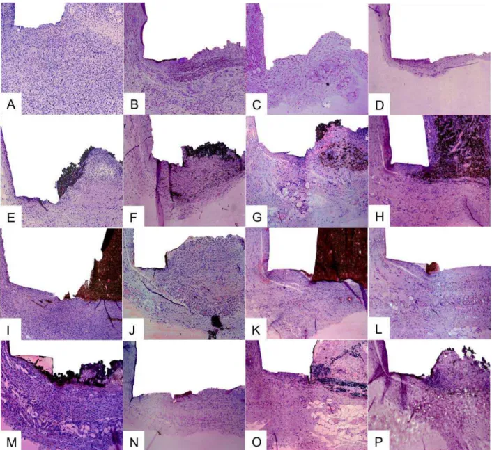
Figure 1 Subcutaneous tissue reactions in the experimental groups. Control group: (A, B) thick fibrous capsule and moderate inflammatory reaction (7 and 15 days HE, 100x);
(C) reduction in the thickness of fibrous capsule and mild inflammatory reaction (30
days HE, 100x); (D) thin fibrous capsule and mild inflammatory reaction (60 days HE,
10x). Sealapex: (E, F) thick fibrous capsule formation and moderate inflammatory cell
infiltration (7 and 15 days HE, 100x); (G) reduction in the thickness of fibrous capsule
formation and mild inflammatory cell infiltration, consisting of macrophages (30 days HE, 100x); and (H) thin fibrous capsule formation and mild inflammatory cell infiltration,
with macrophages phagocyting sealer (60 days HE, 100x). Acroseal: (I,J) thick fibrous
capsule and moderate inflammatory cell infiltration (7 and 15 days HE, 100x); (K,L) the
fibrous capsule surrounding the tube was thin with few chronic inflammatory cells (30
and 60 days HE, 100x); Smarpaste Bio: (M) thick fibrous capsule and moderate
inflammatory reaction (7 days HE, 100x); (N-P) thin fibrous capsule and mild
36
Anexo A
International Endodontic Journal
© International Endodontic Journal. Published by John Wiley & Sons Ltd
Edited By: PMH Dummer__Impact Factor: 2.273__ISI Journal Citation Reports © Ranking: 2013: 16/83 (Dentistry Oral Surgery & Medicine) Online ISSN: 1365-2591
Author Guidelines
Content of Author Guidelines: 1. General, 2. Ethical Guidelines, 3. Manuscript
Submission Procedure, 4. Manuscript Types Accepted, 5. Manuscript Format and Structure, 6. After Acceptance
Useful Websites: Submission Site (http://mc.manuscriptcentral.com/iej),
Articles published in International Endodontic Journal
(http://onlinelibrary.wiley.com/journal/10.1111/(ISSN)1365-2591), Author Services (http://authorservices.wiley.com/bauthor/author.asp), Wiley Blackwell’
s Ethical Guidelines
(http://authorservices.wiley.com/bauthor/publicationethics.asp), Guidelines for Figures (http://authorservices.wiley.com/bauthor/author.asp)
The journal to which you are submitting your manuscript employs a plagiarism detection system. By submitting your manuscript to this journal you accept that your manuscript may be screened for plagiarism against previously published works.
1. GENERAL
International Endodontic Journal publishes original scientific articles, reviews,
clinical articles and case reports in the field of Endodontology; the branch of dental sciences dealing with health, injuries to and diseases of the pulp and periradicular region, and their relationship with systemic well- being and health. Original scientific articles are published in the areas of biomedical science, applied materials science, bioengineering, epidemiology and social science relevant to endodontic disease and its management, and to the restoration of root-treated teeth. In addition, review articles, reports of clinical cases, book reviews, summaries and abstracts of scientific meetings and news items are accepted.
38
2. ETHICAL GUIDELINES
International Endodontic Journal adheres to the below ethical guidelines for
publication and research.
2.1. Authorship and Acknowledgements
Authors submitting a paper do so on the understanding that the manuscript has been read and approved by all authors and that all authors agree to the submission of the manuscript to the Journal. International Endodontic Journal adheres to the definition of authorship set up by The International Committee of Medical Journal Editors (ICMJE). According to the ICMJE, authorship criteria should be based on 1) substantial contributions to conception and design of, or acquisiation of data or analysis and interpretation of data, 2) drafting the article or revising it critically for important intellectual content and 3) final approval of the version to be published. Authors should meet conditions 1, 2 and 3.
Acknowledgements: Under acknowledgements please specify contributors to
the article other than the authors accredited. Please also include specifications of the source of funding for the study and any potential conflict of interests if appropriate.
2.2. Ethical Approvals
Experimentation involving human subjects will only be published if such research has been conducted in full accordance with ethical principles, including the World Medical Association Declaration of Helsinki (http://www.wma.net/en/20activities/10ethics/10helsinki/index.html) (version 2008) and the additional requirements, if any, of the country where the research has been carried out. Manuscripts must be accompanied by a statement that the experiments were undertaken with the understanding and written consent of each subject and according to the above mentioned principles. A statement regarding the fact that the study has been independently reviewed and approved by an ethical board should also be included. Editors reserve the right to reject papers if there are doubts as to whether appropriate procedures have been used.
When experimental animals are used the methods section must clearly indicate that adequate measures were taken to minimize pain or discomfort. Experiments should be carried out in accordance with the Guidelines laid down by the National Institute of Health (NIH) in the USA regarding the care and use of animals for experimental procedures or with the European Communities Council Directive of 24 November 1986 (86/609/EEC) and in accordance with local laws and regulations.
2.3 Clinical Trials
Clinical trials should be reported using the guidelines available at www.consort- statement.org (http://www.consort-statement.org). A CONSORT checklist (http://www.consort-
statement.org/mod_product/uploads/CONSORT%202001%20checklist.doc ) and flow diagram (as a Figure) should also be included in the submission material.
The International Endodontic Journal encourages authors submitting
manuscripts reporting from a clinical trial to register the trials in any of the following free, public clinical trials registries: www.clinicaltrials.gov (http://www.clinicaltrials.gov), http://clinicaltrials.ifpma.org/clinicaltrials/ (http://clinicaltrials.ifpma.org/clinicaltrials/), http://isrctn.org/ (http://isrctn.org/). The clinical trial registration number and name of the trial register will then be published with the paper.
2.4 Systematic Reviews
Systematic reviews should be reported using the PRISMA guidelines available at http://prisma-statement.org/ (http://prisma-statement.org/). A PRISMA checklist and flow diagram (as a Figure) should also be included in the submission material.
2.5 DNA Sequences and Crystallographic Structure Determinations
Papers reporting protein or DNA sequences and crystallographic structure determinations will not be accepted without a Genbank or Brookhaven accession number, respectively. Other supporting data sets must be made available on the publication date from the authors directly.
2.6 Conflict of Interest and Source of Funding
International Endodontic Journal requires that all sources of institutional, private and corporate financial support for the work within the manuscript must be fully acknowledged, and any potential conflicts of interest noted. Grant or contribution numbers may be acknowledged, and principal grant holders should be listed. Please include the information under Acknowledgements.
2.7 Appeal of Decision
The decision on a paper is final and cannot be appealed.
2.8 Permissions
40
2.8 Copyright Assignment
If your paper is accepted, the author identified as the formal corresponding author for the paper will receive an email prompting them to login into Author Services; where via the Wiley Author Licensing Service (WALS) they will be able to complete the license agreement on behalf of all authors on the paper. Your article cannot be published until this has been done.
For authors choosing OnlineOpen
If the OnlineOpen option is selected the corresponding author will have a choice of the following Creative Commons License Open Access Agreements (OAA):__Creative Commons Attribution License OAA__Creative Commons Attribution Non-Commercial License OAA
Creative Commons Attribution Non-Commercial -NoDerivs License OAA
To preview the terms and conditions of these open access agreements please visit the Copyright FAQs hosted on Wiley Author Services http://exchanges.wiley.com/authors/faqs- --copyright-_301.html (http://exchanges.wiley.com/authors/faqs---copyright-_301.html) and visit http://www.wileyopenaccess.com/details/content/12f25db4c87/Copyright-- License.html
(http://www.wileyopenaccess.com/details/content/12f25db4c87/Copyright- - License.html).
If you select the OnlineOpen option and your research is funded by certain funders [e.g. The Wellcome Trust and members of the Research Councils UK (RCUK) or the Austrian Science Fund (FWF)] you will be given the opportunity to publish your article under a CC- BY license supporting you in complying with Wellcome Trust and Research Councils UK requirements. For more information on this policy and the Journal’s compliant self- archiving policy please visit: http://www.wiley.com/go/funderstatement
(http://www.wiley.com/go/funderstatement).
3. OnlineOpen
OnlineOpen is available to authors of primary research articles who wish to make their article available to non-subscribers on publication, or whose funding agency requires grantees to archive the final version of their article. With OnlineOpen, the author, the author's funding agency, or the author's institution pays a fee to ensure that the article is made available to non-subscribers upon publication via Wiley Online Library, as well as deposited in the funding agency's preferred archive. For the full list of terms and conditions, see
http://wileyonlinelibrary.com/onlineopen#OnlineOpen_Terms
(%20http://wileyonlinelibrary.com/onlineopen#OnlineOpen_Terms)
Any authors wishing to send their paper OnlineOpen will be required to complete the payment form available from our website at: https://authorservices.wiley.com/bauthor/onlineopen_order.asp
Prior to acceptance there is no requirement to inform an Editorial Office that you intend to publish your paper OnlineOpen if you do not wish to. All OnlineOpen articles are treated in the same way as any other article. They go through the journal's standard peer-review process and will be accepted or rejected based on their own merit.
3.1 MANUSCRIPT SUBMISSION PROCEDURE
Manuscripts should be submitted electronically via the online submission site http://mc.manuscriptcentral.com/iej (http://mc.manuscriptcentral.com/iej). The use of an online submission and peer review site enables immediate distribution of manuscripts and consequentially speeds up the review process. It also allows authors to track the status of their own manuscripts. Complete instructions for submitting a paper is available online and below. Further assistance can be obtained from iejeditor@cardiff.ac.uk (mailto:iejeditor@cardiff.ac.uk).
3.2. Getting Started
• Launch your web browser (supported browsers include Internet Explorer 5.5 or higher, Safari 1.2.4, or Firefox 1.0.4 or higher) and go to the journal's online Submission Site: http://mc.manuscriptcentral.com/iej (http://mc.manuscriptcentral.com/iej)__• Log-in, or if you are a new user, click on 'register here'.
•If you are registering as a new user.-__ After clicking on 'register here', enter your name and e-mail information and click 'Next'. Your e-mail information is very important.-__ Enter your institution and address information as appropriate, and then click 'Next.'-__ Enter a user ID and password of your choice (we recommend using your e-mail address as your user ID), and then select your areas of expertise. Click 'Finish'.__• If you are registered, but have forgotten your log in details, please enter your e-mail address under 'Password Help'. The system will send you an automatic user ID and a new temporary password.__• Log-in and select 'Author Centre '
3.3. Submitting Your Manuscript
• After you have logged into your 'Author Centre', submit your manuscript by clicking on the submission link under 'Author Resources'.__• Enter data and answer questions as appropriate. You may copy and paste directly from your manuscript and you may upload your pre-prepared covering letter.
• Click the 'Next' button on each screen to save your work and advance to the next screen. • You are required to upload your files.-__ Click on the 'Browse' button and locate the file on your computer.-__ Select the designation of each file in the drop down next to the Browse button.
42
3.4. Manuscript Files Accepted
Manuscripts should be uploaded as Word (.doc) or Rich Text Format (.rft) files (not write- protected) plus separate figure files. GIF, JPEG, PICT or Bitmap files are acceptable for submission, but only high-resolution TIF or EPS files are suitable for printing. The files will be automatically converted to HTML and PDF on upload and will be used for the review process. The text file must contain the abstract, main text, references, tables, and figure legends, but no embedded figures or Title page. The Title page should be uploaded as a separate file. In the main text, please reference figures as for instance 'Figure 1', 'Figure 2' etc to match the tag name you choose for the individual figure files uploaded. Manuscripts should be formatted as described in the Author Guidelines below.
3.5. Blinded Review
Manuscript that do not conform to the general aims and scope of the journal will be returned immediately without review. All other manuscripts will be reviewed by experts in the field (generally two referees). International Endodontic Journal aims to forward referees ́ comments and to inform the corresponding author of the result of the review process. Manuscripts will be considered for fast-track publication under special circumstances after consultation with the Editor.__International Endodontic Journal uses double blinded review. The names of the reviewers will thus not be disclosed to the author submitting a paper and the name(s) of the author(s) will not be disclosed to the reviewers.__To allow double blinded review, please submit (upload) your main manuscript and title page as separate files.__Please upload:__• Your manuscript without title page under the file designation 'main document'__• Figure files under the file designation 'figures'__• The title page and Acknowledgements where applicable, should be uploaded under the file designation 'title page'__All documents uploaded under the file designation 'title page' will not be viewable in the html and pdf format you are asked to review in the end of the submission process. The files viewable in the html and pdf format are the files available to the reviewer in the review process.
3.6. Suspension of Submission Mid-way in the Submission Process
You may suspend a submission at any phase before clicking the 'Submit' button and save it to submit later. The manuscript can then be located under 'Unsubmitted Manuscripts' and you can click on 'Continue Submission' to continue your submission when you choose to.
3.7. E-mail Confirmation of Submission
3.8. Manuscript Status
You can access ScholarOne Manuscripts any time to check your 'Author Centre' for the status of your manuscript. The Journal will inform you by e- mail once a decision has been made.
3.9. Submission of Revised Manuscripts
To submit a revised manuscript, locate your manuscript under 'Manuscripts with Decisions' and click on 'Submit a Revision'. Please remember to delete any old files uploaded when you upload your revised manuscript.
4. MANUSCRIPT TYPES ACCEPTED__Original Scientific Articles: must
describe significant and original experimental observations and provide sufficient detail so that the observations can be critically evaluated and, if necessary, repeated. Original Scientific Articles must conform to the highest international standards in the field.
Review Articles: are accepted for their broad general interest; all are refereed
by experts in the field who are asked to comment on issues such as timeliness, general interest and balanced treatment of controversies, as well as on scientific accuracy. Reviews should
generally include a clearly defined search strategy and take a broad view of the field rather than merely summarizing the authors ́ own previous work. Extensive or unbalanced citation of the authors ́ own publications is discouraged.
Mini Review Articles: are accepted to address current evidence on well-
defined clinical, research or methodological topics. All are refereed by experts in the field who are asked to comment on timeliness, general interest, balanced treatment of controversies, and scientific rigor. A clear research question, search strategy and balanced synthesis of the evidence is expected. Manuscripts are limited in terms of word-length and number of figures.
Clinical Articles: are suited to describe significant improvements in clinical
practice such as the report of a novel technique, a breakthrough in technology or practical approaches to recognised clinical challenges. They should conform to the highest scientific and clinical practice standards.
Case Reports: illustrating unusual and clinically relevant observations are
acceptable but they must be of sufficiently high quality to be considered worthy of publication in the Journal. On rare occasions, completed cases displaying non-obvious solutions to significant clinical challenges will be considered. Illustrative material must be of the highest quality and healing outcomes, if appropriate, should be demonstrated.
Supporting Information: International Endodontic Journal encourages
44
Letters to the Editor: are also acceptable. Meeting Reports: are also
acceptable.
5. MANUSCRIPT FORMAT AND STRUCTURE 5.1. Format
Language: The language of publication is English. It is preferred that
manuscript is professionally edited. A list of independent suppliers of editing
services can be found at
http://authorservices.wiley.com/bauthor/english_language.asp
(http://authorservices.wiley.com/bauthor/english_language.asp). All services are paid for and arranged by the author, and use of one of these services does not guarantee acceptance or preference for publication
Presentation: Authors should pay special attention to the presentation of their
research findings or clinical reports so that they may be communicated clearly. Technical jargon should be avoided as much as possible and clearly explained where its use is unavoidable. Abbreviations should also be kept to a minimum, particularly those that are not standard. The background and hypotheses underlying the study, as well as its main conclusions, should be clearly explained. Titles and abstracts especially should be written in language that will be readily intelligible to any scientist.
Abbreviations: International Endodontic Journal adheres to the conventions
outlined in Units, Symbols and Abbreviations: A Guide for Medical and Scientific Editors and Authors. When non-standard terms appearing 3 or more times in the manuscript are to be abbreviated, they should be written out completely in the text when first used with the abbreviation in parenthesis.
5.2. Structure
All manuscripts submitted to International Endodontic Journal should include Title Page, Abstract, Main Text, References and Acknowledgements, Tables, Figures and Figure Legends as appropriate
Title Page: The title page should bear: (i) Title, which should be concise as well
as descriptive; (ii) Initial(s) and last (family) name of each author; (iii) Name and address of department, hospital or institution to which work should be attributed; (iv) Running title (no more than 30 letters and spaces); (v) No more than six keywords (in alphabetical order); (vi) Name, full postal address, telephone, fax number and e-mail address of author responsible for correspondence.
Abstract for Original Scientific Articles should be no more than 250 words
giving details of what was done using the following structure:
• Results: Give the main results of the study, including the outcome of any statistical analysis. • Conclusions: State the primary conclusions of the study and their implications. Suggest areas for further research, if appropriate.
Abstract for Review Articles should be non-structured of no more than 250
words giving details of what was done including the literature search strategy.
Abstract for Mini Review Articles should be non-structured of no more than
250 words, including a clear research question, details of the literature search strategy and clear conclusions.
Abstract for Case Reports should be no more than 250 words using the
following structure:
• Aim: Give a clear statement of the main aim of the report and the clinical problem which is addressed. • Summary: Describe the methods adopted including, as appropriate, the design of the study, the setting, entry requirements for subjects, use of materials, outcome measures and analysis if any.
• Key learning points: Provide up to 5 short, bullet-pointed statements to highlight the key messages of the report. All points must be fully justified by material presented in the report.
Abstract for Clinical Articles should be no more than 250 words using the
following structure:
• Aim: Give a clear statement of the main aim of the report and the clinical
problem which is addressed.• Methodology: Describe the methods adopted.
• Results: Give the main results of the study. • Conclusions: State the primary
conclusions of the study.
Main Text of Original Scientific Article should include Introduction, Materials
and Methods, Results, Discussion and Conclusion
Introduction: should be focused, outlining the historical or logical origins of the
study and gaps in knowledge. Exhaustive literature reviews are not appropriate. It should close with the explicit statement of the specific aims of the investigation, or hypothesis to be tested.
Material and Methods: must contain sufficient detail such that, in combination
with the references cited, all clinical trials and experiments reported can be fully reproduced.
(i) Clinical Trials should be reported using the CONSORT guidelines available at www.consort-statement.org (http://www.consort- statement.org). A
CONSORT checklist (http://www.consort-
46
(ii) Experimental Subjects: experimentation involving human subjects will only be published if such research has been conducted in full accordance with ethical principles, including the World Medical Association Declaration of Helsinki (http://www.wma.net/en/20activities/10ethics/10helsinki/index.html) (version 2008) and the additional requirements, if any, of the country where the research has been carried out. Manuscripts must be accompanied by a statement that the experiments were undertaken with the understanding and written consent of each subject and according to the above mentioned principles. A statement regarding the fact that the study has been independently reviewed and approved by an ethical board should also be included. Editors reserve the right to reject papers if there are doubts as to whether appropriate procedures have been used.
When experimental animals are used the methods section must clearly indicate that adequate measures were taken to minimize pain or discomfort. Experiments should be carried out in accordance with the Guidelines laid down by the National Institute of Health (NIH) in the USA regarding the care and use of animals for experimental procedures or with the European
Communities Council Directive of 24 November 1986 (86/609/EEC) and in accordance with local laws and regulations.
All studies using human or animal subjects should include an explicit statement in the Material and Methods section identifying the review and ethics committee approval for each study, if applicable. Editors reserve the right to reject papers if there is doubt as to whether appropriate procedures have been used.
(iii) Suppliers: Suppliers of materials should be named and their location (Company, town/city, state, country) included.
Results: should present the observations with minimal reference to earlier
literature or to possible interpretations. Data should not be duplicated in Tables and Figures.
Discussion: may usefully start with a brief summary of the major findings, but
repetition of parts of the abstract or of the results section should be avoided. The Discussion section should progress with a review of the methodology before discussing the results in light of previous work in the field. The Discussion should end with a brief conclusion and a comment on the potential clinical relevance of the findings. Statements and interpretation of the data should be appropriately supported by original references.
Conclusion: should contain a summary of the findings.
Main Text of Review Articles should be divided into Introduction, Review and
reach clear conclusions and/or recommendations on the basis of the evidence presented.
Main Text of Mini Review Articles should be divided into Introduction, Review
and Conclusions. The Introduction section should briefly introduce the subject matter and justify the need and timeliness of the literature review. The Review section should be divided into logical sub-sections to enhance readability and understanding and may be supported by up to 5 tables and figures. Search strategies must be described and the use of state- of-the-art evidence-based systematic approaches is expected. The Conclusions section should present clear statements/recommendations and suggestions for further work. The manuscript, including references and figure legends should not normally exceed 4000 words.
Main Text of Clinical Reports and Clinical Articles should be divided into
Introduction, Report, Discussion and Conclusion,. They should be well illustrated with clinical images, radiographs, diagrams and, where appropriate, supporting tables and graphs. However, all illustrations must be of the highest quality
Acknowledgements: International Endodontic Journal requires that all sources
of institutional, private and corporate financial support for the work within the manuscript must be fully acknowledged, and any potential conflicts of interest noted. Grant or contribution numbers may be acknowledged, and principal grant holders should be listed. Acknowledgments should be brief and should not include thanks to anonymous referees and editors. See also above under Ethical Guidelines.
5.3. References
It is the policy of the Journal to encourage reference to the original papers rather than to literature reviews. Authors should therefore keep citations of reviews to the absolute minimum.
We recommend the use of a tool such as EndNote (http://www.endnote.com) or Reference Manager (http://ww.refman.com) for reference management and formatting. The EndNote reference style can be obtained upon request to the editorial office (iejeditor@cardiff.ac.uk (mailto:iejeditor@cardiff.ac.uk)). Reference Manager reference styles can be searched for here: www.refman.com/support/rmstyles.asp
(http://www.refman.com/support/rmstyles.asp)
In the text: single or double authors should be acknowledged together with the