Evaluation of the use of transbronchial biopsy in
patients with clinical suspicion of interstitial lung disease*
Avaliação da utilização de biópsia transbrônquica em pacientes com suspeita clínica de doença pulmonar intersticial
Cristiano Claudino Oliveira, Alexandre Todorovic Fabro, Sérgio Marrone Ribeiro, Julio Defaveri, Vera Luiza Capelozzi,
Thais Helena Thomaz Queluz, Hugo Hyung Bok Yoo
Abstract
Objective: To study the clinical, radiological, and histopathological patterns of transbronchial biopsy (TBB) used in order to confirm the diagnosis in patients with clinical suspicion of interstitial lung disease (ILD) treated at a tertiary-care university hospital. Methods: We reviewed the medical records, radiology reports, and reports of transbronchial biopsies from all patients with suspected ILD who underwent TBB between January of 1999 and December of 2006 at the Hospital das Clínicas de Botucatu, located in the city of Botucatu, Brazil. Results: The study included 56 patients. Of those, 11 (19.6%) had a definitive diagnosis of idiopathic pulmonary fibrosis (IPF), the rate of which was significantly higher in the patients in which ILD was a possible diagnosis in comparison with those in which ILD was the prime suspect (p = 0.011), demonstrating the contribution of TBB to the diagnostic confirmation of these diseases. The histopathological examination of the biopsies revealed that 27.3% of the patients with IPF showed a pattern of organizing pneumonia, which suggests greater disease severity. The most common histological pattern was the indeterminate pattern, reflecting the peripheral characteristic of IPF. However, the fibrosis pattern showed high specificity and high negative predictive value. For CT scan patterns suggestive of IPF, the ROC curve showed that the best relationship between sensitivity and specificity occurred when five radiological alterations were present. Honeycombing was found to be strongly suggestive of IPF (p = 0.01). Conclusions: For ILDs, chest CT should always be performed, and TBB should be used in specific situations, according to the suspicion and distribution of lesions.
Keywords: Lung diseases, interstitial; Diagnosis, differential; Bronchoscopy.
Resumo
Objetivo: Estudar os padrões clínicos, radiológicos e histopatológicos da biópsia transbrônquica (BTB) utilizados para a confirmação diagnóstica em pacientes com suspeita clinica de doença pulmonar intersticial (DPI) atendidos em um hospital universitário de nível terciário. Métodos: Os prontuários, laudos radiológicos e de biópsias transbrônquicas de todos os pacientes com suspeita de DPI submetidos a BTB entre janeiro de 1999 e dezembro de 2006 no Hospital das Clínicas de Botucatu, localizado na cidade de Botucatu (SP), foram revisados. Resultados: Foram incluídos no estudo 56 pacientes. Desses, 11 (19,6%) apresentaram o diagnóstico definitivo de fibrose pulmonar idiopática (FPI), que foi significativamente maior nos casos nos quais DPI era uma possibilidade diagnóstica em comparação com aqueles nos quais DPI era a principal suspeita (p = 0,011), demonstrando a contribuição da BTB para a definição diagnóstica dessas doenças. O exame histopatológico dessas biópsias revelou que 27,3% dos pacientes com FPI apresentavam o padrão de pneumonia organizante, o que pode sugerir doença mais avançada. O padrão histológico indeterminado foi o mais frequente, refletindo a característica periférica da FPI. Entretanto, o padrão fibrose apresentou alta especificidade e alto valor preditivo negativo. Para os padrões sugestivos de FPI em TC, a curva ROC indicou que a melhor relação entre sensibilidade e especificidade ocorreu com a presença de cinco alterações radiológicas, sendo o aspecto de favo de mel fortemente sugestivo de FPI (p = 0,01). Conclusões: Nas DPIs, a TC de tórax deve ser sempre realizada e a BTB usada em situações individualizadas, conforme a suspeita e distribuição das lesões.
Descritores: Doenças pulmonares intersticiais; Diagnóstico diferencial; Broncoscopia.
* Study carried out at the Faculdade de Medicina de Botucatu, Universidade Estadual Paulista – FMB-UNESP, São Paulo State University Botucatu School of Medicine – Botucatu, Brazil.
Correspondence to: Hugo Hyung Bok Yoo. Departamento de Clínica Médica, Disciplina de Pneumologia, Faculdade de Medicina de Botucatu, UNESP Campus de Botucatu, CEP 18618-970, Botucatu, SP, Brasil.
Tel 55 14 3882-2969. Fax: 55 14 3882-2238. E-mail: hugo@fmb.unesp.br Financial support: None.
Methods
We conducted a retrospective study of patients treated at the ILD outpatient clinic of the Faculdade de Medicina de Botucatu da Universidade Estadual Paulista (FMB-UNESP, São Paulo State University Botucatu School of Medicine) Hospital das Clínicas, located in the city of Botucatu, Brazil. The patients had undergone bronchoscopy for TBB between January of 1999 and December of 2006. The patients included in the study were those in whom there was a high level of clinical suspicion regarding ILD or there was a working diagnosis of ILD. All of the patients with gaps in their clinical history or an incomplete physical examination were excluded, as were those who had not undergone CT evaluation. Clinical suspicion of ILD was defined as the presence of dyspnea or dry cough accompanied by radiological findings of nodules or reticular pattern for at least three months. The final diagnosis was established by correlating the clinical, radiological, and histological criteria, in accordance with the 2002 American Thoracic Society/European Respiratory Society consensus.(1) The present study was
approved by the Research Ethics Committee of the FMB-UNESP.
All of the patients underwent bronchoscopy(13)
with a flexible bronchoscope (model 1T10; Olympus Corp., Tokyo, Japan). The bronchoscope was directed toward the middle lobe/lingula or the most affected site. In general, at least three samples were obtained.
The TBB samples were processed and submitted to histopathological, bacteriological, and cellular analysis in the Pathology Departments of the FMB-UNESP Hospital das Clínicas and of the University of São Paulo School of Medicine Hospital das Clínicas, the latter located in the city of São Paulo, Brazil.
We reviewed the medical records of the patients and tabulated the demographic data (gender and age), as well as the data related to clinical symptoms (dyspnea, cough, chest pain, hemoptysis, wheezing, joint pain, weight loss, and fever) and smoking history. We also investigated the presence of comorbidities that were possibly related to interstitial pulmonary involvement, including tuberculosis, heart failure/other heart diseases, systemic arterial hypertension, COPD, collagen diseases (rheumatoid arthritis, systemic lupus erythematosus, and scleroderma),
Introduction
Interstitial lung diseases (ILDs) constitute a heterogeneous group of diseases with similar clinical, radiological, and pulmonary function profiles. The principal pathological change in ILD is the involvement of the interstitial alveolar structures. However, this does not necessarily indicate that the pathophysiological mechanisms of ILDs are understood, since ILDs can also affect the small airways and pulmonary blood vessels.(1)
The ILD group comprises over 150 diseases.
(2) Among those, idiopathic pulmonary fibrosis
(IPF) is noteworthy due to its frequency and associated mortality, the present study being therefore based on analyses of IPF, as well as on those of sarcoidosis.(2-4) The diagnostic approach
to ILDs is complex, and CT evaluation of patients is indispensable.(5) The histopathological
evaluation of samples obtained by transbronchial biopsy (TBB) can be used in patients suspected of having diseases such as sarcoidosis and hypersensitivity pneumonitis. Although the rates of morbidity and mortality are higher for surgical biopsy than for TBB, the former is the method of choice for histopathological analysis.
(6,7) Therefore, although surgical biopsy is the
gold standard for the diagnosis of ILDs, it is performed in only 12% of cases, whereas TBB is performed in 28% of cases.(8)
Nearly all of the studies comparing the diagnostic power of ancillary tests (CT and TBB) in the identification of ILD were performed at large referral centers for ILDs and might not reflect the diagnostic reality of other hospitals, especially that of those in developing countries.(8)
In cases of clinical and radiological suspicion of ILD encountered at such facilities, the diagnostic procedure is based on CT and TBB,(9,10) surgical
biopsy being only occasionally employed. The principal advantage of TBB is the possibility of ruling out a series of differential diagnoses, such as infectious diseases,(11) with lower rates of
morbidity and mortality than those associated with surgical biopsy.(12)
stroma, forming intraluminal plugs in alveolar ducts and alveoli
• Fibrosis with a diffuse radial pattern,
characterized by proliferation of fibroblasts and small vessels in alveolar septa and around the bronchovascular axis with secondary invasion of the alveolar septa
• Respiratory bronchiolitis, characterized
by macrophage accumulation in the bronchiolar and alveolar lumens
• Normal pattern—lung tissue adjacent to
the lesion (periphery of the lesion)
• Others, characterized by other processes,
including vasculitides and lymphatic dissemination of carcinoma
• Indeterminate pattern, characterized by
histopathological features that are distinct from those of the abovementioned patterns For samples that presented more than one histopathological pattern, the predominant pathological pattern was adopted. The slides were reviewed by three pathologists, all of whom were blinded to the clinical diagnoses.
The frequencies of the histological, radiological, and clinical variables were compared by chi-square test or two-tailed Fisher’s exact test, as appropriate. The level of significance was set at p < 0.05. We calculated, as appropriate, the sensitivity, specificity, positive predictive value (PPV), negative predictive value (NPV), accuracy, and likelihood ratio of the diagnostic tests, adopting the prevalence of IPF in our hospital during the study period as the prevalence of IPF for analysis. In addition, we constructed a ROC curve based on the number of radiological changes suggestive of ILD. The data were analyzed with the Statistical Package for the Social Sciences, version 15.0 (SPSS Inc., Chicago, IL, USA).
Results
Between January of 1999 and December of 2006, 86 patients underwent TBB. Of those, 56 (65.1%) met the criteria for inclusion in the present study, and 11 (19.6%) had a final diagnosis of IPF. Of the 56 patients, 25 (45%) were female and 31 (55%) were male, the median age being 56 years (range: 15-80 years). In addition, 11 (19.6%) were younger than 40 years of age. Of those, 5 (45.4%) were initially suspected of having sarcoidosis. Among those with a final diagnosis of IPF, the median diabetes mellitus, gastroesophageal reflux,
schistosomiasis, renal diseases, hematologic diseases, neoplasia, and AIDS.
The radiology reports, which had been analyzed by two radiologists and attached to the medical records, were reviewed for nodules, ground-glass opacities, reticular pattern, traction bronchiectasis/bronchiolectasis, honeycombing, vascular changes, right ventricular changes, air trapping, cysts, and parenchymal alterations (peribronchial, diffuse, or subpleural).(14)
The TBB samples were fixed in 10% buffered formalin and embedded in paraffin. The samples were cut into serial sections of 5 µm, which were deparaffinized and processed for histology in accordance with laboratory protocol. For histological evaluation, the following staining methods were used: H&E staining; Ziehl-Neelsen staining (for mycobacteria); periodic acid-Schiff staining and the Grocott-Gomori methenamine-silver stain technique (for fungi, Pneumocystis sp., and Nocardia sp.); and Gram staining (for bacteria).
The biopsies were considered representative when the histological section was composed of at least one bronchovascular axis in continuity with interstitial septa, alveolar spaces, and peripheral interstitium. The lung parenchyma was histologically divided into four compartments of reference: interstitium (septal, peripheral, and axial); airways (respiratory and terminal bronchioles); vessels (arteries, veins, and lymphatic vessels); and alveoli/alveolar ducts. The histological changes in the four anatomical compartments were evaluated by the temporal evolution of acute changes (necrosis, degeneration, edema, hemorrhage, congestion, thrombosis, fibrin deposition, hyaline membrane formation, and the appearance of polymorphonuclear cells) and chronic changes (hyperplasia and neoplasia, as well as the presence of mononuclear cells, granulation tissue, granuloma, and proliferation of fiber cells). On the basis of those criteria, seven histological patterns were identified:
• Granuloma, characterized by a focus of
epithelioid cells surrounded by lymphocytes and, occasionally, Langhans/foreign body giant cells or caseous necrosis
• Organizing pneumonia, characterized by
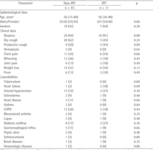
0.08), chest pain (54% vs. 24%; p = 0.06), and productive cough (45% vs. 20%; p = 0.09); however, there were no significant differences between the two groups of patients in terms of the comorbidities (Table 1). The principal clinical hypothesis was specific ILD, which was the clinical hypothesis in 36 (64.3%) of the patients. Of those 36 patients, only 3 were diagnosed with IPF (p = 0.011; Table 2).
The most common radiological findings were alteration of the peribronchial parenchyma, in 69.9% of the patients; nodules, in 64.3%; and diffuse parenchymal alteration, in 55.4%. In patients with a final diagnosis of IPF, the most common radiological findings were parenchymal age was also 56 years (range: 36-80 years), and
only 1 patient was younger than 40 years of age. That patient was clinically suspected of having ILD. The most common symptoms were dyspnea (70%), dry cough (59%), and weight loss (36%). A total of 26 patients (46%) were smokers. The principal comorbidities were hypertension (36%), COPD (25%), and diabetes mellitus (20%). Autoimmune diseases, such as systemic lupus erythematosus, rheumatoid arthritis, and scleroderma, were diagnosed in 14% of the patients. There were significant differences between patients with a final diagnosis of IPF and those with other diagnoses in terms of the frequency of dyspnea (91% vs. 64%; p =
Table 1 - Frequency of the epidemiological data, clinical data, and comorbidities in patients with a diagnosis of idiopathic pulmonary fibrosis and in those with other diagnoses.a
Parameter Non-IPF IPF p
n = 45 n = 11
Epidemiological data
Age, yearsb 56 (15-80) 56 (36-80)
Males/Femalesc 25/20 (55/45) 6/5 (54/46) 0.60
Smokers 19 (42) 7 (64) 0.20
Clinical data
Dyspnea 29 (64) 10 (91) 0.08
Dry cough 28 (62) 5 (45) 0.24
Productive cough 9 (20) 5 (45) 0.09
Hemoptysis 2 (4) 0 (0) 0.64
Chest pain 11 (24) 6 (54) 0.06
Wheezing 12 (26) 2 (18) 0.44
Joint pain 6 (13) 2 (18) 0.49
Weight loss 14 (31) 6 (54) 0.13
Fever 6 (13) 2 (18) 0.49
Comorbidities
Tuberculosis 1 (2) 0 (0) 0.80
Heart failure 1 (2) 2 (18) 0.09
Arterial hypertension 17 (37) 3 (27) 0.39
Scleroderma 2 (4) 1 (9) 0.48
Heart disease 5 (11) 1 (9) 0.66
Asthma 2 (4) 0 (0) 0.64
COPD 12 (26) 2 (18) 0.44
Rheumatoid arthritis 2 (4) 1 (9) 0.35
Lupus 2 (4) 1 (9) 0.48
Diabetes mellitus 8 (17) 3 (27) 0.36
Gastroesophageal reflux 5 (11) 1 (9) 0.66
Peptic ulcer 2 (4) 1 (9) 0.48
Schistosomiasis 1 (2) 0 (0) 0.80
Renal diseases 1 (2) 1 (9) 0.35
Hematologic diseases 1 (2) 0 (0) 0.80
IPF: idiopathic pulmonary fibrosis. aValues expressed as n (%), except where otherwise noted. bValues expressed
Evaluating the IPF patients in comparison with the remaining patients, the fibrotic pattern was less common and the organizing pneumonia pattern was more common.
Of the 56 patients in the sample, 8 (14.3%) had also undergone surgical biopsy for diagnostic investigation. Of those 8 patients, 6 were older than 40 years of age, and 4 had a final diagnosis of IPF. Of those, only 1 was clinically suspected of having a specific ILD (sarcoidosis). The remaining 4 patients had final diagnoses of sarcoidosis (2 patients), ILD (1 patient), and hypersensitivity pneumonia (1 patient). Initially, however, 3 of those patients were suspected of having sarcoidosis.
The radiological and pathological patterns were evaluated in terms of their sensitivity, specificity, PPV, NPV, accuracy, and likelihood ratio. Few radiological findings suggestive of ILD actually indicated the presence of an interstitial disease, as evidenced by the high sensitivity. However, a greater number of changes translated to greater chances of distortion (peribronchial), in 81.8%; diffuse
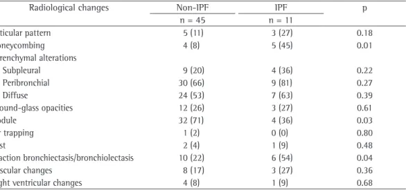
parenchymal alteration, in 63.6%; and traction bronchiectasis, in 54.5% (Table 3). Nodules contributed significantly to the exclusion of the diagnosis of IPF (p = 0.03), whereas honeycombing and traction bronchiectasis aided in confirming the diagnosis of IPF (p = 0.01 and p = 0.043, respectively).
The most common histopathological finding was fibrosis with a diffuse radial pattern (in 32.2%), followed by the indeterminate pattern (in 17.8%), respiratory bronchiolitis (in 14.3%), granuloma (in 12.5%), organizing pneumonia (in 10.7%), the normal pattern (in 7.2%), and other patterns (in 5.3%). Among the patients with a diagnosis of IPF, the indeterminate pattern and the organizing pneumonia pattern were the most common (in 45.4% and 27.3%, respectively). The patterns of granuloma, fibrosis, and others were observed in 9.1% each. There were no cases of respiratory bronchiolitis. The indeterminate pattern was significantly more common among patients with IPF (p = 0.018).
Table 2 - Frequency of patients with clinical suspicion or possible differential diagnosis of interstitial lung disease vs. final diagnosis of idiopathic pulmonary fibrosis.
Type Final diagnosis of IPF Total
Absent Present
Differential diagnosis 12 (21.4) 8 (14.3) 20 (35.7)
Clinical suspicion 33 (58.9) 3 (5.4) 36 (64.3)
Total 45 (80.4) 11 (19.6)* 56 (100)
IPF: idiopathic pulmonary fibrosis. aValues expressed as n (%). *p = 0.011 (Fisher’s exact test).
Table 3 - Frequency of radiological changes in patients with a diagnosis of idiopathic pulmonary fibrosis and in those with other diagnoses.a
Radiological changes Non-IPF IPF p
n = 45 n = 11
Reticular pattern 5 (11) 3 (27) 0.18
Honeycombing 4 (8) 5 (45) 0.01
Parenchymal alterations
Subpleural 9 (20) 4 (36) 0.22
Peribronchial 30 (66) 9 (81) 0.27
Diffuse 24 (53) 7 (63) 0.39
Ground-glass opacities 12 (26) 3 (27) 0.61
Nodule 32 (71) 4 (36) 0.03
Air trapping 1 (2) 0 (0) 0.80
Cyst 2 (4) 1 (9) 0.48
Traction bronchiectasis/bronchiolectasis 10 (22) 6 (54) 0.04
Vascular changes 8 (17) 3 (27) 0.36
Right ventricular changes 4 (8) 1 (9) 0.68
In the present study, honeycombing was the least common radiological finding (14%). However, the presence of honeycombing was highly suggestive of IPF (p = 0.01). Although nodules are extremely common in ILDs, they are uncommon in IPF. Nodules are most common in sarcoidosis, showing high sensitivity (86%). In the absence of this lesion, the possibility of sarcoidosis is more remote (NPV = 90%). The presence of at least six radiological changes had a PPV of 67% for IPF, a value very similar to that found in another study (71%).(18) The PPV
increased as the number of changes increased. However, the ROC curve showed that the best a diagnosis of IPF, as demonstrated by the
increase in specificity (Table 4). The PPV, NPV, accuracy, and likelihood ratio also increased with the number of changes. However, the best relationship between sensitivity and specificity occurred when five radiological changes were present, as shown by the ROC curve (Figure 1). Regarding the histological patterns, fibrosis with a diffuse radial pattern showed high specificity and NPV for IPF. However, a diagnosis of IPF cannot be confirmed by a finding of fibrosis (Table 4).
The study of the diagnostic tests for sarcoidosis revealed that a radiological finding of nodules had high sensitivity (85.7%) and NPV (90%) but low specificity (42.8%). In contrast, the histological pattern of granuloma had low sensitivity (42.8%) and high specificity (97.6%).
Discussion
In the present study, the patients suspected of having ILD presented with a highly prevalent clinical and epidemiological profile, meaning that age, gender, prevalence of dyspnea, and prevalence of chest pain were similar to those reported in studies conducted at other referral centers.(15) The same was observed for the
patients with IPF.(16,17)
Clinical suspicion of ILD is fundamental due to the wide variety of differential diagnoses with similar clinical profiles. Our results show that the frequency of IPF cases was greater when IPF was only a possible diagnosis and lower when ILD was the prime suspect (p = 0.011). This underscores the need for ancillary tests in order to establish a definitive diagnosis.
Table 4 - Correlation of radiological and histological changes with the diagnosis of idiopathic pulmonary fibrosis.
Type Sensitivity, % Specificity, % PPV, % NPV, % Accuracy, % LR Radiological changes, n
≥ 1 90.9 2.2 18.5 50.0 19.6 0.5
≥ 2 90.9 13.3 20.4 85.7 28.6 0.6
≥ 3 72.7 33.3 21.0 83.3 41 0.8
≥ 4 72.7 55.5 28.6 89.3 58.9 0.5
≥ 5 63.0 86.7 53.8 90.7 82.1 0.4
≥ 6 18.1 97.7 66.7 83.0 82.1 0.8
Histological changes
Radial fibrosis 9.0 86.6 14.2 79.6 71.4 1.0
Diffuse fibrosis 9.0 71.1 7.1 76.1 58.9 1.3
PPV: positive predictive value; NPV: negative predictive value; and LR: likelihood ratio.
revealed values that were very similar to those found at large referral centers and should be used in all hospitals at which the required equipment is available and there are qualified, multidisciplinary medical teams working in concert to treat patients with ILDs.
References
1. American Thoracic Society; European Respiratory Society. American Thoracic Society/European Respiratory Society International Multidisciplinary Consensus Classification of the Idiopathic Interstitial Pneumonias. This joint statement of the American Thoracic Society (ATS), and the European Respiratory Society (ERS) was adopted by the ATS board of directors, June 2001 and by the ERS Executive Committee, June 2001. Am J Respir Crit Care Med. 2002;165(2):277-304. Erratum in: Am J Respir Crit Care Med. 2002166(3):426.
2. Swigris JJ, Brown KK. Idiopathic pulmonary fibrosis: a decade of progress. J Bras Pneumol. 2006;32(3):249-60. 3. Fellrath JM, du Bois RM. Idiopathic pulmonary
fibrosis/cryptogenic fibrosing alveolitis. Clin Exp Med. 2003;3(2):65-83.
4. Costabel U, Ohshimo S, Guzman J. Diagnosis of sarcoidosis. Curr Opin Pulm Med. 2008;14(5):455-61. 5. Ishie RT, Cardoso JJ, Silveira RJ, Stocco L. Video-assisted
thoracoscopy for the diagnosis of diffuse parenchymal lung disease. J Bras Pneumol. 2009;35(3):234-41. 6. Gaensler EA, Carrington CB. Open biopsy for
chronic diffuse infiltrative lung disease: clinical, roentgenographic, and physiological correlations in 502 patients. Ann Thorac Surg. 1980;30(5):411-26. 7. Kramer MR, Berkman N, Mintz B, Godfrey S, Saute M,
Amir G. The role of open lung biopsy in the management and outcome of patients with diffuse lung disease. Ann Thorac Surg. 1998;65(1):198-202.
8. Bensard DD, McIntyre RC Jr, Waring BJ, Simon JS. Comparison of video thoracoscopic lung biopsy to open lung biopsy in the diagnosis of interstitial lung disease. Chest. 1993;103(3):765-70.
9. Danel C. Usefulness of transbronchial and surgical biopsies for the management of interstitial lung disease [Article in French]. Rev Pneumol Clin. 2005;61(3):149-57.
10. Balfe D, Mohsenifar Z. Downward trends in bronchoscopies performed between 1991 and 1997. Chest. 1999(1):238-42.
11. Fabro AT, Yoo HH, Queluz TH. Profile of research published in the annals of the Brazilian Pulmonology and Phthisiology Conferences held over the last twenty years. J Bras Pneumol. 2006;32(4):309-15.
12. Wall CP, Gaensler EA, Carrington CB, Hayes JA. Comparison of transbronchial and open biopsies in chronic infiltrative lung diseases. Am Rev Respir Dis. 1981;123(3):280-5.
13. Sackner MA. Bronchofiberscopy. Am Rev Respir Dis. 1975;111(1):62-88.
14. Silva CI, Marchiori E, Souza Júnior AS, Müller NL; Comissão de Imagem da Sociedade Brasileira de Pneumologia e Tisiologia. Illustrated Brazilian consensus of terms and fundamental patterns in chest CT scans. J Bras Pneumol. 2010;36(1):99-123.
relationship between sensitivity and specificity occurred when five changes were present. This difference shows the need for qualifying such changes. Although the number of radiological changes is important, the patterns observed must be those that the literature associates with high specificity for IPF, such as honeycombing.
(18-20)
The predominant histopathological pattern in IPF was the indeterminate pattern. This is due to the peripheral characteristic of IPF(21,22) and
rules out other differential diagnoses. Therefore, the indeterminate pattern was associated with consistent clinical and radiological findings, which aided in establishing a final diagnosis of IPF.(22,23) We find it curious that 27.3%
of the patients presented with a pattern of organizing pneumonia, which can be suggestive of greater disease severity.(24) There is currently
no treatment that can change the prognosis and complications of this type of profile.(9,16) In
general, fibrosis was the most common finding, similarly to what has been reported in other studies.(25) Fibrosis with a radial pattern showed
high specificity and high NPV and might have resulted from the extension of peripheral fibrosis. Although the granulomatous pattern was a common TBB finding, it was shown to be far more specific for sarcoidosis, having high PPV and NPV, in agreement with the findings of a previous study.(26) Therefore, in order to
increase the diagnostic yield, it is fundamental that specialists in ILDs evaluate the clinical, radiological, and histopathological aspects in conjunction.(27)
The patients with ILD investigated in the present study had the same clinical, radiological, and pathological profiles as those of patients treated at other referral centers. For patients with clinical suspicion of ILD and for those in whom ILD is only a possible differential diagnosis, CT is an indispensable ancillary test. In some cases, the use of CT in combination with clinical and functional evaluation is sufficient to establish a diagnosis. The histopathological analysis complements the diagnostic rationale; however, the use of histopathological analysis and the type of biopsy employed (TBB or surgical biopsy) should be considered on a case-by-case basis.
21. Parra ER, Otani LH, de Carvalho EF, Ab’Saber A, Capelozzi VL. Systemic sclerosis and idiopathic interstitial pneumonia: histomorphometric differences in lung biopsies. J Bras Pneumol. 2009;35(6):529-40. 22. Gonçalves JJ, Leão LE, Ferreira RG, Oliveira R, Ota LH,
dos Santos RS. Semiquantitative analysis of surgical biopsies of distinct lung lobes of patients with usual interstitial pneumonia/idiopathic pulmonary fibrosis. J Bras Pneumol. 2009;35(7):676-82.
23. Capelozzi VL. Dificuldades na interpretação de biópsias em doenças pulmonares difusas. J Pneumol. 1998;24(1):30-42
24. Katzenstein AL, Mukhopadhyay S, Myers JL. Diagnosis of usual interstitial pneumonia and distinction from other fibrosing interstitial lung diseases. Hum Pathol. 2008;39(9):1275-94.
25. Berbescu EA, Katzenstein AL, Snow JL, Zisman DA. Transbronchial biopsy in usual interstitial pneumonia. Chest. 2006;129(5):1126-31.
26. Hsu RM, Connors AF Jr, Tomashefski JF Jr. Histologic, microbiologic, and clinical correlates of the diagnosis of sarcoidosis by transbronchial biopsy. Arch Pathol Lab Med. 1996;120(4):364-8.
27. Hunninghake GW, Zimmerman MB, Schwartz DA, King TE Jr, Lynch J, Hegele R, et al. Utility of a lung biopsy for the diagnosis of idiopathic pulmonary fibrosis. Am J Respir Crit Care Med. 2001;164(2):193-6.
15. Raghu G, Mageto YN, Lockhart D, Schmidt RA, Wood DE, Godwin JD. The accuracy of the clinical diagnosis of new-onset idiopathic pulmonary fibrosis and other interstitial lung disease: A prospective study. Chest. 1999;116(5):1168-74.
16. Johnston ID, Prescott RJ, Chalmers JC, Rudd RM. British Thoracic Society study of cryptogenic fibrosing alveolitis: current presentation and initial management. Fibrosing Alveolitis Subcommittee of the Research Committee of the British Thoracic Society. Thorax. 1997;52(1):38-44. 17. Brauner MW, Grenier P, Mompoint D, Lenoir S, de
Crémoux H. Pulmonary sarcoidosis: evaluation with high-resolution CT. Radiology. 1989;172(2):467-71. 18. Johkoh T, Müller NL, Cartier Y, Kavanagh PV, Hartman
TE, Akira M, et al. Idiopathic interstitial pneumonias: diagnostic accuracy of thin-section CT in 129 patients. Radiology. 1999;211(2):555-60.
19. MacDonald SL, Rubens MB, Hansell DM, Copley SJ, Desai SR, du Bois RM, et al. Nonspecific interstitial pneumonia and usual interstitial pneumonia: comparative appearances at and diagnostic accuracy of thin-section CT. Radiology. 2001;221(3):600-5. 20. Lynch DA, Travis WD, Müller NL, Galvin JR, Hansell DM,
Grenier PA, et al. Idiopathic interstitial pneumonias: CT features. Radiology. 2005;236(1):10-21.
About the authors
Cristiano Claudino Oliveira
Medical Student. Faculdade de Medicina de Botucatu, Universidade Estadual Paulista – FMB-UNESP, São Paulo State University Botucatu School of Medicine – Botucatu, Brazil.
Alexandre Todorovic Fabro
Doctoral Student in Pathology. Department of Pathology, Faculdade de Medicina de Botucatu, Universidade Estadual Paulista – FMB-UNESP, São Paulo State University Botucatu School of Medicine – Botucatu, Brazil.
Sérgio Marrone Ribeiro
Assistant Professor. Diagnostic Imaging Section of the Department of Tropical Diseases and Diagnostic Imaging, Faculdade de Medicina de Botucatu, Universidade Estadual Paulista – FMB-UNESP, São Paulo State University Botucatu School of Medicine – Botucatu, Brazil.
Julio Defaveri
Adjunct Professor. Department of Pathology, Faculdade de Medicina de Botucatu, Universidade Estadual Paulista – FMB-UNESP, São Paulo State University Botucatu School of Medicine – Botucatu, Brazil.
Vera Luiza Capelozzi
Adjunct Professor. Department of Pathology, Faculdade de Medicina, Universidade de São Paulo – FMUSP, São Paulo University School of Medicine – São Paulo, Brazil.
Thais Helena Thomaz Queluz
Full Professor. Pulmonology Section of the Department of Clinical Medicine, Faculdade de Medicina de Botucatu, Universidade Estadual Paulista – FMB-UNESP, São Paulo State University Botucatu School of Medicine – Botucatu, Brazil.
Hugo Hyung Bok Yoo