The effect of hypoxia and reoxygenation in the response of mesangial cells to angiotensin II in vitro
Texto
Imagem

Documentos relacionados
The numbers of MAC-2-positive cells in the peribronchial region and distal lung parenchyma were higher in the CS + PPE group than in the other groups, as were the numbers of
vegrandis crude venom, hemorrhagic, and neurotoxic fractions were evaluated on mouse primary renal cells and a continuous cell line of Vero cells maintained in vitro.. Cells
The results about bacterial recovery from cells studied showed that there were fewer bacteria within the Caco-2 cells than in hepatocytes on normoxia and hypoxia conditions
gallinaceum to invade the vector epithelium were in fact an in vitro artifact; the Ross cells are believed to represent cells that have lost their integrity and some of
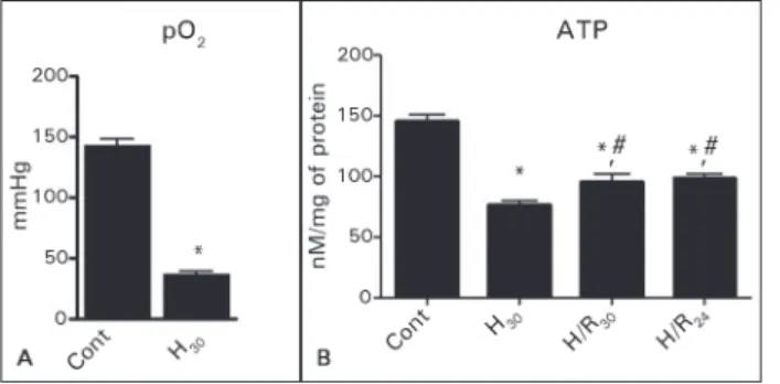
In the present study, cultured hepatocytes were exposed to hypoxia for 2 hours and reoxygenation for 2 hours and compared with simple hypoxia for 4 hours, and also with
However, in experiments where sweet, bitter, and umami stimuli were applied to the same group of cells, a large population of sweet or bitter responsive Type II (Receptor) cells
Comparison of HIF-1 α , PKM2 and ISCU1/2 protein expression in metastatic breast cancer cells and metastatic melanoma cells in the presence and absence of methyl sulfone and
cells have similar levels of Stat1 mRNA (Fig. The expression levels of Mx1 in Stat1 2 / 2 cells and untreated Stat1 ind cells were below detection limits but upon treatment with
