Generation and Characterization of an
Nse
-
CreER
Transgenic Line Suitable for Inducible Gene
Manipulation in Cerebellar Granule Cells
Theresa Pohlkamp1,2*, Laura Steller1, Petra May1,3, Thomas Gu¨nther4, Roland Schu¨le4, Michael Frotscher5, Joachim Herz1,2*., Hans H. Bock1,3
*.
1Center for Neuroscience, Department of Neuroanatomy, Albert-Ludwigs-University, Freiburg, Germany,2Department of Molecular Genetics, University of Texas Southwestern Medical Center, Dallas, Texas, United States of America,3Clinic for Gastroenterology, Hepatology and Infectiology, Heinrich-Heine-University, Du¨sseldorf, Germany,4Department of Urology, University Hospital Freiburg, Freiburg, Germany,5Institute for Structural Neurobiology, Center for Molecular Neurobiology, Hamburg, Germany
Abstract
We created anNse-CreERT2mouse line expressing the tamoxifen-inducible CreERT2recombinase under the control of the
neuron-specific enolase (Nse) promoter. By using Cre reporter lines we could show that this Nse-CreERT2 line has recombination activity in the granule cells of all cerebellar lobules as well as in postmitotic granule cell precursors in the external granular layer of the developing cerebellum. A few hippocampal dentate gyrus granule cells showed Cre-mediated recombination as well. Cre activity could be induced in both the developing and adult mouse brain. The established mouse line constitutes a valuable tool to study the function of genes expressed by cerebellar granule cells in the developing and adult brain. In combination with reporter lines it is a useful model to analyze the development and maintenance of the cerebellar architecture including granule cell distribution, migration, and the extension of granule cell fibersin vivo.
Citation:Pohlkamp T, Steller L, May P, Gu¨nther T, Schu¨le R, et al. (2014) Generation and Characterization of anNse-CreERT2
Transgenic Line Suitable for Inducible Gene Manipulation in Cerebellar Granule Cells. PLoS ONE 9(6): e100384. doi:10.1371/journal.pone.0100384
Editor:Branden Nelson, Seattle Children’s Research Institute, United States of America
ReceivedDecember 20, 2013;AcceptedMay 27, 2014;PublishedJune 20, 2014
Copyright:ß2014 Pohlkamp et al. This is an open-access article distributed under the terms of the Creative Commons Attribution License, which permits unrestricted use, distribution, and reproduction in any medium, provided the original author and source are credited.
Funding:This work was supported by the Deutsche Forschungsgemeinschaft (DFG) (grant numbers SFB 780/TP5 to J.H., H.B. and M.F.), MA2410/1-3 and -4 (P.M.), FR 620/12-1, National Institutes of Health (NIH) grant R37 HL63762 (to J.H.), and Bundesministerium fu¨r Bildung und Forschung (BMBF, e:bio ReelinSys, to H.B.). J.H. was the recipient of a Wolfgang Paul award of the Humboldt Foundation and is further supported by the American Health Assistance Foundation, the Consortium for Frontotemporal Dementia Research, the Bright Focus Foundation, the Lupe Murchison Foundation, and The Ted Nash Long Life Foundation. M.F. is Senior Professor for Neuroscience of the Hertie Foundation. The authors wish to thank Jonathan Go¨ldner, Aileen Koch, Tamara Terrones and Sandy Turner for technical support with genotyping. The funders had no role in study design, data collection and analysis, decision to publish, or preparation of the manuscript.
Competing Interests:The authors have declared that no competing interests exist.
* Email: [email protected] (TP); [email protected] (JH); [email protected] (HHB)
.These authors contributed equally to this work.
Introduction
The cerebellum plays an important role in motor control and is involved in cognitive processing and motor learning [1,2]. The small-sized granule cells (GC) in the cerebellum are the most abundant neurons in the mammalian brain. They are densely packed into a thick layer of the cerebellar cortex, the GC layer (GCL). Next to the GCL of the trilaminated cerebellum, the cell bodies of the Purkinje cells form the Purkinje cell layer. The most superficial layer is the molecular layer (ML), which mainly consists of dendritic and axonal fibers. Amongst these fibers are Purkinje cell dendrites, innervated by climbing fibers (projections from the inferior olivary nucleus of the medulla oblongata) and parallel fibers (the axons of the vast number of the GCs). Moreover, the ML harbors two types of small inhibitory interneurons (basket and stellate cells). During development most cerebellar neurons originate in the ventricular zone of rhombomere 1, except GCs [3]. In the mouse, the vast majority of proliferating GC-precursors (GCPs) start to arise at the rhombic lip between embryonic day 13 (E13) and E16. Some deep cerebellar neurons arise between E9 and E11, even before GCs are generated. From there GCPs migrate tangentially to form the external granular layer (EGL), a
secondary proliferative zone beneath the cerebellar pial surface [4–7]. GCP proliferation in the outer EGL continues until approximately postnatal day 15 [8]. The GCPs exit their cell cycle and start differentiating by migrating into the inner part of the EGL. Postmitotic GCPs in the deep EGL extend bipolar processes, which will become parallel fiber axons, and migrate tangentially – in parallel to the pial surface – before they turn orthogonally, to migrate radially along Bergmann glial fibers. After passing the ML and the Purkinje cell layer, GCs reach the deeper internal granular layer (IGL), which later becomes the mature GCL [3,9].
CreERT2 is a fusion protein of the bacteriophage P1 recombinase (Cre) and the estrogen receptor (ER) whose ligand binding-domain was modified to bind tamoxifen (ERT2 [10]).
Tamoxifen forces the dissociation of the ERT2-bound heat shock protein 90 (HSP90), allowing CreERT2 to translocate into the nucleus. In the nucleus Cre(ERT2) binds to LoxP-sequences and deletes LoxP-flanked genomic parts via recombination. Hence, the Cre(ERT2)/LoxP-system is a useful tool to modify gene expression in a temporally and spatially controlled manner.
transcription. Microinjection of the cloned Nse-CreERT2 cassette yielded one transgenic mouse line (Nse-CreERT2). To characterize Cre recombinase activity after tamoxifen injection, two different reporter lines were used, expressing b-Galactosidase (b-Gal, also known as LacZ) or membrane-tagged GFP (mGFP) after recombination. The results show that CreERT2-recombinase in this mouse line enables a tamoxifen-inducible recombination of LoxP-flanked genomic regions that is specific to the GC population in the cerebellum. In addition, a few hippocampal dentate gyrus granule cells also showed inducible Cre activity. Some fibers crossing the colliculus superior in the midbrain and the medial vestibular nuclei in the medulla were mGFP-postive as well.
To our knowledge, this is the first described mouse line with abundantCreERT2expression in the GCL of all cerebellar lobules, restricted to the GC population. Targeting gene manipulation to cerebellar GCs in a temporally specific manner provides a powerful tool for investigating the function of proteins expressed or secreted by GCs, as well as a means of fate-mapping cerebellar GCs. This will lead to novel insights into the molecular and cellular events of postnatal development and maturation of the cerebellum, as well as GC function in the adult brain.
Material and Methods
Ethics Statement
All experimental procedures were performed according to the approved institutional guidelines for animal care at the University of Freiburg (license no. 35-9185.81; G-08/33) or University of Texas Southwestern Medical Center (IACUC license no. 0701-07-01-4).
Generation of anNse-CreERT2Transgenic Mouse Line
To generate pGS-Nse-CreERT2, the Nse-promoter fragment of pNse-NuKCre[11] was excised using KpnI and NotI. After filling in the restriction sites using Klenow fragment (NEB, DNA Polymer-ase I, Large Klenow Fragment) the promoter was cloned into the SalI-opened, Klenow-treated and dephosphorylated (Alkaline
Phosphatase, NEB) pGS-CreERT2acceptor-vector [12] (Fig. 1A). The orientation of the promoter was verified by SacI and EcoRI restriction digest and PCR (see below).
For microinjection pGS-Nse-CreERT2was cut with NotI and the
Nse-CreERT2cassette was isolated via gel electrophoresis and gel-extraction (Qiagen). The DNA was eluted in 5 mM Tris, pH 7.5 with 0.1 mM EDTA and microinjected into pronuclei of fertilized FVB oocytes using standard procedures. The resulting founder transgenic mouse was transferred to a specific pathogen free (SPF) housing. Breeding of wild type (C57/BL6 Background) and transgenic animals was conducted in accordance with institutional guidelines. Tail biopsies were digested with proteinase K (Applichem) and genotyping was performed byNse-CrePCR (see next section) to screen for transgenic founder animals and for routine genotyping. The line has been deposited at Jackson Laboratories (Jax Stock 022763).
Nse-CrePCR, Genotyping
PCR was performed using transgene-specific primers that bind within theNse-promoter and the Cre-coding regions (forwardNse -primer sequence 59-CGT CAC CAC CGC CAC CGC CAC-39; reverse Cre-primer sequence 59- ACG ACC GGC AAA CGG ACA GAA GCA-39). The reaction mixture contained 67 mM Tris (pH 8.8); 16.6 mM (NH4)2SO4; 6.7 mM MgCl2; 6.7mM EDTA;
0.0348% b-mercaptoethanol; 10% DMSO; 0.1 mg/ml BSA; 1.25 mM dNTPs (each); 1% Taq polymerase; 500 nM primer (each); and template (final reaction volume 20ml). The PCR started with 2 min at 94uC, 2 min at 55uC and 3 min at 67uC, followed by 40 cycles (94uC for 30 s, 55uC for 30 s, and 67uC for 60 s) and a final elongation step of 10 min at 67uC. PCR products (916 bp) were visualized by separation on an 1% agarose gel and staining with ethidium bromide.
Detection ofCreERT2Transcripts via RT-PCR
Whole brain RNA was extracted using the TRIzol method (Invitrogen). After DNase treatment (Fermentas) 2mg of RNA were reverse transcribed (M-MLV-RT, Promega) into cDNA, using random primer mix (Promega). To detect the CreERT2
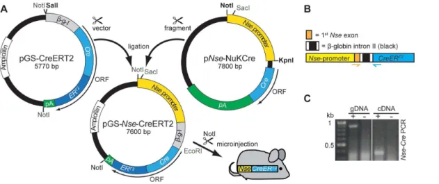
-Figure 1. Cloning of pGS-Nse-CreERT2and transgenic cDNA detection inNse-CreERT2.A: After linearizing (SalI, blunted) the pGS-CreERT2
vector, it was ligated with theNse-promoter fragment of the pNse-NukCreplasmid (cut with NotI and KpnI, blunted). To prove the correct orientation of the fragment, a PCR was performed. TheNse-CreERT2
transgenic cassette (cut with NotI) was used for pronucleus injection. B: Schematic illustration of the transgenic cassette. The chickenb-globin intron II (black, flanked by fragments of theb-globin exons II and III, white) is located between the Nse-promoter (yellow, part of first non-codingNse-Exon in orange) andCreERT2
. To detect the spliced transcript anNse-Cre-PCR was designed where the forward primer is located in theNse-exon and the reverse primer in the Creencoding region (orange and blue arrows, respectively). C: Amplification (+) of genomic DNA (gDNA, 900 bp) and transcript cDNA (350 bp), wt genomic/cDNA was used as negative control (2).
transcript an Nse-Cre-PCR was performed. The Nse-CreERT2
cassette consists of the Nse-promoter containing the entire non-coding exon I, followed by the rabbitb-globin intron II (flanked by parts ofb-globin exon II and III; 39and 59, respectively), and the
CreERT2gene with a poly-A signal. TheNse-Cre-PCR (for protocol see section above) was designed to span the b-globin intron: the forward primer binds to theNse-exon region, the reverse primer to the Cre-coding region (see Fig. 1B). Amplification of genomic DNA yields a 916 bp fragment, the same PCR using cDNA as a template results in a 343 bp fragment (Fig. 1C).
Breeding ofNse-CreERT2Transgenic Mice with Reporter Lines and Tamoxifen Treatment
Mice were crossed with the reporter mouse linesRosa26-LacZ
(abbreviated LacZ: Gt(ROSA)26Sortm1Sor [13]) or R26R-td-tomato-mEGFP(abbreviatedmTmG:Gt(ROSA)26Sortm4(ACTB-tdTomato,-EGFP)Luo
[14]. P3 mice were once intraperitoneally injected with 300mg Tamoxifen (Sigma, T5648, 18 mg/ml in sunflower oil and 10% ethanol) and sacrificed three weeks later (P23) to analyze Cre recombinase activity. Adult mice (.2 months) were intraperito-neally injected with 135mg Tamoxifen/g bodyweight on five subsequent days. Recombination was analyzed not earlier than 10 days after the injection.
Perfusion and Sectioning
Transcardial perfusion started with PB (0.1 M phosphate buffer, pH 7.4) for 5 min (ca. 50 ml), followed by fixative solution with 4% paraformaldehyde in PB (PFA) for an additional 5 min (ca. 50 ml). Removed brains were postfixed in 4% PFA at 4uC overnight and stored in PB with 0.02% NaN3at 4uC. Brains were
embedded in 5% agarose (in PB) and 50mm sections were cut with a Leica VT 1000S Vibratome.
Immunohistochemistry and LacZ Staining
Sections were blocked for 2 h in PB-Tx (PB with 0.1% Triton X) containing 10% donkey serum. To amplify the mGFP (membrane-tagged GFP) signal, sections were incubated with anti-GFP antibody (rabbit, Clontech) at 4uC overnight. After three washing steps with PB-Tx, slices were incubated with the secondary anti-rabbit Alexa Fluor 488-coupled antibody (donkey, Invitrogen) for 2 h at room temperature. Slices were washed once with PB-Tx containing DAPI (1:10.000, Applichem) and twice with PB-Tx, each washing step 20 min at room temperature, and finally mounted with Mowiol.
For co-immunohistochemistry of GFP-positive cells and neuro-nal marker proteins, slices were first blocked for 2 h in PB-Tx containing 10% goat serum and 10% Avidin (Vector Laboratories) at room temperature. The primary antibodies anti-GFP (rabbit, Clontech; for co-detection with GFAP: mouse, Millipore) and anti-NeuN (mouse, Chemicon), anti-GFAP (rabbit, Dako Cytomation), anti-Calbindin (mouse, Swant), or anti-Map2 (mouse, Leinco) were incubated in PB with 10% Biotin (Vector Laboratories) for 48 h at 4uC. After washing in PB-Tx the slices were incubated with secondary biotinylated antibody directed against the neuronal marker antibody (anti-mouse/anti-rabbit) for 2 h at room temperature. Incubation with AMCA-Avidin D (goat, Vector Laboratories; to detect biotin) and the secondary Alexa Fluor 488-coupled antibody (donkey, Invitrogen; directed against anti-mouse/anti-rabbit GFP) in PB-Tx was performed, followed by an additional washing step for 2 h at room temperature. Anti-Parvalbumin antibody (rabbit, Swant) was used for co-immuno-histochemistry with anti-GFP (mouse, Clontech) and detected with
Alexa Fluor 350 and 488 secondary antibodies (goat, Invitrogen). After a final washing step the slices were mounted with Mowiol.
For LacZ staining first the slices were incubated on ice, twice (5 min and 10 min) with 2 mM MgCl2in PB, and once for 10 min
in Detergent Rinse (2 mM MgCl2, 0.01% Natriumdeoxycholate,
and 0.02% NP40 in PB). Afterwards, the slices were incubated in staining solution (Detergent Rinse plus 5 mM Potassium ferricy-anide, 5 mM Potassium ferrocyferricy-anide, and 1 mg/ml x-Gal) for 3 h at 37uC. After washing, the slices were mounted with Mowiol. Pictures were imaged on a Zeiss Axioplan 2 microscope.
Results
Generation of anNse-CreERT2-Line
To enable an inducible, neuron-specific Cre recombination of LoxP-flanked target-regions, anNse-CreERT2cassette was designed and microinjected into oocytes to generateNse-CreERT2transgenic mice. TheNse-promoter contains 1.8 kb DNA of the rat neuron-specific enolase (NSE) gene promoter, described in Cinato et al. [11], including 43 bp of the first noncoding exon. In the Nse-CreERT2
construct the promoter is followed by the rabbitb-globin intronic enhancer sequence (spanning intron II and containing 18 bp of the 59flanking exon II and 53 bp of the 39flanking exon III, seeFig. 1B) to increase expression levels of the downstream-locatedCreERT2-gene.
To verify the function of theNse-CreERT2
constructin vitroprior to the generation of transgenic mice, neuroblastoma cells (N2A) were transiently co-transfected with pGS-Nse-CreERT2and a test-plasmid containing a LoxP-flanked region. Recombination was verified by PCR and only occurred in co-transfected cells that were treated with 4-Hydroxytamoxifen (data not shown).
Pronucleus injection of the linearized construct yielded one founder mouse. To detect theCreERT2transcript by RT-PCR, a primer pair was designed that spans the b-globin intron II, allowing us to distinguish the PCR product of the genomic
CreERT2gene from the cDNA by their transcript sizes (916 versus 343 bp, respectively,Fig. 1C).
Pattern of Inducible Cre Activity in the Generated Mouse Line
To analyze the expression pattern and function of Cre recombinase, different reporter lines are available, carrying LoxP modified marker-genes. Here, the mTmG [14] and the LacZ
(Rosa26-LacZ[13]) reporter strains were used. InmTmGmice the CMVb-actin enhancer-promoter (CAG [15]) and two adjacent marker-genes are targeted into theRosa26locus. The first gene, encoding a membrane-tagged Tomato fluorescence protein (mT or mTomato), is flanked by LoxP sites and will be expressed as long as recombination does not occur. After Cre-mediated excision ofmTomatothe second gene, encoding membrane-tagged GFP (mG or mGFP), will be transcribed, switching the cell from producing a red to a green signal. To amplify the mGFP signal an anti-GFP antibody was used.
were found in the colliculus superior (midbrain, Fig. 3B) and medial vestibular nuclei (medulla,Fig. 3E).
Distribution of Cre Recombinase Activity in the Cerebellum
In adult mice the GFP signal was detected in the entire region of the cerebellar ML and GCL when injected with tamoxifen at P3 or at adult age. In the GCL, the individual GC-somata were visible, whereas the staining in the ML was too intense to differentiate between individual structures (Fig. 5C and D). However, in the P3-injected early postnatal (P8) and juvenile (P23) mice a less intense fluorescence allowed the identification of fibrous structures (Fig. 5A and B) extending from the GCL into the ML. In P8 animals the trailing and/or leading processes of GCs are labeled in the inner EGL, ML, and IGL (Fig. 5A and 6),
demonstrating that some postmitotic GCPs express CreERT2. At P23, fluorescent somata were only seen in the GCL, and in the ML fibrous structures were labeled. In contrast, mGFP fluores-cence of GC somata in the IGL/GCL was of comparable intensity and density in all mice (Fig. 5 A-D; Fig. S1).
Cre-Mediated Recombination is Restricted to Cerebellar GCs
To clarify the identity of the mGFP-labeled fibrous structures, a Calbindin and GFAP co-staining, labeling Purkinje cell dendrites and the Bergmann glial fibers, was performed. Neither Calbindin-nor GFAP-positive structures showed a co-immuCalbindin-noreactivity with mGFP (Fig. 7B,C). NeuN-immunoreactivity was found in the nuclei of mGFP-postive cells (Fig. 7A and Fig. S1). NeuN is an established GC-marker; it is not expressed in Purkinje cells [16],
Figure 2. Tamoxifen-induced CreERT2-activity in the juvenile and mature brain of P3- and adult-injectedNse-CreERT2;mTmGmice.
In control mice not carrying aCreERT2
-tansgene (A) or inCreERT2
-transgenic mice without tamoxifen injection (B), the mGFP reporter was not labeled. C-E: Tamoxifen injection of P3 (C, D) and adult (E, section more lateral, compared to A-D) mice led to a strikingly similar fluorescence pattern of mGFP in the juvenile (P23 in C) or mature (D and E) brain. mGFP fluorescence was found to be most intense in the cerebellum. Less intense staining of cells and fibers was observed in the hippocampus, and some fibers in nuclei of the midbrain and pons showed mGFP-immunoreactivity.
interneurons of the ML, and several other non-GC cerebellar neurons [17]. When injected at P3 and examined at P8 a small fraction of NeuN-positive cells in the EGL expressed mGFP but several GCs in the IGL were co-labeled (Fig. S1A). In the more juvenile and mature brain of P3-injected animals, the majority of NeuN-stained GCs showed mGFP expression (Fig. S1B and C). In the P8 and P23 brain some mGFP-positive structures in the IGL or GCL, respectively, are not surrounding NeuN-positive nuclei (Fig. 7A and Fig. S1A and B) fewer of these structures were found in the adult brain (Fig. S1C). MAP2-immunoreac-tivity, visualizing dendritic structures, overlapped with mGFP in
the GCL (Fig. 7D). Lack of co-immunostaining of mGFP with Parvalbumin, which labels stellate and basket cells in the ML [18,19], excludes the possibility that these interneurons express
CreERT2(Fig. 7E).
To further clarify that recombination is restricted to GCs in the cerebellum, another reporter line was used. In theLacZreporter line aLacZgene, with a preceding floxed stop cassette, is targeted into theRosa26locus, ensuring thatb-Gal is expressed only after recombination. The gene product hydrolyzes X-gal, leading to the precipitation of 4-chloro-3-brom-indigo inLacZ expressing cells. LacZ staining of an adult-injectedNse-CreERT2
mouse carrying the
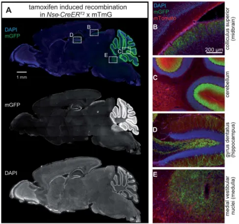
Figure 3. CreERT2-activity inNse-CreERT2;mTmGafter tamoxifen injection.Sagittal section showing mGFP-immunoreactive cells (green, A) indicating that recombination has occurred (mGFP/single channel in the middle) and DAPI-stained nuclei (blue/single channel at the bottom). Enlargements of the framed rectangles in sections B to E are shown on the right: (B) fibers in a region of the colliculus superior in the midbrain, (C) layers of the cerebellum, (D) hippocampal granule cells and their fibers, and (E) fibers in the medial vestibular nuclei (medulla) show green fluorescence. Red (mTomato) fluorescence in 1–4 shows cells where no recombination occurred.
doi:10.1371/journal.pone.0100384.g003
Figure 4. Dentate gyrus ofNse-CreERT2;mTmGafter tamoxifen injection.Left: DAPI staining of dentate gyrus, the red-framed area is shown
enlarged. Middle: mGFP-immunoreactive cells where recombination has taken place are located in the outer part of the GCL. White arrowhead points to the soma, white arrow to an axon, black arrowhead to a dendrite of GFP-immunoreactive granule cells. Right: Merged picture showing nuclei in blue (DAPI), cells without recombination in red (mTomato autofluorescence) and cells where recombination occurred in green (mGFP-immunoreactive cells). Scale bar = 50mm.
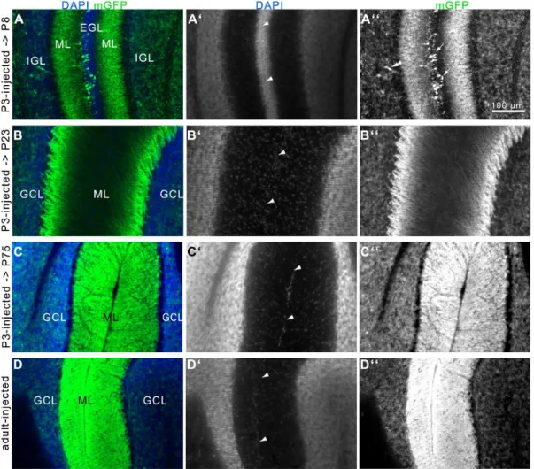
Figure 5. Development of the GCL and ML in the cerebellum ofNse-CreERT2;mTmGmice after tamoxifen-induction.A-C: P3-injected
mice were killed at P8 (A), P23 (B), or P75 (C). Pictures show the EGLs (A) or MLs (B-D) next to a fissure (arrowheads in A9-D9) of two lobules. A: At P8 some mGFP-postive GCPs are found in the EGL (arrows in A0) and a substantial amount of GCs are labeled in the IGL. Double arrowhead points to a presumably migrating cell with leading/trailing processes. Structures in the ML are immunoreactive as well. B: At P23 cells are labeled in the GCL, and structures are labeled in the ML. C: At P75, the labeling of GCs in the GCL is comparable to P8 and P23, but the structures in the ML show a more brightly immunolabeling, filling the entire ML. D: The mGFP labeling in the GCL and ML of an adult-injected mouse is strikingly similar to that of the P3-injected adult mouse in C. A-D = merge, A9-D9 = DAPI, A0-D0 = mGFP. Scale bar = 100mm.
doi:10.1371/journal.pone.0100384.g005
Figure 6. mGFP-labeled, postmitotic migrating GCPs in the EGL with trailing and leading processes at P8. Left: P3-injected P8 cerebellum, mGFP = cells after recombination, mTomato = cells without recombination. Frames A-C are shown enlarged, A9-C9 = further enlargement to show mGFP-immunoreactive GCPs in the inner EGL, A0-C0= mTomato. Arrowheads in A-C point to the fissure between two lobules. Arrows presumable point to tangentially (A9, with leading and trailing processes) or radially (B9, C9, with leading and/or trailing processes) migrating GCPs in the EGL.
LacZreporter demonstrated that the blue precipitate was restricted to the somata of cells in the cerebellar GCL (Fig. 8), whereas in the ML staining was absent. To further ensure that Cre-activity is restricted to the GCL, a co-immunostaining against Parvalbumin and b-Gal was performed:b-Gal labeling was absent in the ML and did not co-stain with Parvalbumin labeled interneurons or with Purkinje cells (Fig S2).
Discussion
The Nse-CreERT2
transgenic mouse line described here was established with the purpose to provide a neuron-specific, temporally inducible recombination of LoxP-flanked genomic regions. To achieve this, a promoter of the neuron-specific enolase (Nse) gene containing a 1.8 kb DNA-fragment upstream of the rat
Nse-coding region was used.Nse-transgenic mouse lines using this
promoter have been described where aCreorLacZtransgene was ubiquitously expressed in neurons [11,20]. However, the expres-sion levels and pattern of Nse-promoter-driven Cre and LacZ
transgenes significantly varied between the founder lines estab-lished by Cinato et al.[11], Forss-Petter et al.[20], and Kwon et al.[21]; and several founder lines showed a dispersed neuronal expression as result of position effects that are common in transgenic animals [22,23]. In our NSE-CreERT2line, CreERT2 was almost exclusively produced in the GCs of the cerebellum.
Pattern of Cre Recombinase Activity After Tamoxifen Injection Outside the Cerebellum
OurNse-CreERT2line shows a distinct expression ofCreERT2in the cerebellum that is strikingly restricted to cells in the GCL. The hippocampus showed restricted CreERT2 mediated recombina-tion, too. Cre recombinase activity was observed in the outer
Figure 7. Cerebellar lobules of tamoxifen-injectedNse-CreERT2;mTmGmice labeled for different neuronal marker proteins.A-E:
mGFP-immunoreactivity is shown in green (mGFP), mGFP-immunoreactivity for different cell type specific markers is shown in the red channel (labeled with blue fluorescence). Arrowheads point to the fissure between two lobules. A: NeuN (in the cerebellum a GC-specific marker), B: GFAP (produced by cells of glial lineage), C: Calbindin (CB, produced by Purkinje cells), D: MAP2 (located in neuronal dendrites), and E: Parvalbumin (PV, expressed by Purkinje cells and ML interneurons). NeuN was mainly found in the GCL and cells were co-immunoreactive for GFP. GFAP-labeled structures showed no GFP-signal, nor did Purkinje cells or the interneurons of the ML. MAP2-positive structures were densely immunostained in the GCL, representing the dendrites of GCs. Scale bar = 50mm.
granule cell layer of the dentate gyrus. Dentate gyrus granule cells in the outermost layer are the most mature neurons, while granule cell progenitors reside in the subgranular zone at the inner side of the granule cell layer [24]. InNse-CreERT2;mTmGmice we could not see any obvious differences in the amount and location of mGFP-postive granule cells in the gyrus dentatus of P3 and adult-injected animals.
In addition, immunoreactive fibers in different brain regions were found, e.g. in the superficial-most layers of the superior colliculus, where retinocollicular projections terminate [25,26]. Some mGFP-postive fibers were also found in the medial vestibular nuclei of the medulla that are traversed by neurons of the vestibular nerve terminating in the cerebellar nuclei. In general, the same pattern of Cre activity was found in adult-injected animals and young (P8), juvenile (P23), or adult (P75) P3-injected animals.
Initiation of CreERT2Expression in Cerebellar GCPs and Mature GCs
The proliferation of GCP in mice starts shortly before birth and lasts until P15 [5]. Migration and differentiation continues until adulthood [6]. Depending on the age of the animal at the time of sacrifice a difference in the appearance of mGFP-postive cerebellar fibers was noted, whereas the density of mGFP-postive GC-somata in the GCL was comparable. In P3- or adult-injected mice, analyzed at two months or older, the immunoreactivity of fibers filled the entire ML, whereas in P3-injected mice sacrificed at P8 and P23 the fibrous structures extending from the inner to the outer region of the ML thinned out. Whereas in the P23 cerebellum all GCs are located in the mature GCL, at P8 GCPs are still present in the EGL, before migrating into the IGL. In P3-injected P8 animals GCPs are labeled in the EGL, demonstrating that transcription initiation of the CreERT2 transgene starts in GCPs. As only a few GCPs in the inner EGL are labeled,CreERT2
expression is not yet active in proliferating GCPs. mGFP-labeled cells in the inner EGL extend radial leading processes which are used by GCPs to migrate along radial Bergmann glial fibers [27,28]. GCPs that seem to pursue a tangential route of migration were seen, too (Fig. 6A). The low amount of mGFP-labeled GCPs in the EGL at P8 can be the result of the restricted effective period of tamoxifen action (injected at P3): due to different metabolic activity, tamoxifen-induced recombination events in the embryo are restricted to a short time frame (up to 48 h), compared to several days/up to weeks in the adult brain [29]. With these data one can estimate that P3-injected tamoxifen loses its effect hours/ days before P8. Thus, it can be assumed that at P8 moreCreERT2
expressing postmitotic GCPs are present than are effectively labeled by the CreERT2 reporter gene product.
Depending on the age of the mice the appearance of mGFP-labeled GC processes ascending into the ML varies. GCs ascend their axons into the ML, where they bifurcate orthogonally into
parallel fibers. Parallel fibers are oriented transversally and are therefore cut orthogonally in sagittal sections. They innervate the dendritic trees of Purkinje cells that in turn are orientated in the sagittal plane. The developmental organization of the ML is well described, especially with regard to the maturation of the Purkinje cell dendrites. The dendritic tree of Purkinje cells expands massively between P7 and P30, reaching its lateral boundaries at P12–15 and expanding to the borders of the pia mater until P30 [30]. Recent findings show that this organization is strictly confined to sagittal planes until the fourth postnatal week [31]. Furthermore, dendritic arborization is dependent on GCs and their parallel fibers [32–34]. Finally the dendritic tree at P23 does not yet fully expand to the pia mater, as do the innervating parallel fibers. In the P8 and P23 cerebellum, we observe fiber structures in the ML that can be related to leading processes and/or ascending axons from migrating and/or matured GCs. We could not clearly define mGFP-labeled parallel fibers in the ML, which can either be due to the orientation of the sections (sagittal) or due to the distribution of mGFP in the axons. At P8 mGFP is highly present in the leading and trailing processes of migrating GCs. However, in the adult cerebellum the cell bodies of mature GCs are restricted to the GCL and only their ascending axons and parallel fibers extend into the ML, which is brightly labeled with mGFP at this point. Hence the ascending axons of GCs and/or their parallel fibers are mGFP-labeled in the mature cerebellum. For a better visualization of cell morphology, reporter strains expressing non-membrane tagged fluorescent proteins like the Ai6- (ZsGreen) or Ai14- (tdTomato) reporter [35] can be considered.
Coverage of GCs ExpressingCreERT2
With both reporters we could detect that abundant recombi-nation occurred in the GCL in all lobules. NeuN immunoreac-tivity labels the nucleus of neurons. We used NeuN as an established GC-specific marker [17]: Co-immunohistochemistry of mGFP and NeuN – even though tamoxifen was injected at P3 – showed an extensive overlap in the IGL/GCL of animals sacrificed at different ages (Fig. S1). Some mGFP-positive structures in the IGL and GCL are not surrounding NeuN-positive nuclei. Fewer of these structures are found in the adult (P75) compared to the pup (P8) and juvenile (P23) cerebellum. Proliferation of GCs lasts until P15, then they migrate to their final position in the IGL where the dendritic development occurs to establish synaptic connections to mossy fiber axons. During this transformation, there is a phase of production of provisional dendrites, followed by retraction and pruning of processes that last until adulthood where the distal ends of the remaining dendrites finally have a claw-like morphology [36,37]. These claw-like structures are part of synaptic glomeruli that are located in the GCL and are composed of large mossy fiber axon terminals, surrounded by dozens of GC dendrites, as well as Golgi cell axons and dendrites [37–39]. mGluR2 Immunostaining of Golgi cells
Figure 8. Cerebellar LacZ-staining ofNse-CreERT2;LacZmice.A: Schematic overview, B: LacZ staining (blue) is restricted to the GCL, one lobule is shown enlarged in (C). WM = white matter.
illustrates their cell bodies and their processes terminating in glomeruli [40]. Whereas the Golgi cells are larger in size (around 20mm), the diameter of a glomerulus (5–10mm) is similar to the GC diameter (6–8mm). The mGFP-positive structures not surrounding NeuN-labeled nuclei can be attributed to synaptic glomeruli and (in the developing cerebellum) provisional dendrites. In contrast, the size and shape of mGFP-labeled structures clearly resembles somata of GCs (Fig. S1).
While the GC morphology with the membrane-tagged fluores-cent reporter protein mGFP was difficult to track, the LacZ reporter clearly showed that CreERT2-positive cells were restricted to the GCL. However, at this point we cannot clearly say if all GCs express CreERT2 since GCs are very tiny and densely packed, which makes it difficult to differentiate between single cells to quantify the coverage.
Of note, other transgenic mouse lines with an inducible Cre recombinase that is active in cerebellar GCs have been described before; however, none of them cause a specific and inducible recombination in the GCs of all lobules in both the developing and adult cerebellum [41–43].
Conclusion
The describedNse-CreERT2transgenic mouse is a powerful tool to address different aspects of cerebellar development. The line has been deposited at the Jackson laboratories (Jax Stock 022763). Mature GCs and their fibers as well as GCPs in the inner EGL can be fate-tracked at different developmental time points by using different reporter lines. Additionally individual floxed genes can be
knocked out to study their function in a time-dependent and GC-specific manner. This will allow to tackle questions concerning maturation of the cerebellum and the assembly of the network of GC ascending axons and parallel fibers in the ML. In addition, the role of distinct presynaptic proteins in synaptic plasticity can be addressed using this mouse model, since long term depression is a prominent type of synaptic plasticity at the parallel fiber-Purkinje cell dendrite synapses and a widely accepted model for storing motor memories [44].
Supporting Information
Figure S1 Cerebellar lobules of P3-injectedNse-CreERT2;mTmG
mice co-labeled for NeuN and mGFP. (TIF)
Figure S2 b-Gal immunoreacitivity was absent in the ML and did not co-label with Parvalbumin.
(TIF)
Text S1 Supplementary Materials and Methods, Figure Legends S1 and S2.
(DOCX)
Author Contributions
Conceived and designed the experiments: JH. Performed the experiments: TP LS TG. Analyzed the data: TP HHB JH. Contributed reagents/ materials/analysis tools: HHB JH MF PM RS. Wrote the paper: TP.
References
1. Chizhikov V, Millen KJ (2003) Development and malformations of the cerebellum in mice. Mol Genet Metab 80: 54–65.
2. Schmahmann JD (2004) Disorders of the cerebellum: ataxia, dysmetria of thought, and the cerebellar cognitive affective syndrome. J Neuropsychiatry Clin Neurosci 16: 367–378.
3. Hatten ME (1999) Central nervous system neuronal migration. Annu Rev Neurosci 22: 511–539.
4. Hanaway J (1967) Formation and differentiation of the external granular layer of the chick cerebellum. J Comp Neurol 131: 1–14.
5. Miale IL, Sidman RL (1961) An autoradiographic analysis of histogenesis in the mouse cerebellum. Exp Neurol 4: 277–296.
6. Fujita S, Shimada M, Nakamura T (1966) H3-thymidine autoradiographic studies on the cell proliferation and differentiation in the external and the internal granular layers of the mouse cerebellum. J Comp Neurol 128: 191–208. 7. Machold R, Fishell G (2005) Math1 is expressed in temporally discrete pools of
cerebellar rhombic-lip neural progenitors. Neuron 48: 17–24.
8. Fujita S (1967) Quantitative analysis of cell proliferation and differentiation in the cortex of the postnatal mouse cerebellum. J Cell Biol 32: 277–287. 9. Herrup K, Kuemerle B (1997) The compartmentalization of the cerebellum.
Annu Rev Neurosci 20: 61–90.
10. Indra AK, Warot X, Brocard J, Bornert JM, Xiao JH, et al. (1999) Temporally-controlled site-specific mutagenesis in the basal layer of the epidermis: comparison of the recombinase activity of the tamoxifen-inducible Cre-ER(T) and Cre-ER(T2) recombinases. Nucleic Acids Res 27: 4324–4327.
11. Cinato E, Mirotsou M, Sablitzky F (2001) Cre-mediated transgene activation in the developing and adult mouse brain. Genesis 31: 118–125.
12. Feil R, Wagner J, Metzger D, Chambon P (1997) Regulation of Cre recombinase activity by mutated estrogen receptor ligand-binding domains. Biochem Biophys Res Commun 237: 752–757.
13. Soriano P (1999) Generalized lacZ expression with the ROSA26 Cre reporter strain. Nat Genet 21: 70–71.
14. Muzumdar MD, Tasic B, Miyamichi K, Li L, Luo L (2007) A global double-fluorescent Cre reporter mouse. Genesis 45: 593–605.
15. Niwa H, Yamamura K, Miyazaki J (1991) Efficient selection for high-expression transfectants with a novel eukaryotic vector. Gene 108: 193–199.
16. Wolf HK, Buslei R, Schmidt-Kastner R, Schmidt-Kastner PK, Pietsch T, et al. (1996) NeuN: a useful neuronal marker for diagnostic histopathology. J Histochem Cytochem 44: 1167–1171.
17. Weyer A, Schilling K (2003) Developmental and cell type-specific expression of the neuronal marker NeuN in the murine cerebellum. J Neurosci Res 73: 400– 409.
18. Zilla P, Celio MR, Fasol R, Zenker W (1985) Ectopic parvalbumin-positive cells in the cerebellum of the adult mutant mouse ’nervous’. Acta Anat (Basel) 124: 181–187.
19. Kosaka T, Kosaka K, Nakayama T, Hunziker W, Heizmann CW (1993) Axons and axon terminals of cerebellar Purkinje cells and basket cells have higher levels of parvalbumin immunoreactivity than somata and dendrites: quantitative analysis by immunogold labeling. Exp Brain Res 93: 483–491.
20. Forss-Petter S, Danielson PE, Catsicas S, Battenberg E, Price J, et al. (1990) Transgenic mice expressing beta-galactosidase in mature neurons under neuron-specific enolase promoter control. Neuron 5: 187–197.
21. Kwon CH, Zhou J, Li Y, Kim KW, Hensley LL, et al. (2006) Neuron-specific enolase-cre mouse line with cre activity in specific neuronal populations. Genesis 44: 130–135.
22. Allen ND, Cran DG, Barton SC, Hettle S, Reik W, et al. (1988) Transgenes as probes for active chromosomal domains in mouse development. Nature 333: 852–855.
23. Kioussis D, Festenstein R (1997) Locus control regions: overcoming hetero-chromatin-induced gene inactivation in mammals. Curr Opin Genet Dev 7: 614–619.
24. Bayer SA (1980) Development of the hippocampal region in the rat. I. Neurogenesis examined with 3H-thymidine autoradiography. J Comp Neurol 190: 87–114.
25. Edwards MA, Schneider GE, Caviness VS Jr (1986) Development of the crossed retinocollicular projection in the mouse. J Comp Neurol 248: 410–421. 26. Weimann JM, Zhang YA, Levin ME, Devine WP, Brulet P, et al. (1999) Cortical
neurons require Otx1 for the refinement of exuberant axonal projections to subcortical targets. Neuron 24: 819–831.
27. Rakic P (1971) Neuron-glia relationship during granule cell migration in developing cerebellar cortex. A Golgi and electronmicroscopic study in Macacus Rhesus. J Comp Neurol 141: 283–312.
28. Komuro H, Rakic P (1995) Dynamics of granule cell migration: a confocal microscopic study in acute cerebellar slice preparations. J Neurosci 15: 1110– 1120.
29. Reinert RB, Kantz J, Misfeldt AA, Poffenberger G, Gannon M, et al. (2012) Tamoxifen-Induced Cre-loxP Recombination Is Prolonged in Pancreatic Islets of Adult Mice. PLoS One 7: e33529.
30. Sadler M, Berry M (1984) Remodelling during development of the Purkinje cell dendritic tree in the mouse. Proc R Soc Lond B Biol Sci 221: 349–367. 31. Kaneko M, Yamaguchi K, Eiraku M, Sato M, Takata N, et al. (2011)
32. Hirai H, Launey T (2000) The regulatory connection between the activity of granule cell NMDA receptors and dendritic differentiation of cerebellar Purkinje cells. J Neurosci 20: 5217–5224.
33. Baptista CA, Hatten ME, Blazeski R, Mason CA (1994) Cell-cell interactions influence survival and differentiation of purified Purkinje cells in vitro. Neuron 12: 243–260.
34. Morrison ME, Mason CA (1998) Granule neuron regulation of Purkinje cell development: striking a balance between neurotrophin and glutamate signaling. J Neurosci 18: 3563–3573.
35. Madisen L, Zwingman TA, Sunkin SM, Oh SW, Zariwala HA, et al. (2010) A robust and high-throughput Cre reporting and characterization system for the whole mouse brain. Nat Neurosci 13: 133–140.
36. Ramon y Cajal S (1995) Histology of the Nervous System of Man and Vertebrates. Oxford University Press. 1672 p.
37. Hamori J, Somogyi J (1983) Differentiation of cerebellar mossy fiber synapses in the rat: a quantitative electron microscope study. J Comp Neurol 220: 365–377. 38. Szentagothai J (1963) New data on the functional anatomy of synapses. Magy
Tud Akad Biol Orv Tud Osztal Kozl 6: 217–227.
39. Eccles JC, Llinas R, Sasaki K (1966) The mossy fibre-granule cell relay of the cerebellum and its inhibitory control by Golgi cells. Exp Brain Res 1: 82–101. 40. Ohishi H, Ogawa-Meguro R, Shigemoto R, Kaneko T, Nakanishi S, et al.
(1994) Immunohistochemical localization of metabotropic glutamate receptors, mGluR2 and mGluR3, in rat cerebellar cortex. Neuron 13: 55–66. 41. Tsujita M, Mori H, Watanabe M, Suzuki M, Miyazaki J, et al. (1999) Cerebellar
granule cell-specific and inducible expression of Cre recombinase in the mouse. J Neurosci 19: 10318–10323.
42. Sgaier SK, Millet S, Villanueva MP, Berenshteyn F, Song C, et al. (2005) Morphogenetic and cellular movements that shape the mouse cerebellum; insights from genetic fate mapping. Neuron 45: 27–40.
43. Chow LM, Tian Y, Weber T, Corbett M, Zuo J, et al. (2006) Inducible Cre recombinase activity in mouse cerebellar granule cell precursors and inner ear hair cells. Dev Dyn 235: 2991–2998.