Clinical Study
The Effect of a Community-Based, Primary Health Care Exercise
Program on Inflammatory Biomarkers and Hormone Levels
Camila Bosquiero Papini,
1Priscila M. Nakamura,
1Lucas P. Zorzetto,
1Janice L. Thompson,
2Anna C. Phillips,
2and Eduardo Kokubun
11Department of Physical Education, S˜ao Paulo State University, Avenida 24-A, 1515 Bela Vista, 13506-900 Rio Claro, SP, Brazil
2School of Sport, Exercise and Rehabilitation Sciences, University of Birmingham, Edgbaston, Birmingham, UK
Correspondence should be addressed to Camila Bosquiero Papini; mila papini@yahoo.com.br
Received 29 May 2014; Accepted 30 June 2014; Published 17 July 2014
Academic Editor: F´abio Santos de Lira
Copyright © 2014 Camila Bosquiero Papini et al. his is an open access article distributed under the Creative Commons Attribution License, which permits unrestricted use, distribution, and reproduction in any medium, provided the original work is properly cited.
he aim of this study was to analyze the impact of a community-based exercise program in primary care on inlammatory biomarkers and hormone levels. he 1-year quasiexperimental study involved 13 women (mean age = 56.8±11.4 years) and it was developed in two basic health care units in Rio Claro City, Brazil. he physical exercise intervention was comprised of two, 60-minute sessions/week. he inlammatory biomarkers were measured at baseline, 6 months, and 1 year. Repeated measures ANOVA analyses indicated that the intervention was efective in reducing CRP and TNF�ater 1 year compared to baseline and 6 months (� < 0.05). here were no changes in IL10, IL6, and insulin ater 1 year. However, leptin signiicantly increased at 1 year(� = 0.016). he major inding of this study is that a community-based exercise program can result in a decrease or maintenance of inlammatory biomarkers ater 1 year, and thus has the potential to be a viable public health approach for chronic disease prevention.
1. Introduction
It is well established that chronic diseases are the leading cause of mortality in the world. According to the World
Health Organization [1] 60% of all death is attributed
to cardiovascular diseases, diabetes, cancers, and chronic respiratory diseases. he inlammatory process related to chronic diseases, characterized by dysregulation in the balance between pro- and anti-inlammatory processes, is linked with several complications such as insulin resistance, endothelial dysfunction, atherosclerosis, and vascular and
metabolic disorders [2–5].
Regular physical exercise has been increasingly viewed as an efective therapeutic strategy for the management of
chronic diseases [6]. It has long been known that regular
physical activity induces multiple adaptations within skeletal muscles and the cardiorespiratory system, providing posi-tive outcomes for the prevention and treatment of chronic
diseases [7, 8]. Some studies have indicated that regular
physical activity has anti-inlammatory efects and is asso-ciated with improvement in inlammatory biomarkers such as a reduction in levels of the proinlammatory cytokines
[9–14]. According to Pedersen [8], the anti-inlammatory
processes provided by physical exercise play important roles in the protection against diseases associated with low-grade inlammation such as cardiovascular diseases and type 2 diabetes.
Considering that physical inactivity is the fourth leading
cause of death worldwide [15] and causes 6–10% of the
major noncommunicable diseases [6], it is necessary to
induce social, economic, and environmental changes and multiple strategies that promote public policies related to physical active life style. “Sa´ude Ativa Rio Claro” (SARC) is a community-based exercise intervention in primary care designed to promote and maintain physical activity levels of residents in Rio Claro City, Brazil. Since 2001, SARC operates in basic health care units and reaches approximately
400 low-income adults aged 35 years or older [16]. Evidence
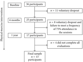
36 participants
25 participants
17 participants
Final sample
participants Baseline
6 months
1 year
Ph
ysical
e
xe
rc
is
e i
n
te
rv
en
ti
on
n = 13
n = 11voluntary dropout
n = 4did not complete all evaluations
n = 8voluntary dropout and
failure to meet a frequency of75% attendance in
the sessions
Figure 1: Recruitment of participants for the study. Evaluations were done at baseline, 6 months, and 1 year of SARC intervention.
suggests that this program improves blood cholesterol, LDL,
HDL, and glucose proiles [17,18]. However, it is unknown
whether the SARC intervention can improve inlammatory biomarkers and thus potentially aid in the prevention of chronic disease and associated complications. herefore, the aim of this study was to assess the impact of SARC on a range of common inlammatory biomarkers and hormone levels in adult women, including leptin, insulin, C-reactive protein (CRP), interleukin 6 (IL6), tumor necrosis factor
alpha (TNF�), and interleukin 10 (IL10). It was hypothesized
that there would be an increase in IL10 and a decrease in
inlammatory markers (CRP, IL6, and TNF�) and hormone
(leptin and insulin) levels ater 1 year of SARC intervention.
2. Methods
2.1. Participants. his 1-year quasiexperimental study was
developed in two basic health care units in Rio Claro City, Brazil. Adult females were recruited via lyers and newspaper advertisements. Participants were assigned to the interven-tion group based upon proximity from their residence. hirty-six participants were recruited at the beginning of intervention. As a result of either voluntary dropout or failure to meet the inclusion criterion for the study (frequency of 75% attendance in the sessions), 25 participants remained in the intervention ater 6 months. Although 17 participants completed the 1 year intervention, four participants did not complete all evaluations; thus the inal sample size was 13
women (mean age =56.8 ± 11.4years,Figure 1). he study
was approved by the Human Research Ethics Committee of Biosciences Institute, UNESP, protocol number: 2308.
2.2. Physical Exercise Intervention. SARC is a
community-based exercise intervention comprised of two 60-minute sessions/week of physical exercises. he sessions were divided in warm-up activities (5 minutes), moderate intensity aer-obic exercise (30 minutes), strength-training exercises (20 minutes), and cool-down activities (5 minutes). Furthermore,
during each session, the participants received counseling designed to increase daily physical activity levels and encour-age participation in physical exercise outside of the interven-tion.
he warm-up and cool-down activities included static stretching exercises and articular movements. Static stretch-ing was maintained for a minimum period of 15 to 30 seconds, twice for each muscle group. he participants were advised to
sustain a muscle stretch that did not cause pain [19,20]. he
aerobic exercises consisted of walking at moderate intensity (60–70% of peak heart rate). he target zone for exercise was
calculated using the equation HRpeak = 206−(0.88×age), as
suggested by Gulati et al. [21]. All participants were instructed
to maintain a subjective efort between 13 and 15 [22] on
the Borg scale [23] during walking. Four participants were
randomly selected to measure the intensity of their activity twice a month using a cardiac rate monitor (Polar, FS1) and the subjective efort scale. he strength-training exercises were performed using free weights, exercise mats, and latex exercise bands. Exercises included all major muscle groups and were performed in 3 sets of 30 seconds, followed by one minute of recovery.
2.3. Inlammatory Biomarkers Measures. A 10 mL venous
blood sample was collected at baseline, ater 6 months, and ater 1 year of intervention, in the morning ater 12 hours of fasting. he blood sample was transported under refrig-eration to the laboratory within 30 minutes, centrifuged for 10 minutes with the serum immediately separated following centrifugation. he inlammatory biomarkers were analyzed in duplicate using commercial kits. C-reactive protein (CRP) was analyzed using an enzyme-linked immunosorbent assay (ELISA). Interleukin 10 (IL10), interleukin 6 (IL6), tumor
necrosis factor alpha (TNF�), leptin, and insulin were
ana-lyzed using Luminex technology assay (Luminex).
Intra-assay coeicients were all <10%. To minimize analytical
variations, the same technician tested all samples without changing reagent lots, standards, or control materials.
2.4. Statistical Analyses. Descriptive data are reported as
means and standard deviations. he ratio of IL-10 to TNF-�
(IL10/TNF-�) was calculated and compared in 1 year.
A repeated measures ANOVA was used to analyze the changes in anthropometric variables, inlammatory biomark-ers, and hormones levels over time (baseline, 6 months, and 1 year). Signiicant diferences were determined by Bonferroni post hoc tests. Statistical analyses were conducted using SPSS
20.0, with the alpha level set at� < 0.05.
3. Results
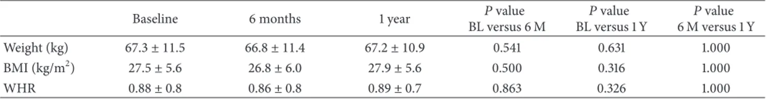
Table 1shows the anthropometric characteristics of the
par-ticipants (� = 13, mean age of 56.8 ± 11.4) at baseline, 6
months, and 1 year. No changes in weight, body mass index
(BMI), or waist to hip ratio (WHR) were seen over time (� >
0.05). he prevalence of diseases was 7.7% (� = 1) for diabetes,
Table 1: Anthropometric characteristics (mean, standard deviation) of participants at baseline, ater 6 months, and ater 1 year of exercise intervention.
Baseline 6 months 1 year BL versus 6 M�value BL versus 1 Y�value 6 M versus 1 Y�value
Weight (kg) 67.3 ± 11.5 66.8 ± 11.4 67.2 ± 10.9 0.541 0.631 1.000
BMI (kg/m2) 27.5 ± 5.6 26.8 ± 6.0 27.9 ± 5.6 0.500 0.316 1.000
WHR 0.88 ± 0.8 0.86 ± 0.8 0.89 ± 0.7 0.863 0.326 1.000
BL: baseline; 6 M: 6 months; 1 Y: 1 year; BMI: body mass index; WHR: waist and hip ratio.
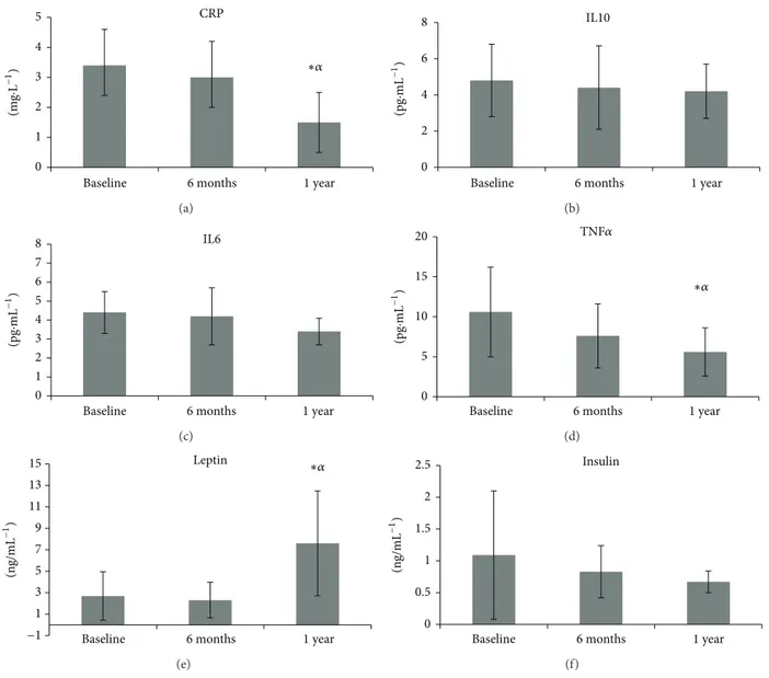
Table 2: Inlammatory biomarkers and hormone concentration levels (mean, standard deviation) at baseline, ater 6 months, and ater 1 year of exercise intervention.
Biomarker BL 6 M 1 Y �value
BL versus 6 M �
value
BL versus 1 Y � value 6 M versus 1 Y
CRP (mg⋅L−1) 3.4 ± 1.2 3.0 ± 1.2 1.5 ± 1.0∗� 0.999 0.001 0.003
IL10 (pg⋅mL−1) 4.8 ± 2.0 4.4 ± 2.3 4.2 ± 1.5 0.988 0.681 0.602
IL6 (pg⋅mL−1) 4.4 ± 1.1 4.2 ± 1.5 3.4 ± 0.7 0.999 0.236 0.163
TNF�(pg⋅mL−1) 10.6 ± 5.6 7.6 ± 4.0 5.6 ± 3.0∗� 0.082 0.001 0.004
Leptin (ng/mL−1) 2.69 ± 2.25 2.30 ± 1.66 7.60 ± 4.89∗� 0.999 0.016 0.003
Insulin (ng/mL−1) 1.09 ± 1.01 0.83 ± 0.41 0.67 ± 0.17 0.898 0.405 0.642
IL10/TNF� 0.59 ± 0.4 0.64 ± 0.2 0.85 ± 0.3 — — —
BL: baseline; 6 M: 6 months; 1 Y: 1 year; CRP: C-reactive protein; IL10: interleukin 10; IL6: interleukin 6; TNF�: tumor necrosis factor alpha.
∗Statistically signiicant diference from baseline.
�Statistically signiicant diference ater 6 months.
he prevalence of participants having at least 1 disease was
46.1% (� = 6).
Table 2andFigure 2illustrate the inlammatory biomark-ers and hormone concentration levels and indicate the outcomes of statistical analyses between time at baseline, 6 months, and 1 year. CRP levels signiicantly decreased ater 1
year of intervention (1.5 ± 1.0mg⋅L−1) compared to baseline
(3.4 ± 1.2mg⋅L−1,� = 0.001) and 6 months (3.0 ± 1.2mg⋅L−1,
� = 0.003). A signiicant decrease in TNF�levels was shown
ater 1 year of intervention (56.6 ± 3.0pg⋅mL−1) compared
to baseline (10.6 ± 5.6pg⋅mL−1,� = 0.001) and 6 months
(7.6 ± 4.0pg⋅mL−1,� = 0.004). IL10, IL6, and insulin did not
change over 1 year (� > 0.05). Leptin levels were signiicantly
increased ater 1 year (7.6 ± 4.89pg⋅mL−1) of intervention
compared to baseline (2.69 ± 2.25pg⋅mL−1,� = 0.016) and 6
months (2.3±1.66pg⋅mL−1,� = 0.003). he IL10/TNF�ratio
increased ater 1 year of intervention (BL =0.59 ± 0.4; 6 M =
0.64 ± 0.2; 1 Y =0.85 ± 0.3).
4. Discussion
Chronic inlammation is an important pathophysiological factor in the development of several diseases and complica-tions, through the efects of proinlammatory cytokines such
as TNF�and IL6, among others [2–5]. On the contrary,
anti-inlammatory cytokines, such as adiponectin and IL-10, seem
to be protective against pathological conditions [24,25].
Analyses indicate that the SACR intervention was
efec-tive in decreasing CRP and TNF� levels and maintaining
IL10, IL6, and insulin levels over 1 year. However, lep-tin levels increased over 1 year. Several studies show that inlammatory biomarkers are reduced following longer term lifestyle modiication involving reduced food intake and
increased physical activity [9]. hus, the efects of regular
physical activity on basal levels of inlammatory markers have been used to recommend exercise as an anti-inlammatory
therapy. According to Soares and de Souza [14] integrative
interventions, including diet, moderate aerobic exercise (60%
to 80% of HRmax or 50% to 60% of VO2max) and circuit
resistance training (8 to 10 exercises, 8 to 12 repetitions), health education, and counseling, used together, appeared to be efective strategies to improve inlammatory biomarkers in women.
Our results (Table 2and Figure 2) indicated that SARC
was efective in decreasing CRP levels ater 1 year compared to baseline and 6 months. hese indings are in agreement with other studies in the literature indicating that a physical
lifestyle can reduce CRP levels [13,26–29]. CRP has a long
plasma half-life (>96 h), no variation of diurnal or seasonal,
and no age or gender dependence [30,31]. It plays a pivotal
role in the innate immune response, is released in response to a variety of proinlammatory cytokines, and is triggered by many factors such as cardiovascular diseases, trauma,
malignancy, and chronic arthritis [32]. In our study, the
56% decrease in CRP is clinically relevant because the value changed from a level considered “high risk” for cardiovascu-lar disease at baseline (above 3.0 mg/L) to an “average risk” (1.0 to 3.0 mgL) ater 1 year of the SARC intervention.
According to You et al. [33], indings about the
0 1 2 3 4 5
Baseline 6 months 1 year
CRP
∗�
(m
g
·
L
−1
)
(a)
0 2 4 6 8
Baseline 6 months 1 year
IL10
(pg
·
mL
−1
)
(b)
0 1 2 3 4 5 6 7 8
Baseline 6 months 1 year
IL6
(pg
·
mL
−1
)
(c)
0 5 10 15 20
Baseline 6 months 1 year
TNF�
∗�
(pg
·
mL
−1
)
(d)
1 3 5 7 9 11 13 15
Baseline 6 months 1 year
Leptin
(n
g/mL
−1
)
−1
∗�
(e)
0 0.5 1 1.5 2 2.5
Baseline 6 months 1 year
Insulin
(n
g/mL
−1
)
(f)
Figure 2: Levels of C-reactive protein (CRP), interleukin 10 (IL10), interleukin 6 (IL6), tumor necrosis factor alpha (TNF�), leptin, and insulin at baseline, ater 6 months, and ater 1 year of exercise intervention.∗Statistically signiicant diference from baseline.�Statistically signiicant diference from 6 months.
are more consistent for CRP than for other biomarkers. However, the SARC intervention was efective in reducing TNF�(Table 2andFigure 2) ater 1 year compared to baseline and ater 6 months. Studies have indicated that regular physical activity is associated with a reduction or no change
in TNF�[27,28,34,35]. TNF-�is a cytokine with a varied
range of proinlammatory activities, such as inluencing the atherosclerotic process both by causing metabolic perturba-tions and by increasing the expression of cellular adhesion
molecules [36].
No changes were detected in IL10 following 1 year of
inter-vention (Table 2). IL10 has multifaceted anti-inlammatory
properties. It is able to reduce serum levels of TNF�and IL6
and plays a protective role against atherosclerosis [24, 25].
here is lack of consensus in the literature as to whether
physical activity can improve IL10 levels. Kadoglou et al. [28]
demonstrated in their study that a higher volume of aerobic
exercise (four times/week, 45–60 min/session) was efective in increasing IL10 levels ater 6 months. Similarly, Jankord
and Jemiolo [37] compared groups performing diferent
amounts of physical activity volume and concluded that the
higher volume was associated with an increase of IL-10 [37].
hus, it appears that 2 sessions per week of physical exercise delivered by SARC may be insuicient to improve IL10
levels. However, the IL10/TNF�ratio increased ater 1 year of
intervention. his result indicates that physical exercise was able to improve the proportion of anti- to proinlammatory cytokines ater 1 year.
he SARC intervention did not change IL6 levels
fol-lowing 1 year of intervention (Table 2). Some studies have
reported that physical exercise is correlated with lower
IL6 levels [13, 29, 34, 37–40]. However, our results are in
agreement with other studies. Olson et al. [41] found that
per week was not efective in reducing IL6 levels ater 1 year.
Campbell et al. [42] and Donges et al. did not also ind
lower levels of IL6 following physical exercise interventions
[43]. Diferent cells produce IL6 and this cytokine plays both
“good” and “bad” roles depending on the circumstances. It has been suggested that an elevation in IL6 in response to physical exercise can exert an anti-inlammatory role. Myokine, the IL6 from muscle, can increase during physical exercise. It wields metabolic efects on liver and adipose tissues (activating glycogenolysis and lipolysis) and inhibits
the production of TNF�[44,45].On the other hand, IL6 is
also secreted by macrophages and lymphocytes in response
to injury or infection [46] and has been associated with
several pathological conditions as a marker of low-grade
inlammation [47,48]. hus, the maintenance of IL6 levels
during a 1-year intervention could be considered a positive outcome.
It is currently well accepted that regular physical exercise is an efective therapeutic intervention to reduce the risk of developing insulin resistance by improving glucose tolerance and insulin action in individuals predisposed to developing
type 2 diabetes [7]. It has been hypothesized that insulin
resistance increases with age due to increased adiposity, decreased lean muscle mass, changes in dietary habits, and
reduced physical activity [49]. Although there was not a
statistically signiicant change in insulin in the present study (Table 2), insulin levels decreased by 38.4% ater 1 year of the intervention, suggesting that insulin sensitivity may have improved, although an insulin sensitivity test in participants would be needed to conirm this.
In the present study, leptin levels were maintained until 6 months and then increased signiicantly ater 1 year of the
intervention (Table 2 and Figure 2). Despite these changes,
leptin levels remained in the normal range (2.5–21.8 ng/mL).
According to Mota and Zanesco [50], the relationship
between physical activity and plasma leptin is unclear, with some studies showing a reduction in their levels while
others fail to ind any change. Recently, Akbarpour [13]
demonstrated that 12 weeks of physical exercise was able to reduce leptin levels, BMI, and IL6, and in contrast to our
indings they did not ind any changes in TNF�.
Plasma levels of leptin can increase as the result of
obesity [51]; in the present study we saw no changes in
body weight, BMI, or WHR ater 1 year. In addition,
TNF-� and CRP have been shown to be related to high levels
in adipose tissue, and its level in the circulation indicates
the production of these biomarkers in adipose tissue [51].
In the present study, although no decrease in BMI and
weight was observed, the levels of TNF� and CRP were
decreased, supporting the efect of exercise on these biomark-ers independent of weight loss. Current evidence supports that exercise training reduces chronic inlammation and this efect is independent of the exercise induced weight loss
[33].
he mechanisms related to physical exercise as a therapy in changing inlammatory biomarkers are not clear, despite studies showing positive outcomes. he discrepancy between the results from various studies in the literature can be attributed to the diferences among the groups studied,
training period, volume, intensity, duration, and type of training.
his study has a number of limitations that should be considered. he small sample size that resulted in the study has low statistical power and was a result of the diiculty in maintaining a 75% participation rate in the intervention sessions over the 1-year intervention period. We attempted to reduce dropout by assigning participants to an intervention groups geographically near their home. In addition, this study employed a quasiexperimental design, and thus we are not able to state with conidence that the changes in inlammatory markers are due to participation in the SARC intervention. We attempted to include a control group (doing no physical exercise over 1 year) to allow us to conduct a controlled trial, but the university ethics committee would not approve this study design.
Considering the fact that 46.1% of participants already had at least 1 disease related with the inlammation process, this study illustrates that a public health exercise intervention delivered in low-income communities has the potential to exert a beneicial efect and improve or maintain inlam-matory biomarkers proiles, assisting in the prevention of chronic diseases. However, a larger randomized controlled trial needs to be conducted to conirm or refute these suggestive indings.
5. Conclusion
he major inding of the present study was that a public health exercise intervention was efective in decreasing CRP and
TNF� levels and maintaining IL10, IL6, and insulin levels
over 1 year. Developing and delivering a community-based, public health exercise intervention like SARC could be a viable initiative to promote health at the public health level.
Conflict of Interests
he authors declare that there is no conlict of interests regarding the publication of this paper.
Acknowledgments
References
[1] WHO, Noncommunicable Diseases Country Proiles, World Health Organization, 2011.
[2] G. King, “he role of inlammatory cytokines in diabetes and its complications,”Journal of Periodontology, vol. 79, no. 8, pp. 1527–1534, 2008.
[3] R. B. Goldberg, “Cytokine and cytokine-like inlammation markers, endothelial dysfunction, and imbalanced coagulation in development of diabetes and its complications,”Journal of Clinical Endocrinology and Metabolism, vol. 94, no. 9, pp. 3171– 3182, 2009.
[4] D. Fern´andez-Berg´es, L. Consuegra-S´anchez, J. Pe˜naiel et al., “Metabolic and inlammatory proiles of biomarkers in obesity, metabolic syndrome, and diabetes in a Mediterranean population: DARIOS inlammatory study,”Revista Espa˜nola de Cardiologia, 2014.
[5] I. Vinagre, J. L. S´anchez-Quesada, J. S´anchez-Hern´andez et al., “Inlammatory biomarkers in type 2 diabetic patients: efect of glycemic control and inpact of LDL subfraction phenotype,” Cardiovascular Diabetology, vol. 13, article 34, 2014.
[6] I. Lee, E. J. Shiroma, F. Lobelo et al., “Efect of physical inactivity on major non-communicable diseases worldwide: an analysis of burden of disease and life expectancy,”he Lancet, vol. 380, no. 9838, pp. 219–229, 2012.
[7] J. A. Hawley, “Exercise as a therapeutic intervention for the prevention and treatment of insulin resistance,” Dia-betes/Metabolism Research and Reviews, vol. 20, no. 5, pp. 383– 393, 2004.
[8] B. K. Pedersen, “he anti-inlammatory efect of exercise: its role in diabetes and cardiovascular disease control,”Essays in Biochemistry, vol. 42, pp. 105–117, 2006.
[9] U. N. Das, “Anti-inlammatory nature of exercise,”Nutrition, vol. 20, no. 3, pp. 323–326, 2004.
[10] A. M. W. Petersen and B. K. Pedersen, “he anti-inlammatory efect of exercise,”Journal of Applied Physiology, vol. 98, no. 4, pp. 1154–1162, 2005.
[11] L. K. Stewart, M. G. Flynn, W. W. Campbell et al., “he inluence of exercise training on inlammatory cytokines and C-reactive protein,”Medicine and Science in Sports and Exercise, vol. 39, no. 10, pp. 1714–1719, 2007.
[12] K. L. Timmerman, M. G. Flynn, P. M. Coen, M. M. Markof-ski, and B. D. Pence, “Exercise training-induced lowering of inlammatory (CD14+CD16+) monocytes: a role in the anti-inlammatory inluence of exercise?”Journal of Leukocyte Biol-ogy, vol. 84, no. 5, pp. 1271–1278, 2008.
[13] M. Akbarpour, “he efect of aerobic training on serum adiponectin and leptin levels and inlammatory markers of coronary heart disease in obese men,”Biology of Sport, vol. 30, no. 1, pp. 21–27, 2013.
[14] F. H. R. Soares and M. B. C. de Sousa, “Diferent types of physical activity on inlammatory biomarkers in women with or without metabolic disorders: a systematic review,”Women and Health, vol. 53, no. 3, pp. 298–316, 2013.
[15] H. W. Kohl III, C. L. Craig, E. V. Lambert et al., “he pandemic of physical inactivity: global action for public health,” he Lancet, vol. 380, no. 9838, pp. 294–305, 2012.
[16] P. M. Nakamura, C. B. Papini, I. P. Teixeira et al., “Efect of a 10-year physical activity intervention in primary health care settings on physical itness,”Journal of Physical Activity and Health, 2014.
[17] E. Kokubun, E. Luciano, C. Y. Sibuya et al., “Programa de ativi-dade f´ısica em uniativi-dades b´asicas de sa´ude: relato de experiˆencia no munic´ıpio de Rio Claro-SP,”Revista Brasileira de Atividade F´ısica e Sa´ude, vol. 12, no. 1, pp. 45–53, 2007.
[18] L. P. Zorzetto,Comparac¸˜ao entre modelos de intervenc¸˜ao de exerc´ıcio f´ısico em unidades de sa´ude e suas inluˆencias na aderˆencia e vari´aveis relacionadas `a sa´ude [Ph.D. dissertation], Biosciences Institute of Rio Claro Campus, S˜ao Paulo State University, 2013, as part the requirements for obtaining the Master title of Science in Motricity, in area concentration of Physical Activity and Health.
[19] ACSM,ACSM's Guidelines for Exercise Testing and Prescription, American College of Sports Medicine, Lippincott Williams & Wilkins, London, UK, 2005.
[20] A. A. Junior,Exerc´ıcios de alongamento: Anatomia e Fisiologia, chapter 7, Manole, 3rd edition, 2010.
[21] M. Gulati, L. J. Shaw, R. A. histed, H. R. Black, C. Noel Bairey Merz, and M. F. Arnsdorf, “Heart rate response to exercise stress testing in asymptomatic women: the St. James women take heart project,”Circulation, vol. 122, no. 2, pp. 130–137, 2010.
[22] C. E. Garber, B. Blissmer, M. R. Deschenes et al., “Quantity and quality of exercise for developing and maintaining cardiorespi-ratory, musculoskeletal, and neuromotor itness in apparently healthy adults: guidance for prescribing exercise,”Medicine and Science in Sports and Exercise, vol. 43, no. 7, pp. 1334–1359, 2011. [23] G. A. V. Borg, “Psychophysical bases of perceived exertion,” Medicine and Science in Sports and Exercise, vol. 14, no. 5, pp. 377–381, 1982.
[24] P. Wang, P. Wu, M. I. Siegel, R. W. Egan, and M. M. Billah, “Inter-leukin (IL)-10 inhibits nuclear factor�B (NF�B) activation in human monocytes. IL-10 and IL-4 suppress cytokine synthesis by diferent mechanisms,”he Journal of Biological Chemistry, vol. 270, no. 16, pp. 9558–9563, 1995.
[25] D. A. Smith, S. D. Irving, J. Sheldon, D. Cole, and J. C. Kaski, “Serum levels of the antiinlammatory cytokine interleukin-10 are decreased in patients with unstable angina,”Circulation, vol. 104, no. 7, pp. 746–749, 2001.
[26] E. S. Ford, “Does exercise reduce inlammation? Physical activ-ity and C-reactive protein among U.S. adults,”Epidemiology, vol. 13, no. 5, pp. 561–568, 2002.
[27] R. Elosua, B. Bartali, J. M. Ordovas, A. M. Corsi, F. Lauretani, and L. Ferrucci, “Association between physical activity, phys-ical performance, and inlammatory biomarkers in an elderly population: the InCHIANTI study,”Journals of Gerontology A: Biological Sciences and Medical Sciences, vol. 60, no. 6, pp. 760– 767, 2005.
[28] N. P. E. Kadoglou, F. Iliadis, N. Angelopoulou et al., “he anti-inlammatory efects of exercise training in patients with type 2 diabetes mellitus,”European Journal of Cardiovascular Prevention and Rehabilitation, vol. 14, no. 6, pp. 837–843, 2007. [29] R. E. Walter, J. B. Wilk, M. G. Larson et al., “Systemic
inlamma-tion and COPD: the Framingham heart study,”Chest Journal, vol. 133, no. 1, pp. 19–25, 2008.
[30] H. K. Meier-Ewert, P. M. Ridker, N. Rifai, N. Price, D. F. Dinges, and J. M. Mullington, “Absence of diurnal variation of C-reactive protein concentrations in healthy human subjects,” Clinical Chemistry, vol. 47, no. 3, pp. 426–430, 2001.
[32] S. Fichtlscherer, C. Heeschen, and A. M. Zeiher, “Inlammatory markers and coronary artery disease,” Current Opinion in Pharmacology, vol. 4, no. 2, pp. 124–131, 2004.
[33] T. You, N. C. Arsenis, B. L. Disanzo, and M. J. Lamonte, “Efects of exercise training on chronic inlammation in obesity: current evidence and potential mechanisms,”Sports Medicine, vol. 43, no. 4, pp. 243–256, 2013.
[34] L. H. Colbert, M. Visser, E. M. Simonsick et al., “Physical activity, exercise, and inlammatory markers in older adults: indings from the health, aging and body composition study,” Journal of the American Geriatrics Society, vol. 52, no. 7, pp. 1098–1104, 2004.
[35] S. Balducci, S. Zanuso, A. Nicolucci et al., “Anti-inlammatory efect of exercise training in subjects with type 2 diabetes and the metabolic syndrome is dependent on exercise modalities and independent of weight loss,” Nutrition, Metabolism and Cardiovascular Diseases, vol. 20, no. 8, pp. 608–617, 2010. [36] P. Vassalli, “he pathophysiology of tumor necrosis factors,”
Annual Review of Immunology, vol. 10, pp. 411–452, 1992. [37] R. Jankord and B. Jemiolo, “Inluence of physical activity on
serum IL-6 and IL-10 levels in healthy older men,”Medicine and Science in Sports and Exercise, vol. 36, no. 6, pp. 960–964, 2004. [38] D. R. Taafe, T. B. Harris, L. Ferrucci, J. Rowe, and T. E. Seeman, “Cross-sectional and prospective relationships of interleukin-6 and C-reactive protein with physical performance in elderly persons: MacArthur studies of successful aging,”Journals of Gerontology A: Biological Sciences and Medical Sciences, vol. 55, no. 12, pp. M709–M715, 2000.
[39] D. B. Reuben, L. Judd-Hamilton, T. B. Harris, and T. E. Seeman, “he associations between physical activity and inlammatory markers in high-functioning older persons: macArthur studies of successful aging,”Journal of the American Geriatrics Society, vol. 51, no. 8, pp. 1125–1130, 2003.
[40] B. J. Nicklas, F. Hsu, T. J. Brinkley et al., “Exercise training and plasma C-reactive protein and interleukin-6 in elderly people,” Journal of the American Geriatrics Society, vol. 56, no. 11, pp. 2045–2052, 2008.
[41] T. P. Olson, D. R. Dengel, A. S. Leon, and K. H. Schmitz, “Changes in inlammatory biomarkers following one-year of moderate resistance training in overweight women,” Interna-tional Journal of Obesity, vol. 31, no. 6, pp. 996–1003, 2007. [42] P. T. Campbell, K. L. Campbell, M. H. Wener et al., “A
yearlong exercise intervention decreases CRP among obese postmenopausal women,”Medicine and Science in Sports and Exercise, vol. 41, no. 8, pp. 1533–1539, 2009.
[43] C. E. Donges, R. Duield, and E. J. Drinkwater, “Efects of resistance or aerobic exercise training on interleukin-6, C-reactive protein, and body composition.,”Medicine & Science in Sports Exercise, vol. 42, no. 2, pp. 304–313, 2010.
[44] M. Pedersen, H. Bruunsgaard, N. Weis et al., “Circulating levels of TNF-alpha and IL-6-relation to truncal fat mass and muscle mass in healthy elderly individuals and in patients with type-2 diabetes,”Mechanisms of Ageing and Development, vol. 124, no. 4, pp. 495–502, 2003.
[45] J. Prestes, F. F. Donato, and R. Dias, “Papel da interleucina-6 como um sinalizador de diferentes tecidos durante o exerc´ıcio f´ısico,”Fitness Performance Journal, vol. 5, no. 6, pp. 348–353, 2006.
[46] B. K. Pendersen and A. D. Tot, “Efects of exercise on lympho-cytes and cytokines,”British Journal of Sports Medicine, vol. 34, no. 4, pp. 246–251, 2000.
[47] B. K. Pedersen, “IL-6 signalling in exercise and disease,” Bio-chemical Society Transactions, vol. 35, no. 5, pp. 1295–1297, 2007. [48] E. Z. Fisman and A. Tenenbaum, “he ubiquitous interleukin-6: a time for reappraisal,”Cardiovascular Diabetology, vol. 9, article 62, 2010.
[49] A. J. Scheen, “Diabetes mellitus in the elderly: insulin resistance and/or impaired insulin secretion?”Diabetes and Metabolism, vol. 31, no. 2, pp. 5S27–5S34, 2005.
[50] G. Mota and A. Zanesco, “Leptina, Ghrelina e Exerc´ıcio F´ısico,” Arquivos Brasileiros de Endocrinologia & Metabologia, vol. 51, no. 1, pp. 25–33, 2007.
Submit your manuscripts at
http://www.hindawi.com
Stem Cells
International
Hindawi Publishing Corporation
http://www.hindawi.com Volume 2014
Hindawi Publishing Corporation
http://www.hindawi.com Volume 2014
INFLAMMATION
Hindawi Publishing Corporation
http://www.hindawi.com Volume 2014
Behavioural
Neurology
Endocrinology
International Journal ofHindawi Publishing Corporation
http://www.hindawi.com Volume 2014
Hindawi Publishing Corporation
http://www.hindawi.com Volume 2014
Disease Markers
Hindawi Publishing Corporation
http://www.hindawi.com Volume 2014
BioMed
Research International
Oncology
Journal of Hindawi Publishing Corporationhttp://www.hindawi.com Volume 2014
Hindawi Publishing Corporation
http://www.hindawi.com Volume 2014
Oxidative Medicine and Cellular Longevity
Hindawi Publishing Corporation
http://www.hindawi.com Volume 2014
PPAR Research
The Scientiic
World Journal
Hindawi Publishing Corporation
http://www.hindawi.com Volume 2014
Immunology Research
Hindawi Publishing Corporation
http://www.hindawi.com Volume 2014 Journal of
Obesity
Journal ofHindawi Publishing Corporation
http://www.hindawi.com Volume 2014
Hindawi Publishing Corporation
http://www.hindawi.com Volume 2014
Computational and Mathematical Methods in Medicine
Ophthalmology
Journal ofHindawi Publishing Corporation
http://www.hindawi.com Volume 2014
Diabetes Research
Journal ofHindawi Publishing Corporation
http://www.hindawi.com Volume 2014
Hindawi Publishing Corporation
http://www.hindawi.com Volume 2014
Research and Treatment
AIDS
Hindawi Publishing Corporationhttp://www.hindawi.com Volume 2014
Gastroenterology Research and Practice
Hindawi Publishing Corporation
http://www.hindawi.com Volume 2014
Parkinson’s
Disease
Evidence-Based Complementary and Alternative Medicine
Volume 2014