Review
Strongyloidiasis—An Insight into Its Global Prevalence
and Management
Santhosh Puthiyakunnon1, Swapna Boddu1, Yiji Li1, Xiaohong Zhou1, Chunmei Wang1, Juan Li2,
Xiaoguang Chen1*
1Key Laboratory of Prevention and Control for Emerging Infectious Diseases of Guangdong Higher Institutes, Department of Pathogen Biology, School of Public Health and Tropical Medicine, Southern Medical University, Guangzhou, Guangdong, China,2Department of Traditional Chinese Medicine, Nanfang Hospital, Southern Medical University, Guangzhou, Guangdong, China
Abstract
Background: Strongyloides stercoralis, an intestinal para-sitic nematode, infects more than 100 million people worldwide. Strongyloides are unique in their ability to exist as a free-living and autoinfective cycle. Strongyloi-diasis can occur without any symptoms or as a potentially fatal hyperinfection or disseminated infection. The most common risk factors for these complications are immu-nosuppression caused by corticosteroids and infection with human T-lymphotropic virus or human immunode-ficiency virus. Even though the diagnosis of strongyloidi-asis is improved by advanced instrumentation techniques in isolated and complicated cases of hyperinfection or dissemination, efficient guidelines for screening the population in epidemiological surveys are lacking.
Methodology and Results: In this review, we have discussed various conventional methods for the diagnosis and management of this disease, with an emphasis on recently developed molecular and serological methods that could be implemented to establish guidelines for precise diagnosis of infection in patients and screening in epidemi-ological surveys. A comprehensive analysis of various cases reported worldwide from different endemic and nonen-demic foci of the disease for the last 40 years was evaluated in an effort to delineate the global prevalence of this disease. We also updated the current knowledge of the various clinical spectrum of this parasitic disease, with an emphasis on newer molecular diagnostic methods, treatment, and management of cases in immunosuppressed patients.
Conclusion: Strongyloidiasis is considered a neglected tropical disease and is probably an underdiagnosed parasitic disease due to its low parasitic load and uncertain clinical symptoms. Increased infectivity rates in many developed countries and nonendemic regions nearing those in the most prevalent endemic regions of this parasite and the increasing transmission potential to immigrants, travelers, and immunosuppressed populations are indications for initiating an integrated approach towards prompt diagnosis and control of this parasitic disease.
Introduction
Strongyloides stercoralis, one of the most common and globally distributed human pathogens of clinical importance, infects 30– 100 million people worldwide [1].S. fuelleborni, another species of the same genus, is found sporadically in Africa and Papua New Guinea [2]. Strongyloidiasis is endemic in Southeast Asia, Latin America, sub-Saharan Africa, and parts of the Southeast United
States [3]. Unique characteristics of this nematode are its immense ability to persist and replicate within a host for decades while producing minimal or no symptoms and its potential to cause life-threatening infections by dissemination and hyperinfection in debilitated and immune-compromised patients [4].
After its first report in 1876 from the feces of French soldiers with diarrhea who were returning from the old Indochina region, the disease was known for many years as ‘‘Cochin-China diarrhea’’ [5], which describes the most common gastrointestinal manifestations, such as epigastric pain and watery diarrhea, of this parasitic infection [6]. It took more than a century to trace most of the basic biology of this nematode and its extravagant ability to disseminate in host tissues, thereby leading to a spectrum of clinical complications.
In this review, we analyze the various case reports since 1970 from different parts of endemic and nonendemic foci of S. stercoralis to delineate a comprehensive global survey of this parasitic infection. We have focused more details on the different diagnostic methods followed by the investigators in various case reports and discussed some recent novel methods in serological and molecular diagnosis towards the aim of establishing guidelines for diagnosis to decipher the global prevalence of this disease.
Epidemiology and Global Prevalence of Strongyloidiasis
Strongyloidiasis is an emerging global infection that is underestimated in many countries [7]. The prevalence of this disease has been on the increase, especially in southern, eastern and central Europe, the islands of the Caribbean, Southeast Asia, Latin America, and sub-Saharan Africa. In nonendemic regions of the world, it is mainly diagnosed in individuals who were prisoners during World War II and in immigrants from endemic countries [8]. Males, people working with soil (such as in coal mines and
Citation:Puthiyakunnon S, Boddu S, Li Y, Zhou X, Wang C, et al. (2014) Strongyloidiasis— An Insight into Its Global Prevalence and Management. PLoS Negl Trop Dis 8(8): e3018. doi:10.1371/journal.pntd.0003018
Editor:Gary L. Simon, George Washington University, United States of America
PublishedAugust 14, 2014
Copyright:ß2014 Puthiyakunnon et al. This is an open-access article distributed under the terms of the Creative Commons Attribution License, which permits unrestricted use, distribution, and reproduction in any medium, provided the original author and source are credited.
Funding:This study was supported by grants from the National Natural Science Foundation of China (31030066) and GPUPS (2009) to XC. The funders had no role in study design, data collection and analysis, decision to publish, or preparation of the manuscript.
Competing Interests:The authors have declared that no competing interests exist.
farms), people of white race, patients with altered cellular immunity (especially those on long-term steroid therapy), patients with lymphoma, allograft transplant recipients, travelers to areas of endemicity, and other institutionalized individuals are at the greatest risk of acquiring this disease [9,10]. A strong association is seen between strongyloidiasis and concurrent immunosuppressive diseases such as human T cell lymphotropic virus-1 (HTLV-1) [11], human immunodeficiency virus (HIV) infection, and hematological malignancies [12,13].
Global prevalence ofS. stercoralishas been on the increase in the past few years, especially in many known endemic areas of the disease (Figure 1). The continued increase in infection rate is solely attributed to poor personal hygiene, insufficient drinking water supply, unsatisfactory sanitary measures, and lack of knowledge about the disease in high-risk populations. Many isolated case reports on the emergence of the disease in different parts of the world that are nonendemic for the disease are being published. Most of these case studies are associated with patients with immunosuppressive diseases, those on corticosteroid therapy, organ transplant recipients, and patients with hematological malignancies or other debilitating diseases. Newer diagnostics and endoscopies are being implemented widely to diagnose strongyloidiasis in many complicated clinical cases. Serological screening and molecular methods like polymerase chain reaction (PCR) are slowly becoming popular and are used in parallel with routine diagnostic screening methods. A comprehensive analysis of the case reports from different areas of endemicity and none-ndemicity was carried out in an effort to highlight the importance of implementing the most appropriate diagnostic methods to delineate the global prevalence of this disease (Table 1).
A statistical analysis carried out in our lab [14] showed a total of 106 detailed cases reported from China since the first documented
case from Guangxi Province in 1973 up until 2012. A total of 67 cases were reported in the past 10 years (2001 to 2011), which exceeds the cumulative cases reported in the 30 years before this period and indicates the increasing rate of emergence of strongyloidiasis in China.
Globally, prevalence rates of strongyloidiasis are as high as 50% in certain areas where moist soil and improper disposal of human waste coexist, especially in West Africa, the Caribbean, Southeast Asia, tropical regions of Brazil, Cambodia, and temperate regions of Spain [15]. Southeast Asia appears to have the highest endemic percentage, and it is highly prevalent in some tropical aboriginal communities in Australia [16]. Although strongyloidiasis is uncommon in the United States, endemic foci exist in rural areas of the southeastern states and the Appalachian region (especially in eastern Tennessee, Kentucky, and west Virginia) and in Puerto Rico [17]. A higher prevalence rate is seen among patients in long-term institutionalized care (mental health facilities and prisons), in immigrants and refugees from tropical and subtropical countries [18], and in veterans of World War II and the Vietnam War [19]. Among the immigrant population, a high prevalence rate of 38% was reported in Southeast Asian immigrants in Washington, D.C. [20]. A Canadian epidemiological study revealed 11.8% incidence of infection in the Vietnamese population and a much higher seroprevalence of 76.6% in Cambodian immigrants [21]. Sudanese Lost Boys and Girls and Somali Bantu refugees demonstrated 46% and 23% seropositive rates, respectively [22]. High rates of larva currens are reported in Latin America. A stool serosurvey conducted in a community in the Peruvian Amazon region found an 8.7% incidence rate of S. stercoralis [23]. Although strongyloides infection is represented in all ages, infection initially occurs in childhood, as children are more likely to play outdoors in contaminated soil with bare feet [24].
Figure 1. Map showing the global prevalence ofS. stercoralisinfection.The map was constructed using the data from the table, with the highest percentage prevalence of reported case studies and screening among populations from different countries that are endemic and nonendemic regions of the disease.
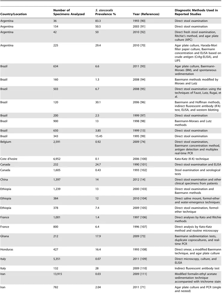
Table 1.Global survey of prevalence ofS. stercoralisin endemic and nonendemic regions of the disease.
Country/Location
Number of Specimens Analyzed
S. stercoralis
Prevalence % Year (References)
Diagnostic Methods Used in Reported Studies
Argentina 36 83.3 1993 [90] Direct stool examination
Argentina 154 50.5 2003 [91] Direct stool examination
Argentina 42 50 2010 [92] Direct fresh stool examination,
Ritchie’s method, and agar plate culture (APC)
Argentina 225 29.4 2010 [70] Agar plate culture, Harada-Mori
filter paper culture, Baermann concentration and ELISA based on crude antigen (CrAg-ELISA), and LIPS
Brazil 634 6.6 2011 [93] Agar plate culture,
Baermann-Moraes (BM), and spontaneous sedimentation
Brazil 160 1.3 2008 [94] Baermann methods modified by
Moraes and Lutz
Brazil 503 6.7 2008 [95] Direct stool examination using the
techniques of Faust, Lutz, Rugai, et al.
Brazil 120 30.1 2006 [96] Baermann and Hoffman methods,
indirect fluorescent antibody (IFA) test, ELISA, and western blotting
Brazil 200 2.5 1999 [97] Direct stool examination
Brazil 900 13 1998 [98] Baermann-Moraes and Lutz
methods
Brazil 650 3.85 1999 [13] Direct stool examination
Brazil 343 15.45 1995 [99] Direct stool examination
Belgium 2,591 0.92 2009 [74] Direct stool examination,
Baermann concentration method, antigen detection and multiplex real-time PCR
Cote d’Ivoire 6,952 0.1 2006 [100] Kato-Katz (K-K) technique
Canada 232 24.7 1990 [101] Direct stool examination and ELISA
Canada 1,605 0.43 1993 [102] Stool examination and serological tests
China 1,397 14 2012 [14] Direct stool examination and other
clinical specimens from patients
Ethiopia 1,239 13 2000 [103] Direct stool examination and
Baermann methods
Ethiopia 384 12 2010 [104] Direct saline mount, formol-ether
and water-emergence techniques
Ethiopia 378 7.4 2009 [105] Direct stool examination,
formol-ether technique
France 1,001 1.4 1997 [106] Direct analyses by Kato and Ritchie methods
France 800 6.4 1996 [107] Direct analysis by Kato-Katz
method and routine microscopy
Ghana 212 17.9 2009 [73] Baermann sedimentation tests,
duplicate coprocultures, and real-time PCR
Honduras 427 16.4 1993 [108] Direct smear, a modified Baermann technique, and agar plate culture
Italy 5,351 0.07 2011 [109] Direct microscopy, culture, and
ELISA
Italy 132 28 2009 [110] Indirect fluorescent antibody test
Iran 13,915 0.03 2009 [111] Modified formalin-ethyl acetate
sedimentation technique accompanied with trichrome stain
Iran 782 2.04 2011 [71] Agar plate culture and PCR (single
Table 1.Cont.
Country/Location
Number of Specimens Analyzed
S. stercoralis
Prevalence % Year (References)
Diagnostic Methods Used in Reported Studies
Iran 150 42 2010 [112] Stool examination by
formalin-ether and Kato-Katz techniques
Israel 106 0.9 1992 [113] Direct stool examination and ELISA
Jamaica 312 24.2 1995 [86] Stool examination and serology
Kuwait 11,230 0.46 2004 [114] Direct stool examination and ELISA
Laos PDR 664 19 1998 [115] Agar plate culture and Kato-Katz
thick smear method
Mexico 200 1 1997 [116] Direct stool examination
Nigeria 4,470 35.2 1997 [117] Direct stool examination
Nigeria 227 5.3 2004 [118] Stool wet preparation and
formol-ether concentration methods
Peru 83 28.91 1999 [119] Baermann concentration
technique modified by Lumbreras
Peru 256 0.87 2009 [120] Direct microscopy of feces and by
rapid sedimentation technique
Romania 294 6.9 1995 [121] Direct stool examination
Romania 35 30 1989 [122] Direct microscopy, culture, and
Jejunal biopsy
Spain 250 12.4 2003 [123] Detection of larvae of triple stool
samples
Spain 16,607 0.9 2001 [124] Direct microscopy, agar plate
culture, and tissue biopsy Sierra Leone 1,164 3.8 1995 [125] Direct stool examination
Sudan 275 3.3 1998 [126] Formol-ether concentration
techniques
Thailand 491 11.2 1989 [127] Direct stool examination
Thailand 332 28.9 2003 [128] Agar plate culture technique and
modified formalin-ethyl acetate concentration technique (MFECT)
Thailand 100 28 2010 [49] Formalin-ether concentration
technique (FECT)
Thailand 1,085 22.2 1999 [129] Direct smear, agar plate culture, formalin-ether sedimentation technique, and the filter-paper method
Tanzania 368 10.2 2009 [130] Kato-Katz, Koga agar plate, and
Baermann techniques
Tanzania 342 10.5 2008 [131] Kato-Katz, Koga agar plate, and
Baermann techniques
Tanzania 1,078 1.6 2009 [132] Direct stool examination and
formalin-ether concentration methods
United Kingdom -Liverpool 2,072 12 2004 [19] Microscopy and culture of stool or duodenal fluid and ELISA US–North Carolina 172 7.5 2009 [133] Direct stool microscopy and
serology
US–Tennessee 225 8.4 1992 [134] Agar plate and formalin-ethyl acetate concentration method
US–Kentucky 125 4 1965 [135] Stool examination
US–Kentucky 561 3 1982 [10] Stool examination
US–Kentucky 3,271 2.5 1982 [10] Stool examination
US–Tennessee 575 4 1987 [32] Stool examination
US–West Virginia 4,566 0.4 2000 [136] Concentration techniques followed by iodine-stained smear examination and sputum culture
Most of the epidemiological studies to assess the prevalence of infection in a population were carried out by microscopic screening of the stool samples (either single or multiple) to find larvae or by one of the concentration and culture methods. The discovery of positive cases has been increased in many studies after implementing serological screening or by using molecular methods in immunocompromised patients or high-risk groups. Recently, a real-time PCR method targeting the small subunit of a ribosomal RNA (rRNA) gene was developed for the detection of strongyloi-des DNA in fecal samples, including an internal control to detect inhibition of the amplification process. Novel methods of diagnosis like luciferase immunoprecipitation system assays based on recombinant antigens that are 100% sensitive may be a promising alternative to routine diagnostic methods which are less sensitive. These newer methods may hopefully enhance routine diagnosis of S. stercoralisinfection in the future.
Biology of the Parasite
GenusStrongyloidesis classified in the order Rhabditida, and most of the 52 species are soil-dwelling, microbiverous nematodes that do not infect human beings. Other thanS. stercoralisandS. fuelleborni, two more species,S. myopotamiandS. procyonis, are reported to infect animal hosts and may be responsible for zoonotic infections [25]. The adult female worm is a slender, almost transparent worm that measures 2.2 to 2.5 mm long, has a diameter of 50mm, and lives in tunnels between enterocyte in the small intestine. Parasitic males, although they do exist, do not have any role in human infections and are easily eliminated from the intestine [26,27].
Pathophysiology and Life Cycle
S. stercoralis exhibits a complex and unique developmental phase with two distinct life cycles: a free-living heterogonic cycle and a parasitic life cycle completed in the same host [28]. In the free-living phase, during the development of nonparasitic adults, both males and females occur in soil, which maintains infestation of the soil (Figure 2). The parasitic phase allows noninfective new larvae to molt in the human host into infective filariform larvae,
which then penetrate the intestine and set up a new cycle, leading to autoinfection or hyperinfection to increase the worm burden without exogenous reinfection [29]. This autoinfective phase is responsible for the decade-long persistence of infection in untreated hosts.
Human infection is mainly acquired by the filariform larvae penetrating the skin or mucous membranes either from autoin-fection or from infected soil by a fecal-oral route [30]. At their portal of entry, larvae usually cause petechial hemorrhagic rash, which is followed by intense pruritis, edema, and congestion [31]. Larvae migrating through lymphatics and venules reach pulmo-nary circulation and produce hemorrhages in pulmopulmo-nary capil-laries. The larvae make their way farther into alveolar spaces and cause inflammatory responses associated with eosinophilic infil-tration terminating in pneumonitis [32]. Finally, larvae crawl up the respiratory tract and are swallowed, thereby reaching the intestine.
Maturation of larvae into adult females occurs in the small bowels after two molts, and the emerged females produce eggs via parthenogenesis [33]. These parasitic females may live up to five years to continue their reproductive cycle. The eggs hatch in the intestine into noninfective rhabditiform larvae, which may be passed through stool into the environment to continue the heterogonic free-living phase in the soil. The excreted larvae in stool thus form the mainstay of laboratory diagnosis of infection and are also responsible for autoinfection after transforming into infective filariform larvae [34].
1. Autoinfection
Autoinfection is unique to S. stercoraliswithin the genus and also among other genera of intestinal parasites of vertebrates and accounts for it being a serious pathogen of humans. Premature transformation of noninfective larvae into infective larvae estab-lishes a parasitic developmental phase within the host, and it can be maintained throughout the host’s life by repeated migratory cycles without exit from the primary host [30]. Infective filariform larvae reenter circulation by penetrating the mucosa of the colon or small intestine and cause internal autoinfection or the larvae penetrate the perianal skin and cause external autoinfection [28]. External autoinfection in most cases leads to the development of
Table 1.Cont.
Country/Location
Number of Specimens Analyzed
S. stercoralis
Prevalence % Year (References)
Diagnostic Methods Used in Reported Studies
US–Maryland 339 0.6 1986 [138] Single direct stool examination US–Louisiana 8,458 0.4 1974 [139] Stool examination by direct and
concentration methods US–Chicago, Illinois 358 1.7 1975 [140] Single direct stool examination US–New York 10,072 1 1975 [141] Stool concentration method and
sputum examination for larvae US–Seattle, Washington 201 2.5 1995 [142] Direct stool examination US–Atlanta, Georgia 150 46 2007 [143] Direct stool examination and
serology
US–Ohio 700 3.71 1987 [144] ELISA and fresh stool examination
for larvae
US–Maryland 128 38 1987 [145] Stool examination and Serology
US–Minneapolis, Minnesota 1,291 11.69 2007 [39] Direct stool examination
Vietnam 3,197 0.84 1970 [146] Direct stool examination and
sedimentation technique
larva currens. After entering the circulation, larvae are carried to the lungs and repeat the cycle, which accounts for the frequent recurrence and chronicity of disease in migrants to endemic areas of disease [35]. Autoinfection, which is otherwise kept in check by the host immune response in healthy individuals, usually occurs in patients with impaired cell-mediated immunity [36]. Autoinfection gives rise to the two most severe forms of strongyloidiasis: hyperinfection syndrome (HIS) and disseminated strongyloidiasis (DS).
2. Hyperinfection syndrome
Hyperinfection syndrome denotes a phenomenon in which a tremendous increase in the number of worms leads to excessive worm burden without the spread of larvae outside the usual migration pattern. The worms are detectable in extraintestinal regions, especially in the lungs, and the detection of larvae in stool and/or sputum is the hallmark in diagnosis of hyperinfection [37]. Generally, the hyperinfection ensues from the enormous multipli-cation of infective larvae and their migration in the immunosup-pressed state, but it is not always true, as some authors have also described hyperinfection syndrome in immunocompetent patients [38].
The clinical manifestation of hyperinfection syndrome is classified, based on the origin, into gastrointestinal and extrain-testinal disease mainly involving the respiratory tract. Risk factors for developing hyperinfection include corticosteroid therapy, stem-cell transplantation, alcoholism, HIV, and HTLV-1 infection. Pulmonary symptoms such as pulmonary infiltrates, diffuse alveolar hemorrhage (DAH), and respiratory failure develop in patients, which, if not treated, turn out to be fatal. The high mortality in hyperinfection is often due to negligence and lack of familiarity of health care providers in recognizing the need for parasite screening before advocating empirical corticosteroid therapy [39].
3. Disseminated strongyloidiasis
Disseminated strongyloidiasis involves widespread dissemina-tion of larvae to extraintestinal organs that are outside the realm of the parasite’s ordinary life cycle. Multiple organs are affected, including the lungs, liver, heart, kidneys, endocrine organs, and central nervous system [40]. In severe disseminated disease, translocation of enteric bacteria may lead to polymicrobial bacteremia and occasionally meningitis with enteric pathogens. The bacteria may be carried on the migrating filariform larvae or may enter the circulation through intestinal ulcers. Major complaints are fever, abdominal pain and distension, weight loss, vomiting, cough, anemia, and hemoptysis [41]. Disseminated strongyloidiasis may not always occur as a fatal outcome of hyperinfection syndrome. Disseminated infection with bloody pericardial effusion has been reported in a nonimmunosuppressed patient without manifestations of hyperinfection syndrome [42]. This clinical finding is relatively common in high-risk populations, which are frequently misdiagnosed with gram-negative septicemia or acute respiratory distress syndrome.
4. Acute and chronic strongyloidiasis
In acute strongyloidiasis, patients become symptomatic imme-diately after exposure, and the symptoms may last up to several weeks [43]. Acute infection is generally characterized by gastrointestinal (GI) and pulmonary symptoms. GI symptoms such as diarrhea, anorexia, and abdominal pain begin about 2 weeks after infection, with larvae detectable in stool after 3 to 4 weeks. Pulmonary symptoms such as tracheal irritation, cough, and bronchitis begin much earlier than GI symptoms as larvae
migrate through the lungs within a few days after exposure. Chronic infection withS. stercoralisis often asymptomatic [44]. In symptomatic patients, major complications are due to chronic gastrointestinal manifestations such as diarrhea, constipation, and intermittent vomiting. Dermatological manifestations like urticar-ial, petechurticar-ial, and purpuric rashes and larva currens are also common.
Diagnostic Challenges
The diagnosis of strongyloidiasis requires a high degree of suspicion, as most of the patients with the infection do not show distinctive clinical features and laboratory and imaging findings often turn out to be nonspecific [45]. Multiple clinical findings appear with fatal outcomes after treatment with steroids for a severe disease of unknown etiology and are later confirmed as a case of disseminated strongyloidiasis. Clinicians should be aware that the clinical spectrum of infection may lead to pulmonary infiltrates, acute respiratory distress syndrome, small bowel obstruction, and multisystem organ failure. Clinical correlation of the symptoms with travel and residence history and imported strongyloidiasis should be considered in travelers to and immigrants from endemic areas. Furthermore, the possibility of strongyloidiasis should always be considered in any immunocom-promised patients who suddenly deteriorate; delay in diagnosis frequently results in death, despite intense treatment [16].
Diagnosis of strongyloides hyperinfection is relatively easy because of the high number of larvae that are seen in stool smears and usually seen in sputum. Many have reported unexpected findings of the larvae in ascites fluids and blood smears [46]. Direct stool examination in saline and Lugol’s iodine stain has been used for mass screening of a population in many epidemiological surveys, but a single direct stool examination alone is always inadequate, as evidenced by many reports of hyperinfection with negative stool screening exams. As the egg output compared to other parasitic helminths is too low, a single stool exam is only about 50% sensitive for making diagnosis and even lower in chronic asymptomatic patients [47].
Other methods of diagnosis include Baermann’s and formalin-ethyl acetate concentration techniques, with improved sensitivity of stool exams. Blood agar plate culture method is also preferred due to high sensitivity and ease of implementation. In a comparative study carried out to determine the efficacy of diagnostic methods, agar plate culture method (APC) has shown high sensitivity, more than 96% compared to direct fecal smear, formalin-ether concentration techniques, and Harada Mori’s filter paper culture methods [48]. It is essential to examine stool samples repeatedly to achieve correct diagnosis, and a negative result does not always indicate the absence of infection [47,48]. Peripheral eosinophilia (.600/mL) represents an immune response to larvae migrating through host tissues and is common during acute infection (as high as 75% to 80%), intermittent during chronic infection (often the only abnormal laboratory test), and frequently absent in severe strongyloidiasis and in the immunocompromised host [44].
1. Microscopy and culture of stool
feces and is easily diagnosed using microscopic techniques. Direct smear examination of stool in saline and Lugol’s iodine stain has been used to discern larvae in stool and is performed as a definitive diagnostic test. Most epidemiological screenings rely only on this method even though direct wet mounts yield very low larvae (sensitivity up to 30%) on many occasions. Hence, it is mandatory to screen multiple samples, and this should be performed with one of the concentration methods, which may increase sensitivity up to 70% to 80%. More than 90% sensitivity for stool samples is seen if seven or more samples are examined [50].
Concentration methods like formalin-ethyl acetate, Harada-Mori techniques, and Baermann concentration increase the yield and are much more sensitive than single stool smears [51]. A modified formalin-ether concentration technique using fresh stool without a preservative, a short-term formalin exposure, use of wire mesh instead of gauze, and a five-minute centrifugation has shown more efficiency in larval yield compared to the conventional method with the usual parameters [49]. In the Harada-Mori technique, filter paper containing fresh fecal material is placed in a test tube with water that soaks the filter paper by capillary action. Incubation at 30uC provides conditions suitable for the develop-ment of larvae, which migrate to the filter paper [47].
Baermann concentration method exploits the tendency of worms and larvae to migrate from a solid into a surrounding liquid medium (hydrotropism) when stimulated by a slightly elevated temperature (thermotropism) and then to settle to the substratum. In this technique, a comparatively large amount of sample can be screened, and the chances of finding different larval stages as well as adults are higher [52]. In the agar culture method, the stool sample is placed on the nutrient agar or blood agar plate and incubated for 48 hours. Serpiginious tracks of bacterial
growth along the paths of crawling larvae become apparent after 1 to 2 days of incubation, and motile larvae can be easily visualized with the aid of a dissecting microscope [47,53]. Agar plate culture method is preferred due to its high sensitivity and ease of implementation in standard microbiological laboratories and also for detection of larvae from samples like sputum, bronchial aspirates, and other body fluids [54]. Although laborious and time consuming, it is proven to be more than four times as efficient as direct smear procedures for detection of larvae in stool [48].
String test (Entero test), once popular, is seldom used for collection of larvae from the patient’s duodenum [55]. The reported sensitivity ranges from 40% to 90%. This method, which creates inconvenience for the patient, is gradually being replaced by more sensitive duodenal aspiration or histological examination of duodenal or jejunal biopsy [54,56].
2. Endoscopy and histological features
Gastrointestinal endoscopy may range from normal-appearing mucosa to severe duodenitis and colitis showing edematous mucosa, white villi, and erythematous mucosa. In most cases of pulmonary hyperinfection, the larvae are identified with duodenal biopsy. Duodenal biopsy and histopathologic examination iden-tified larvae in 71.4% of immunosuppressed patients [57]. Thus, in addition to stool analysis, endoscopic observation and biopsies are very important [58]. In disseminated strongyloides infection, larvae can be recovered from extraintestinal sites, including sputum, bronchoalveolar fluid [59], cerebrospinal fluid (CSF) [60], urine [61], ascites, gastroesophageal biopsy, and skin biopsy [62,63]. CSF analysis shows elevated protein levels, decreased glucose levels, and pleocytosis with neutrophilic predominance, and a gram stain performed may exhibit various bacterial florae Figure 2. Life cycle ofS. stercoraliswith the heterogonic and parasitic phases.
[62,64]. A wet mount preparation of the CSF will usually revealS. stercoralislarvae.
3. Serological testing
Serological assays are now widely available; moreover, due to increased sensitivity, they have the potential to be used in multiple helminthic infections. Several immunodiagnostic methods have been developed over the years with limited success, including skin testing with larval extracts, indirect immunofluorescence using fixed larvae, filarial complement fixation testing, radioallergosor-bent testing for specific immunoglobulin E (IgE), gelatin particle indirect agglutination (GPIA), western blot analysis, and enzyme-linked immunosorbent assay (ELISA) for immunoglobulin G (IgG) antibodies [47]. Strongyloides-specific antibodies may be used for serologic follow-up, which may indicate seroconversion after successful therapy. ELISA testing has been shown to detect the disease in approximately 85% to 90% of patients (sensitivity of 82% to 95%) [65]; however, its sensitivity may be lower in severely immunocompromised patients and does not distinguish past and present infection in endemic areas of disease.
The strongyloides antibody test shows cross-reactivity with other helminth infections such as filariasis, ascariasis, and acute schistosomiasis. These antibodies can persist for a long time in the host; hence, a differential diagnosis of symptomatic strongy-loidiasis is unavoidable in most endemic areas of this disease [66]. An enzyme immune assay for anti-strongyloides IgG may be a good option for rapid diagnosis when a stool examination result is negative as well as in immunosuppressed patients.
A luciferase immunoprecipitation assay described recently may eventually prove to be more accurate than ELISA testing [67]. This luciferase immunoprecipitation system (LIPS) was developed against an antibody to a recombinant strongyloides antigen (NIE). When compared with NIE-ELISA, LIPS showed improved specificity and after a second antigen,S. stercoralis immunoreac-tive antigen (SsIR), was used in combination, it resulted in a 7-fold difference between positive and negative values. Moreover, it did not show any signs of cross-reactivity with serum from filarial infected patients, which accounts for a major drawback in most serological assays. If made cost-effective and widely available, LIPS could be a substitute used in the effective screening of patients, performing more rapidly and specifically than ELISA. In a comparative evaluation study conducted in endemic regions of Thailand, the gelatin particle agglutination test also was judged to be more practical as a screening test than conventional ELISA [68].
4. Molecular diagnosis
Polymerase chain reaction assays for intestinal parasites, including strongyloides, are increasingly being used on fecal DNA samples, with enhanced specificity and sensitivity of detection. In a recently conducted study, multiplex PCR reactions with specific primers for protozoa and another primer set for helminthes were used. The PCR products obtained were hybridized to beads linked to internal oligonucleotide probes and detected on a Luminex platform. This multiplex PCR bead assay showed 83% and 100% sensitivity and specificity compared with parent multiplex real-time PCR assays and provides a sensitive diagnostic screen for a large panel of intestinal parasites [69].
Most conventional serological diagnosis, like ELISA, is based on crude antigen from parasite extracts. Newer techniques such as the luciferase immunoprecipitation system assays based on recombi-nant antigens were devised and showed the highest specificity (97.8%) and 100% sensitivity [70] when compared with
conventional ELISA. Molecular diagnosis based on detection of specific copro-DNA in stool by a single PCR method amplifying a short (100 base pair) target showed greater efficiency (100%) for detection ofS. stercoralisin fecal samples compared to agar plate culture and nested PCR, which amplifies a larger target [71]. In most PCR-based assays, a frequently observed factor that decreases the efficiency of detection is the presence of PCR inhibitors, which are relatively common in stool samples; this is more critical for assays of samples with lower parasite DNA [72]. This is resolved by increasing the amount of feces used for the PCR assay by concentration of stool by acid-ether prior to DNA extraction [71].
A real-time PCR method targeting the small subunit of the rRNA gene was developed for detection ofS. stercoralisDNA in fecal samples. The use of this assay could facilitate monitoring the prevalence and intensity of infection in helminth interventional programs, as this real-time PCR also includes an internal control to detect inhibition of the amplification process by fecal contaminants [73]. This assay showed a 2-fold increase in the detection rate when compared with the Baermann sedimentation method and could be an alternative to less sensitive conventional screening methods.
In another recent study, a multiplex real-time PCR, when compared with microscopic examination and antigen detection in travelers and migrants, showed increased detection rates from 0.1% to 0.8% [74]. A growing number of routine diagnostic laboratories are implementing multiplex real-time PCR for detection of intestinal parasites, and these assays can be extended specifically to screening travelers and migrants forS. stercoralis. If implemented effectively, these emerging new technologies certain-ly may enhance the diagnosis of strongyloides infection in the future.
Prevention and Treatment Strategies
The aim of pharmacotherapy in strongyloidiasis is to eradicate the infection, reduce morbidity and mortality, and prevent hyperinfection and disseminated complications. Several antihel-minthic drugs are available for this purpose, though few are recommended for established infection. Most of these drugs allow selective interference in the biological and metabolic pathways of the adults in relatively small doses. The effectiveness of many of these antihelminthic agents against larvae is poor, and they are more effective only when established infection occurs.
Thiabendazole, albendazole, and mebendazole are used for the treatment of acute and chronic strongyloidiasis [75] but showed varied results in many drug trials [76]. Albendazole has a high-affinity binding capacity to free beta-tubulin in parasite cells, thereby inhibiting tubulin polymerization. This eventually results in loss of cytoplasmic microtubules and thus decreases adenosine triphosphate (ATP) production in worms, ultimately leading to energy depletion, immobilization, and death. Mebendazole inhibits microtubule formation and causes worm glucose depletion but shows variable efficacy against strongyloides. Even though used successfully to treat a number of patients withS. stercoralis hyperinfection, decreased accessibility due to poor absorption of the drug by migrating larvae may be the reason for its variable efficacy [77]. These benzimidazoles not only kill adult gut-dwelling stages of the parasite but also sterilize the larvae and eggs to some extent. Thiabendazole was a therapeutic option for strongyloidi-asis for quite a long time but has been discontinued due to its unfavorable side effects [75,78].
syndrome, and disseminated strongyloidiasis [79]. It is a semisyn-thetic derivative of the macrolide mold product avermectin, which binds selectively to glutamate-gated chloride ion channels of invertebrate nerve and muscle cells, thereby increasing the cell membrane permeability with hyperpolarization and causing paralysis and cell death. In patients who are too sick to tolerate or absorb oral (PO) ivermectin, rectal (PR) or subcutaneous (SC) dosing may be effective [80,81]. The eradication rates with ivermectin in many drug trials showed remarkable results and up to a 97% cure rate with even a 2-day course in asymptomatic cases, but in patients with hyperinfection and dissemination, daily drug administration until symptoms resolve with negative labora-tory tests for larvae for at least two weeks is recommended [82]. The World Health Organization (WHO)-recommended treatment for strongyloidiasis is either ivermectin (200mg/kg bodyweight in a
single dose) or albendazole (400 mg daily for 3 days) [83]. In a study carried out in Zanzibar, the effect of the two regimens was compared: a cure rate of 82.9% was achieved with ivermectin, while three doses of albendazole cured only 45% of infected individuals [79].
Combination therapy including ivermectin and thiabendazole has been shown to be superior to albendazole alone, and ivermectin is becoming the drug of choice because it has fewer unfavorable side effects [79,84] and a better rate of larval clearance from stool [75]. A newer drug, tribendimidine, remains under investigation in China and shows some promise in the treatment of strongyloidiasis [85].
Patients with hyperinfection are highly infectious and should be treated in isolation because sputum, stool, vomitus, and other body excreta may contain infective (filariform) larvae. Screening family members [86] and the use of universal precautionary measures to prevent spread of infection should be followed by all close associates of the patient, including health care workers [87]. Antibiotic therapy directed towards enteric pathogens should be provided if bacteremia or meningitis is present. Steroids and the use of leukotriene synthesis inhibitors should be avoided; they will worsen the infection because leukotrienes play a potential role in immunity against strongyloides infection [88]. Empiric corticoste-roid administration may cause life-threatening hyperinfections, usually in immunosuppressed patients; hence, it should be avoided to the extent that it is possible [39]. Surgical interventions may be required in rare instances of acute abdominal symptoms
(perito-nitis) due to bowel obstruction or infarction in severe strongyloi-diasis. Patients with hyperinfection syndrome often have compli-cations of sepsis, shock, and acute respiratory distress syndrome (ARDS) and hence should receive care in a facility properly equipped for intensive management [89].
Discussion
A definitive diagnosis of strongyloidiasis is usually made by the detection of larvae in stool. However, diagnosis is difficult because of low parasite load and irregular larvae output in the majority of subclinical infections; thus, the true prevalence is often underes-timated. Though endemic in some developing countries, strongy-loidiasis still poses a threat to the developed world. Increased infectivity rates almost nearing endemic rates of infection in many developed countries among immigrants and travelers and in veterans of war made the spread of infection imminent. In
Top Five Papers in the Field
1. Olsen A, van Lieshout L, Marti H, Polderman T, Polman K, et al. (2009) Strongyloidiasis—The Most Neglected of the Neglected Tropical Diseases? Trans R Soc Trop Med Hyg 103: 967–972.
2. Keiser PB, Nutman TB (2004)Strongyloides stercoralisin the Immunocompromised Population. Clin Microbiol Rev 17: 208–217.
3. Siddiqui AA, Berk SL (2001) Diagnosis of Strongyloides stercoralisInfection. Clin Infect Dis 33: 1040–1047. 4. Montes M, Sawhney C, Barros N (2010) Strongylodies
stercoralis: There but Not Seen. Curr Opin Infect Dis 23: 500–504.
5. Knopp S, Mgeni AF, Khamis IS, Steinmann P, Stothard JR, et al. (2008) Diagnosis of Soil-Transmitted Helminths in the Era of Preventive Chemotherapy: Effect of Multiple Stool Sampling and Use of Different Diagnostic Tech-niques. PLoS Negl Trop Dis 2: e331.
Key Learning Points in Risk Factors and Management of Strongyloidiasis
N
Patients with hyperinfection syndrome are highlyinfectious, as their secretions and body excreta maycontain many infective filariform larvae. In rural areas, the disease has been reported, showing gathering in the family and a relation to living and working environment and poor personal hygiene. Occupationally related risks are more common in farmers, gardeners, coal mine workers, and health care workers.
N
Patients with debilitating diseases such as diabetes,nephrotic syndrome, systemic lupus erythematosis (SLE),chronic obstructive pulmonary diseases, and carcinoma and recipients of organ transplantations have a high risk of developing fatal clinical forms of strongyloidiasis. Immunosuppressive patients with HTLV-1, HIV, or many hematological malignancies have shown concurrent strongyloides infection. Travelers and migrants to endemic areas and prisoners of wars in nonendemic areas are at greatest risk of acquiring the disease.
N
Screening family members of patients, treatment inisolation, and following universal precautionarymea-sures to prevent spread of infection to all close associates and to health care workers are mandatory. Improving the living conditions of rural population in areas of endemicity, providing a safe drinking water supply and good sanitary measures, and inculcating knowledge about the disease in high-risk populations help to reduce the infectivity rate in rural populations.
N
Patients with bacteremia or meningitis following dis-seminated strongyloidiasis and hyperinfection should betreated with effective antibiotic therapy, and those developing complications of sepsis, shock, and acute respiratory distress syndrome should receive intensive care and critical support. Surgical interventions are suggested in acute cases with peritonitis, bowel obstruction, or infarction in severe disseminated stron-gyloidiasis.
N
Empiric corticosteroid therapy in immunosuppressedpatients often leads to life-threatening hyperinfectionaddition to natural methods of transmission in rural populations caused by poor personal hygiene and contemptible sanitary measures, the zoonotic transmission capacity makes the situation more serious, as domesticated small ruminants act as reservoir hosts of S. stercoralis. Novel diagnostic methods and treatment strategies with newer effective drugs are expected to improve epidemiological studies and control efforts for the prevention and treatment of strongyloidiasis. As most cases of hyperinfection syndrome and disseminated strongyloidiasis happen in immuno-compromised individuals, especially those taking systemic steroids, clinicians should be aware of the risk factors associated with strongyloides infection prior to administering corticosteroid therapy. Awareness of increased predisposition to strongyloides infection is indispensable when gastrointestinal or pulmonary symptoms are observed in immunocompromised patients.
Many efficient integrated approaches are essential and, depending on the source of infection, infectivity rate in the population, and transmission capacity, should be implemented in
the diagnosis, treatment, and management of this disease. This could be possible through a comprehensive analysis with an aim of understanding the infection in endemic regions of the disease and also by a thorough analysis of this emerging infection in immunocompromised populations and other risk groups in nonendemic regions of the disease.
Supporting Information
File S1 Search strategy and selection criteria. (DOCX)
File S2 Endemicity of Strongyloides infections in China. (DOCX)
Acknowledgments
We thank Noel Roberts for his critical review of the manuscript.
References
1. Bethony J, Brooker S, Albonico M, Geiger SM, Loukas A, et al. (2006) Soil-transmitted helminth infections: ascariasis, trichuriasis, and hookworm. Lancet 367: 1521–1532.
2. Ashford RW, Barnish GB (1989) Strongyloides fuelleborni and similar parasites in animals and man. In: Grove DI, editor. Strongyloidiasis: a major roundworm infection of man. London: Taylor & Francis. pp. 271–286. 3. Genta RM (1989) Global prevalence of strongyloidiasis: critical review with
epidemiologic insights into the prevention of disseminated disease. Rev Infect Dis 11: 755–767.
4. Olsen A, van Lieshout L, Marti H, Polderman T, Polman K, et al. (2009) Strongyloidiasis—the most neglected of the neglected tropical diseases? Trans R Soc Trop Med Hyg 103: 967–972.
5. Normand A (1983) Sur la maladie dite diar-rh.e de Cochinchine. C R Acad Sci (Paris) 83: 316.
6. Genta RM (1995)Strongyloides stercoralis. In: Blaser MJ, Smith PD, Ravdin JI, Greenberg HB, Guerrant RL, editors. Infections of the gastrointestinal tract. New York: Raven Press. pp. 1197–1207.
7. Montes M, Sawhney C, Barros N (2010) Strongylodies stercoralis: there but not seen. Curr Opin Infect Dis 23: 500–504.
8. Coker AO, Isokpehi RD, Thomas BN, Fagbenro-Beyioku AF, Omilabu SA (2000) Zoonotic infections in Nigeria: overview from a medical perspective. Acta Trop 76: 59–63.
9. Marnell F, Guillet A, Holland C (1992) A survey of the intestinal helminths of refugees in Juba, Sudan. Ann Trop Med Parasitol 86: 387–393.
10. Walzer PD, Milder JE, Banwell JG, Kilgore G, Klein M, et al. (1982) Epidemiologic features of Strongyloides stercoralis infection in an endemic area of the United States. Am J Trop Med Hyg 31: 313–319.
11. Evering T, Weiss LM (2006) The immunology of parasite infections in immunocompromised hosts. Parasite Immunol 28: 549–565.
12. Sarangarajan R, Ranganathan A, Belmonte AH, Tchertkoff V (1997) Strongyloides stercoralis infection in AIDS. AIDS Patients Care STDS 11: 407–414.
13. Ferreira MS, Nishioka Sde A, Borges AS, Costa-Cruz JM, Rossin IR, et al. (1999) Strongyloidiasis and infection due to human immunodeficiency virus: 25 cases at a Brazilian teaching hospital, including seven cases of hyperinfection syndrome. Clin Infect Dis 28: 154–155.
14. Wang C, Xu J, Zhou X, Li J, Yan G, et al. (2013) Strongyloidiasis: an emerging infectious disease in China. Am J Trop Med Hyg 88: 420–425.
15. Glinz D, Silue KD, Knopp S, Lohourignon LK, Yao KP, et al. (2010) Comparing diagnostic accuracy of Kato-Katz, Koga agar plate, ether-concentration, and FLOTAC for Schistosoma mansoni and soil-transmitted helminths. PLoS Negl Trop Dis 4: e754.
16. Johnston FH, Morris PS, Speare R, McCarthy J, Currie B, et al. (2005) Strongyloidiasis: a review of the evidence for Australian practitioners. Aust J Rural Health 13: 247–254.
17. Croker C, Reporter R, Redelings M, Mascola L (2010) Strongyloidiasis-related deaths in the United States, 1991–2006. Am J Trop Med Hyg 83: 422–426. 18. Dawson-Hahn EE, Greenberg SL, Domachowske JB, Olson BG (2010)
Eosinophilia and the seroprevalence of schistosomiasis and strongyloidiasis in newly arrived pediatric refugees: an examination of Centers for Disease Control and Prevention screening guidelines. J Pediatr 156: 1016–1018, 1018.e1.
19. Gill GV, Welch E, Bailey JW, Bell DR, Beeching NJ (2004) Chronic Strongyloides stercoralis infection in former British Far East prisoners of war. QJM 97: 789–795.
20. Nuesch R, Zimmerli L, Stockli R, Gyr N, Christoph Hatz FR (2005) Imported strongyloidosis: a longitudinal analysis of 31 cases. J Travel Med 12: 80–84.
21. Lim S, Katz K, Krajden S, Fuksa M, Keystone JS, et al. (2004) Complicated and fatal Strongyloides infection in Canadians: risk factors, diagnosis and management. CMAJ 171: 479–484.
22. Keystone JS (2007) Can one afford not to screen for parasites in high-risk immigrant populations? Clin Infect Dis 45: 1316–1318.
23. Yori PP, Kosek M, Gilman RH, Cordova J, Bern C, et al. (2006) Seroepidemiology of strongyloidiasis in the Peruvian Amazon. Am J Trop Med Hyg 74: 97–102.
24. Moon TD, Oberhelman RA (2005) Antiparasitic therapy in children. Pediatr Clin North Am 52: 917–948.
25. Goncalves AL, Machado GA, Goncalves-Pires MR, Ferreira-Junior A, Silva DA, et al. (2007) Evaluation of strongyloidiasis in kennel dogs and keepers by parasitological and serological assays. Vet Parasitol 147: 132–139.
26. Speare R (1989) Identification of species of Strongyloides. In: Grove DI, editor. Strongyloidiasis: a major roundworm infection of man. London: Taylor and Francis.
27. Neva FA (1994) Intestinal nematodes of human beings. In: Neva FA, editor. Basic Clinical Parasitology. Norwalk (Connecticut): Appleton & Lange. pp. 123–128.
28. Viney ME, Lok JB (2007) Strongyloides spp. WormBook, ed. The C. elegans Research Community, WormBook. doi/10.1895/wormbook.1.141.1 29. Viney ME (1996) Developmental switching in the parasitic nematode
Strongyloides ratti. Proc Biol Sci 263: 201–208.
30. Grove DI (1989) Historical introduction. In: Grove DI, editor. Strongyloidiasis: A Major Roundworm Infection of Man. Philadelphia (Pennsylvania): Taylor & Francis.
31. Ronan SG, Reddy RL, Manaligod JR, Alexander J, Fu T (1989) Disseminated strongyloidiasis presenting as purpura. J Am Acad Dermatol 21: 1123–1125. 32. Berk SL, Verghese A, Alvarez S, Hall K, Smith B (1987) Clinical and
epidemiologic features of strongyloidiasis. A prospective study in rural Tennessee. Arch Intern Med 147: 1257–1261.
33. Woodring JH, Halfhill H 2nd, Reed JC (1994) Pulmonary strongyloidiasis: clinical and imaging features. AJR Am J Roentgenol 162: 537–542. 34. Mansfield LS, Niamatali S, Bhopale V, Volk S, Smith G, et al. (1996)
Strongyloides stercoralis: maintenance of exceedingly chronic infections. Am J Trop Med Hyg 55: 617–624.
35. Grove DI (1996) Human strongyloidiasis. Adv Parasitol 38: 251–309. 36. Vadlamudi RS, Chi DS, Krishnaswamy G (2006) Intestinal strongyloidiasis
and hyperinfection syndrome. Clin Mol Allergy 4: 8.
37. Keiser PB, Nutman TB (2004) Strongyloides stercoralis in the Immunocom-promised Population. Clin Microbiol Rev 17: 208–217.
38. Husni RN, Gordon SM, Longworth DL, Adal KA (1996) Disseminated Strongyloides stercoralis infection in an immunocompetent patient. Clin Infect Dis 23: 663.
39. Boulware DR, Stauffer WM, Hendel-Paterson BR, Rocha JL, Seet RC, et al. (2007) Maltreatment of Strongyloides infection: case series and worldwide physicians-in-training survey. Am J Med 120: 545, e541–548.
40. Hong IS, Zaidi SY, McEvoy P, Neafie RC (2004) Diagnosis of Strongyloides stercoralis in a peritoneal effusion from an HIV-seropositive man. A case report. Acta Cytol 48: 211–214.
41. Ramdial PK, Hlatshwayo NH, Singh B (2006) Strongyloides stercoralis mesenteric lymphadenopathy: clue to the etiopathogenesis of intestinal pseudo-obstruction in HIV-infected patients. Ann Diagn Pathol 10: 209–214. 42. Galimberti R, Ponton A, Zaputovich FA, Velasquez L, Galimberti G, et al.
43. Freedman DO (1991) Experimental infection of human subject with Strongyloides species. Rev Infect Dis 13: 1221–1226.
44. Grove DI (1989) Clinical manifestations. In: Grove DI, editor. Strongyloidiasis: a major roundworm infection of man. Philadelphia (Pennsylvania): Taylor & Francis. pp. 155–173.
45. Pirisi M, Salvador E, Bisoffi Z, Gobbo M, Smirne C, et al. (2006) Unsuspected strongyloidiasis in hospitalised elderly patients with and without eosinophilia. Clin Microbiol Infect 12: 787–792.
46. Wong TY, Szeto CC, Lai FF, Mak CK, Li PK (1998) Nephrotic syndrome in strongyloidiasis: remission after eradication with anthelmintic agents. Nephron 79: 333–336.
47. Siddiqui AA, Berk SL (2001) Diagnosis of Strongyloides stercoralis infection. Clin Infect Dis 33: 1040–1047.
48. Sato Y, Kobayashi J, Toma H, Shiroma Y (1995) Efficacy of stool examination for detection of Strongyloides infection. Am J Trop Med Hyg 53: 248–250. 49. Anamnart W, Pattanawongsa A, Intapan PM, Maleewong W (2010) Factors
affecting recovery of Strongyloides stercoralis larvae: an approach to a newly modified formalin-ether concentration technique for diagnosis of strongyloidi-asis. J Clin Microbiol 48: 97–100.
50. Nielsen PB, Mojon M (1987) Improved diagnosis of strongyloides stercoralis by seven consecutive stool specimens. Zentralbl Bakteriol Mikrobiol Hyg A 263: 616–618.
51. Kemp L, Hawley T (1996) Clinical pathology rounds. Strongyloidiasis in a hyperinfected patient. Lab Med 27: 237–240.
52. Lok JB (2007) Strongyloides stercoralis: a model for translational research on parasitic nematode biology. WormBook: 1–18. doi: 10.1895/worm-book.1.134.1
53. Conway DJ, Lindo JF, Robinson RD, Bundy DA (1995) Towards effective control of Strongyloides stercoralis. Parasitol Today: 420–424.
54. Siddiqui AA, Gutierrez C, Berk SL (1999) Diagnosis of Strongyloides stercoralis by acid-fast staining. J Helminthol 73: 187–188.
55. Beal CB, Viens P, Grant RG, Hughes JM (1970) A new technique for sampling duodenal contents: demonstration of upper small-bowel pathogens. Am J Trop Med Hyg 19: 349–352.
56. Goka AK, Rolston DD, Mathan VI, Farthing MJ (1990) Diagnosis of Strongyloides and hookworm infections: comparison of faecal and duodenal fluid microscopy. Trans R Soc Trop Med Hyg 84: 829–831.
57. Kishimoto K, Hokama A, Hirata T, Ihama Y, Nakamoto M, et al. (2008) Endoscopic and histopathological study on the duodenum of Strongyloides stercoralis hyperinfection. World J Gastroenterol 14: 1768–1773.
58. Mittal S, Sagi SV, Hawari R (2009) Strongyloidiasis: endoscopic diagnosis. Clin Gastroenterol Hepatol 7: e8.
59. Cirioni O, Giacometti A, Burzacchini F, Balducci M, Scalise G (1996) Strongyloides stercoralis first-stage larvae in the lungs of a patient with AIDS: primary localization or a noninvasive form of dissemination? Clin Infect Dis 22: 737.
60. Dutcher JP, Marcus SL, Tanowitz HB, Wittner M, Fuks JZ, et al. (1990) Disseminated strongyloidiasis with central nervous system involvement diagnosed antemortem in a patient with acquired immunodeficiency syndrome and Burkitts lymphoma. Cancer 66: 2417–2420.
61. Fowler CG, Lindsay I, Levin J, Sweny P, Fernando ON, et al. (1982) Recurrent hyperinfestation with Strongyloides stercoralis in a renal allograft recipient. Br Med J (Clin Res Ed) 285: 1394.
62. Jain AK, Agarwal SK, el-Sadr W (1994) Streptococcus bovis bacteremia and meningitis associated with Strongyloides stercoralis colitis in a patient infected with human immunodeficiency virus. Clin Infect Dis 18: 253–254. 63. Gordon SM, Gal AA, Solomon AR, Bryan JA (1994) Disseminated
strongyloidiasis with cutaneous manifestations in an immunocompromised host. J Am Acad Dermatol 31: 255–259.
64. Celedon JC, Mathur-Wagh U, Fox J, Garcia R, Wiest PM (1994) Systemic strongyloidiasis in patients infected with the human immunodeficiency virus. A report of 3 cases and review of the literature. Medicine (Baltimore) 73: 256– 263.
65. Rodrigues RM, de Oliveira MC, Sopelete MC, Silva DA, Campos DM, et al. (2007) IgG1, IgG4, and IgE antibody responses in human strongyloidiasis by ELISA using Strongyloides ratti saline extract as heterologous antigen. Parasitol Res 101: 1209–1214.
66. Gam AA, Neva FA, Krotoski WA (1987) Comparative sensitivity and specificity of ELISA and IHA for serodiagnosis of strongyloidiasis with larval antigens. Am J Trop Med Hyg 37: 157–161.
67. Ramanathan R, Burbelo PD, Groot S, Iadarola MJ, Neva FA, et al. (2008) A luciferase immunoprecipitation systems assay enhances the sensitivity and specificity of diagnosis of Strongyloides stercoralis infection. J Infect Dis 198: 444–451.
68. Sato Y, Toma H, Kiyuna S, Shiroma Y (1991) Gelatin particle indirect agglutination test for mass examination for strongyloidiasis. Trans R Soc Trop Med Hyg 85: 515–518.
69. Taniuchi M, Verweij JJ, Noor Z, Sobuz SU, Lieshout L, et al. (2011) High throughput multiplex PCR and probe-based detection with Luminex beads for seven intestinal parasites. Am J Trop Med Hyg 84: 332–337.
70. Krolewiecki AJ, Ramanathan R, Fink V, McAuliffe I, Cajal SP, et al. (2010) Improved diagnosis of Strongyloides stercoralis using recombinant antigen-based serologies in a community-wide study in northern Argentina. Clin Vaccine Immunol 17: 1624–1630.
71. Moghaddassani H, Mirhendi H, Hosseini M, Rokni M, Mowlavi G, et al. (2011) Molecular Diagnosis of Strongyloides stercoralis Infection by PCR Detection of Specific DNA in Human Stool Samples. Iran J Parasitol 6: 23–30. 72. Verweij JJ, Brienen EA, Ziem J, Yelifari L, Polderman AM, et al. (2007) Simultaneous detection and quantification of Ancylostoma duodenale, Necator americanus, and Oesophagostomum bifurcum in fecal samples using multiplex real-time PCR. Am J Trop Med Hyg 77: 685–690.
73. Verweij JJ, Canales M, Polman K, Ziem J, Brienen EA, et al. (2009) Molecular diagnosis of Strongyloides stercoralis in faecal samples using real-time PCR. Trans R Soc Trop Med Hyg 103: 342–346.
74. ten Hove RJ, van Esbroeck M, Vervoort T, van den Ende J, van Lieshout L, et al. (2009) Molecular diagnostics of intestinal parasites in returning travellers. Eur J Clin Microbiol Infect Dis 28: 1045–1053.
75. Gann PH, Neva FA, Gam AA (1994) A randomized trial of single- and two-dose ivermectin versus thiabendazole for treatment of strongyloidiasis. J Infect Dis 169: 1076–1079.
76. Liu LX, Weller PF (1993) Strongyloidiasis and other intestinal nematode infections. Infect Dis Clin North Am 7: 655–682.
77. Venturi M, Viliotti WM (1984) [Disseminated strongyloidiasis in a diabetic patient]. Rev Paul Med 102: 283.
78. Zaha O, Hirata T, Kinjo F, Saito A (2000) Strongyloidiasis–progress in diagnosis and treatment. Intern Med 39: 695–700.
79. Marti H, Haji HJ, Savioli L, Chwaya HM, Mgeni AF, et al. (1996) A comparative trial of a single-dose ivermectin versus three days of albendazole for treatment of Strongyloides stercoralis and other soil-transmitted helminth infections in children. Am J Trop Med Hyg 55: 477–481.
80. Fusco DN, Downs JA, Satlin MJ, Pahuja M, Ramos L, et al. (2010) Non-oral treatment with ivermectin for disseminated strongyloidiasis. Am J Trop Med Hyg 83: 879–883.
81. Boken DJ, Leoni PA, Preheim LC (1993) Treatment of Strongyloides stercoralis hyperinfection syndrome with thiabendazole administered per rectum. Clin Infect Dis 16: 123–126.
82. Shikiya K, Zaha O, Niimura S, Uehara T, Ohshiro J, et al. (1994) [Clinical study on ivermectin against 125 strongyloidiasis patients]. Kansenshogaku Zasshi 68: 13–20.
83. WHO (2002) Prevention and control of schistosomiasis and soil transmitted helminthiasis. Geneva, Switzerland: World Health Organization. Available: http://whqlibdoc.who.int/trs/WHO_TRS_912.pdf. Accessed 14 July 2014. 84. Datry A, Hilmarsdottir I, Mayorga-Sagastume R, Lyagoubi M, Gaxotte P, et
al. (1994) Treatment of Strongyloides stercoralis infection with ivermectin compared with albendazole: results of an open study of 60 cases. Trans R Soc Trop Med Hyg 88: 344–345.
85. Steinmann P, Zhou XN, Du ZW, Jiang JY, Xiao SH, et al. (2008) Tribendimidine and albendazole for treating soil-transmitted helminths, Strongyloides stercoralis and Taenia spp.: open-label randomized trial. PLoS Negl Trop Dis 2: e322.
86. Lindo JF, Robinson RD, Terry SI, Vogel P, Gam AA, et al. (1995) Age-prevalence and household clustering of Strongyloides stercoralis infection in Jamaica. Parasitology 110 (Pt 1): 97–102.
87. Maraha B, Buiting AG, Hol C, Pelgrom R, Blotkamp C, et al. (2001) The risk of Strongyloides stercoralis transmission from patients with disseminated strongyloidiasis to the medical staff. J Hosp Infect 49: 222–224.
88. Fardet L, Genereau T, Cabane J, Kettaneh A (2006) Severe strongyloidiasis in corticosteroid-treated patients. Clin Microbiol Infect 12: 945–947. 89. Thompson JR, Berger R (1991) Fatal adult respiratory distress syndrome
following successful treatment of pulmonary strongyloidiasis. Chest 99: 772– 774.
90. Taranto NJ, Bonomi de Filippi H, Orione O (1993) [Prevalence of Strongyloides stercoralis infection in childhood. Oran, Salta, Argentina]. Bol Chil Parasitol 48: 49–51.
91. Taranto NJ, Cajal SP, De Marzi MC, Fernandez MM, Frank FM, et al. (2003) Clinical status and parasitic infection in a Wichi Aboriginal community in Salta, Argentina. Trans R Soc Trop Med Hyg 97: 554–558.
92. Repetto SA, Duran PA, Lasala MB, Gonzalez-Cappa SM (2010) High rate of strongyloidosis infection, out of endemic area, in patients with eosinophilia and without risk of exogenous reinfections. Am J Trop Med Hyg 82: 1088–1093. 93. Ines Ede J, Souza JN, Santos RC, Souza ES, Santos FL, et al. (2011) Efficacy of parasitological methods for the diagnosis of Strongyloides stercoralis and hookworm in faecal specimens. Acta Trop 120: 206–210.
94. Machado ER, Santos DS, Costa-Cruz JM (2008) Enteroparasites and commensals among children in four peripheral districts of Uberlandia, State of Minas Gerais. Rev Soc Bras Med Trop 41: 581–585.
95. Mine JC, Rosa JA (2008) Frequency of Blastocystis hominis and other intestinal parasites in stool samples examined at the Parasitology Laboratory of the School of Pharmaceutical Sciences at the Sao Paulo State University, Araraquara. Rev Soc Bras Med Trop 41: 565–569.
96. Mendonca SC, Goncalves-Pires Mdo R, Rodrigues RM, Ferreira A Jr, Costa-Cruz JM (2006) Is there an association between positive Strongyloides stercoralis serology and diabetes mellitus? Acta Trop 99: 102–105. 97. Cimerman S, Cimerman B, Lewi DS (1999) Prevalence of intestinal parasitic
98. Machado ER, Costa-Cruz JM (1998) Strongyloides stercoralis and other enteroparasites in children at Uberlandia city, state of Minas Gerais, Brazil. Mem Inst Oswaldo Cruz 93: 161–164.
99. Nucci M, Portugal R, Pulcheri W, Spector N, Ferreira SB, et al. (1995) Strongyloidiasis in patients with hematologic malignancies. Clin Infect Dis 21: 675–677.
100. Yapi YG, Briet OJ, Vounatsou P (2006) Prevalence of geohelminths in savana and forest areas of Cote d’Ivoire. West Afr J Med 25: 124–125.
101. Gyorkos TW, Genta RM, Viens P, MacLean JD (1990) Seroepidemiology of Strongyloides infection in the Southeast Asian refugee population in Canada. Am J Epidemiol 132: 257–264.
102. Libman MD, MacLean JD, Gyorkos TW (1993) Screening for schistosomiasis, filariasis, and strongyloidiasis among expatriates returning from the tropics. Clin Infect Dis 17: 353–359.
103. Fontanet AL, Sahlu T, Rinke de Wit T, Messele T, Masho W, et al. (2000) Epidemiology of infections with intestinal parasites and human immunodefi-ciency virus (HIV) among sugar-estate residents in Ethiopia. Ann Trop Med Parasitol 94: 269–278.
104. Getaneh A, Medhin G, Shimelis T (2010) Cryptosporidium and Strongyloides stercoralis infections among people with and without HIV infection and efficiency of diagnostic methods for Strongyloides in Yirgalem Hospital, southern Ethiopia. BMC Res Notes 3: 90.
105. Assefa S, Erko B, Medhin G, Assefa Z, Shimelis T (2009) Intestinal parasitic infections in relation to HIV/AIDS status, diarrhea and CD4 T-cell count. BMC Infect Dis 9: 155.
106. Menan EI, Nebavi NG, Adjetey TA, Assavo NN, Kiki-Barro PC, et al. (1997) [Profile of intestinal helminthiases in school aged children in the city of Abidjan]. Bull Soc Pathol Exot 90: 51–54.
107. Gyorkos TW, Camara B, Kokoskin E, Carabin H, Prouty R (1996) [Survey of parasitic prevalence in school-aged children in Guinea (1995)]. Sante 6: 377– 381.
108. de Kaminsky RG (1993) Evaluation of three methods for laboratory diagnosis of Strongyloides stercoralis infection. J Parasitol 79: 277–280.
109. Masucci L, Graffeo R, Bani S, Bugli F, Boccia S, et al. (2011) Intestinal parasites isolated in a large teaching hospital, Italy, 1 May 2006 to 31 December 2008. Euro Surveill 16: pii: 19891.
110. Abrescia FF, Falda A, Caramaschi G, Scalzini A, Gobbi F, et al. (2009) Reemergence of strongyloidiasis, northern Italy. Emerg Infect Dis 15: 1531– 1533.
111. Nasiri V, Esmailnia K, Karim G, Nasir M, Akhavan O (2009) Intestinal parasitic infections among inhabitants of Karaj City, Tehran province, Iran in 2006–2008. Korean J Parasitol 47: 265–268.
112. Ashrafi K, Tahbaz A, Rahmati B (2010) Strongyloides stercoralis: The Most Prevalent Parasitic Cause of Eosinophilia in Gilan Province, Northern Iran. Iran J Parasitol 5: 40–47.
113. Huminer D, Symon K, Groskopf I, Pietrushka D, Kremer I, et al. (1992) Seroepidemiologic study of toxocariasis and strongyloidiasis in institutionalized mentally retarded adults. Am J Trop Med Hyg 46: 278–281.
114. Hira PR, Al-Ali F, Shweiki HM, Abdella NA, Johny M, et al. (2004) Strongyloidiasis: challenges in diagnosis and management in non-endemic Kuwait. Ann Trop Med Parasitol 98: 261–270.
115. Vannachone B, Kobayashi J, Nambanya S, Manivong K, Inthakone S, et al. (1998) An epidemiological survey on intestinal parasite infection in Kham-mouane Province, Lao PDR, with special reference to Strongyloides infection. Southeast Asian J Trop Med Public Health 29: 717–722.
116. Guarner J, Matilde-Nava T, Villasenor-Flores R, Sanchez-Mejorada G (1997) Frequency of intestinal parasites in adult cancer patients in Mexico. Arch Med Res 28: 219–222.
117. Agi PI (1997) Comparative helminth infections of man in two rural communities of the Niger Delta, Nigeria. West Afr J Med 16: 232–236. 118. Dada-Adegbola HO, Bakare RA (2004) Strongyloidiasis in children five years
and below. West Afr J Med 23: 194–197.
119. Gotuzzo E, Terashima A, Alvarez H, Tello R, Infante R, et al. (1999) Strongyloides stercoralis hyperinfection associated with human T cell lymphotropic virus type-1 infection in Peru. Am J Trop Med Hyg 60: 146– 149.
120. Roldan WH, Espinoza YA, Huapaya PE, Huiza AF, Sevilla CR, et al. (2009) Frequency of human toxocariasis in a rural population from Cajamarca, Peru determined by DOT-ELISA test. Rev Inst Med Trop Sao Paulo 51: 67–71. 121. Panaitescu D, Capraru T, Bugarin V (1995) Study of the incidence of intestinal
and systemic parasitoses in a group of children with handicaps. Roum Arch Microbiol Immunol 54: 65–74.
122. Gherman I, Oproiu A, Aposteanu G, Constantinescu M, Cociasu S, et al. (1989) [Observations on 35 cases of strongyloidiasis hospitalized at a clinical
digestive disease unit]. Rev Med Interna Neurol Psihiatr Neurochir Dermatovenerol Med Interna 41: 169–178.
123. Roman-Sanchez P, Pastor-Guzman A, Moreno-Guillen S, Igual-Adell R, Suner-Generoso S, et al. (2003) High prevalence of Strongyloides stercoralis among farm workers on the Mediterranean coast of Spain: analysis of the predictive factors of infection in developed countries. Am J Trop Med Hyg 69: 336–340.
124. Sanchez PR, Guzman AP, Guillen SM, Adell RI, Estruch AM, et al. (2001) Endemic strongyloidiasis on the Spanish Mediterranean coast. QJM 94: 357– 363.
125. Gbakima AA, Sahr F (1995) Intestinal parasitic infections among rural farming communities in eastern Sierra Leone. Afr J Med Med Sci 24: 195–200. 126. Magambo JK, Zeyhle E, Wachira TM (1998) Prevalence of intestinal parasites
among children in southern Sudan. East Afr Med J 75: 288–290.
127. Kasuya S, Khamboonruang C, Amano K, Murase T, Araki H, et al. (1989) Intestinal parasitic infections among schoolchildren in Chiang Mai, northern Thailand: an analysis of the present situation. J Trop Med Hyg 92: 360–364. 128. Sithithaworn P, Srisawangwong T, Tesana S, Daenseekaew W, Sithithaworn J, et al. (2003) Epidemiology of Strongyloides stercoralis in north-east Thailand: application of the agar plate culture technique compared with the enzyme-linked immunosorbent assay. Trans R Soc Trop Med Hyg 97: 398–402. 129. Jongwutiwes S, Charoenkorn M, Sitthichareonchai P, Akaraborvorn P,
Putaporntip C (1999) Increased sensitivity of routine laboratory detection of Strongyloides stercoralis and hookworm by agar-plate culture. Trans R Soc Trop Med Hyg 93: 398–400.
130. Knopp S, Mohammed KA, Rollinson D, Stothard JR, Khamis IS, et al. (2009) Changing patterns of soil-transmitted helminthiases in Zanzibar in the context of national helminth control programs. Am J Trop Med Hyg 81: 1071–1078. 131. Knopp S, Mgeni AF, Khamis IS, Steinmann P, Stothard JR, et al. (2008) Diagnosis of soil-transmitted helminths in the era of preventive chemotherapy: effect of multiple stool sampling and use of different diagnostic techniques. PLoS Negl Trop Dis 2: e331.
132. Kawai K, Saathoff E, Antelman G, Msamanga G, Fawzi WW (2009) Geophagy (Soil-eating) in relation to Anemia and Helminth infection among HIV-infected pregnant women in Tanzania. Am J Trop Med Hyg 80: 36–43. 133. Goswami ND, Shah JJ, Corey GR, Stout JE (2009) Persistent eosinophilia and Strongyloides infection in Montagnard refugees after presumptive albendazole therapy. Am J Trop Med Hyg 81: 302–304.
134. Salazar SA, Gutierrez C, Berk SL (1995) Value of the agar plate method for the diagnosis of intestinal strongyloidiasis. Diagn Microbiol Infect Dis 23: 141–145. 135. Fulmer HS, Huempfner HR (1965) Intestinal helminths in eastern kentucky: a
survey in three rural counties. Am J Trop Med Hyg 14: 269–275. 136. Kitchen LW, Tu KK, Kerns FT (2000) Strongyloides-infected patients at
Charleston area medical center, West Virginia, 1997–1998. Clin Infect Dis 31: E5–E6.
137. Phillips SC, Mildvan D, William DC, Gelb AM, White MC (1981) Sexual transmission of enteric protozoa and helminths in a venereal-disease-clinic population. N Engl J Med 305: 603–606.
138. Ungar BL, Iscoe E, Cutler J, Bartlett JG (1986) Intestinal parasites in a migrant farmworker population. Arch Intern Med 146: 513–515.
139. Hubbard DW, Morgan PM, Yaeger RG, Unglaub WG, Hood MW, et al. (1974) Intestinal parasite survey of kindergarten children in New Orleans. Pediatr Res 8: 652–658.
140. Winsberg GR, Sonnenschein E, Dyer AR, Schnadig V, Bonilla E (1975) Prevalence of intestinal parasites in Latino residents of Chicago. Am J Epide-miol 102: 526–532.
141. Eveland LK, Kenney M, Yermakov V (1975) Laboratory diagnosis of autoinfection in strongyloidiasis. Am J Clin Pathol 63: 421–425.
142. Buchwald D, Lam M, Hooton TM (1995) Prevalence of intestinal parasites and association with symptoms in Southeast Asian refugees. J Clin Pharm Ther 20: 271–275.
143. Franco-Paredes C, Dismukes R, Nicolls D, Hidron A, Workowski K, et al. (2007) Persistent and untreated tropical infectious diseases among Sudanese refugees in the United States. Am J Trop Med Hyg 77: 633–635.
144. Genta RM, Weesner R, Douce RW, Huitger-O’Connor T, Walzer PD (1987) Strongyloidiasis in US veterans of the Vietnam and other wars. JAMA 258: 49– 52.
145. Nutman TB, Ottesen EA, Ieng S, Samuels J, Kimball E, et al. (1987) Eosinophilia in Southeast Asian refugees: evaluation at a referral center. J Infect Dis 155: 309–313.