676
Correspondence to: Vladimir RADLOVIĆ University Children’s Hospital Tiršova 10, 11000 Belgrade Serbia
Srp Arh Celok Lek. 2013 Sep-Oct;141(9-10):676-679 DOI: 10.2298/SARH1310676R
ПРИКАЗ БОЛЕСНИКА / CASE REPORT UDC: 616.7-053.2
SUMMARY
Introduction Pseudoachondroplasia (PSACH) is an autosomal dominant osteochondrodysplasia due to mutations in the gene encoding cartilage oligomeric matrix protein. It is characterized by rhizomelic dwarfism, limb and vertebral deformity, joint laxity and early onset osteoarthrosis. We present the girl with the early expressed and severe PSACH born to clinically and radiographically unaffected parents.
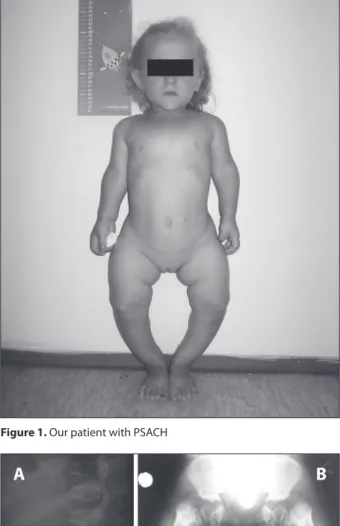
Case Outline A 6.5-year-old girl presented with short-limbed dwarfism (body height 79.5 cm, <P5; -32%) and normal craniofacial appearance and intelligence. The girl was normal until 3 months of age when she expressed growth retardation with apparently shorter extremities in relation to the torso. With age, her rhizomelic dwarfism became increasingly visible, and since completed 15 months of age, when she started to walk, the disease was complicated with genu varum, lumbar lordosis and abnormal gait. Beside visibly short forearms, short, broad and ulnar deviation of the hands, brachydactyly and joint hyperlaxity, the radiographic picture showed markedly flared metaphyses, small and irregular epiphyses and poorly formed acetabulum.
Conclusion PSACH is an achondroplasia-like rhizomelic dwarfism recognized by the absence of abnor-mality at birth, normal craniofacial appearance, characteristic epiphyseal and metaphyseal radiographic finding and joint hyperlaxity.
Keywords: pseudoachondroplasia, rhizomelic dwarfism, osteochondrodysplasias
Pseudoachondroplasia: A Case Report
Vladimir Radlović1, Željko Smoljanić1, Nedeljko Radlović1,2, Miroslav Jakovljević3, Zoran Leković1, Siniša Dučić1,2, Polina Pavićević1,2
1University Children’s Hospital, Belgrade, Serbia;
2School of Medicine, University of Belgrade, Belgrade, Serbia; 3Child and Youth Health Care Institute of Vojvodina, Novi Sad, Serbia
INTRODUCTION
Pseudoachondroplasia (PSACH) is a form of osteochondrodysplasia with an estimated prevalence of approximately 1/20,000-1/30,000 individuals [1-4]. The basis of the disease forms an autosomal dominant defect in the expression of cartilage oligomeric matrix protein (COMP) caused by a structural mutation in exons 8-19 of COMP gene located on the chromosome 19p12-13.1 [5-8]. The majority of COMP muta-tions are missense mutamuta-tions and small in-frame deletions and duplications found in exons 8-14 of this gene encoding the eight calmodulin-like calcium-binding repeats and exons 15-19 en-coding the carboxyl-terminal globular domain [9]. The consequence of this defect, character-ized by penetrance, is a development of the osteogenic disorder, i.e. the transformation of cartilage into bone tissue followed by typical metaphyseal, epiphyseal and vertebral abnor-malities [1, 10]. The main clinical characteris-tics of the disease are a disproportionate short stature similar to achondroplasia, brachydactyly, short, broad and ulnar deviation of the hands, forearms and lower limbs anomalies, joint hy-perlaxity, abnormal gait, lumbar lordosis, scolio-sis and early onset osteoarthroscolio-sis [1, 7-10]. The affected individual has normal length at birth, and generally, growth retardation will not be recognized until late infancy or more frequently between two to three years of age [7-10]. No-tably, all patients have normal craniofacial
ap-pearance and intelligence [2, 10, 11]. Here, we present a girl with an early expressed and severe PSACH born to clinically and radiographically unaffected parents.
CASE REPORT
A 6.5-year-old girl presented with dispropor-tionate short stature (79.5 cm, <P5; -32% from the average height for the age and gender), short and genu varum lower limbs, abnormal gait, exaggerated lumbar lordosis, short forearms, short, broad and ulnar deviation of the hands, brachydactyly and joint hyperlaxity (Figure 1). Craniofacial appearance and intelligence as well all other physical findings were normal.
677
Srp Arh Celok Lek. 2013 Sep-Oct;141(9-10):676-679
www.srp-arh.rs
started to walk, the disease was complicated with the genu valgum plus lumbar lordosis and abnormal gait. With age, her longitudinal growth showed the following values: at 17 months 61 cm (-28.5%), at 25 months 65 cm (-29%) and at 4 years 75 cm (-26%). Having in mind a progressive and diagnostically unclear disorder, the child was referred to our institution for additional investigations.
Based on the abovementioned facts, as well as the char-acteristic radiographic findings, the diagnosis of PSACH was verified (Figures 2A and 2B). The evident signs of osteoarthritis were not seen. Except for decreased serum concentration of 25(OH)D (64 nmol/L) and increased
PTH (91.5 pg/ml) at first examination, all other labora-tory findings, also including serum calcium, phosphorus and alkaline phosphatase, were normal. With 400 IU of vitamin D and additional intake of milk and milk products, the serum levels of 25(OH)D and PTH were normalized after 3 months. In order to maintain a normal balance of calcium and phosphorus, other than daily consumption of about 500 ml of milk or yogurt, the intake of vitamin D, 400 IU and 200 IU daily during colder and warmer weather periods, respectively, was recommended. Besides the corresponding paediatric and orthopaedic follow-up and adequate physical therapy, the parents were advised not to expose their child to exaggerated physical activities.
DISCUSSION
PSACH is a part of osteochondrodysplasias, a group of more than 150 distinct disorders characterized by bone and cartilage maldevelopment [2, 10]. In almost all of pa-tients, it appears to be secondary to autosomal dominant structural mutation within the genes encoding for COMP on the chromosome 19p12-13.1, and is most closely related to multiple epiphyseal dysplasia (MED/EDM1), a disorder also characterized by the mutation of the COMP [7-12].
COMP, also known as thrombospondin 5, is a 757-ami-no acid or 550 k-Da homopentameric glycoprotein present in the extracellular matrix (ECM) of cartilage, tendon, liga-ment and smooth muscle [13, 14]. Its presence in the ECM of these tissues is of essential significance for their growth, development and maintenance. As the consequence of this defect, both PSACH and MED chondrocytes retain lamel-lar and granulamel-lar appearing material in lamel-large rough endo-plasmic reticulum cisternae [11]. This material is com-posed of COMP and other ECM proteins including types II and IX collagens and matrilin-3 [15, 16]. By the method of fluorescence deconvolution analysis, these retained pro-teins have been recently shown to be organized into the matrix network suggesting that the stalled mutant COMP inappropriately interacts intracellularly with the matrix protein partners [15, 16, 17]. The retention of the intra-cellular matrix is toxic to the chondrocytes, and induces apoptosis causing the chondrocyte death [17]. The loss of chondrocytes in the growth plate translates, resulting into decreased long bone growth and the disproportion-ate short stature in PSACH. Additionally, the intracellular retention of the COMP and death of chondrocytes result in loss of these proteins in the ECM [18, 19, 20]. The respec-tive consequence is a disorganized type II collagen network most likely due to the absence of type IX collagen which is needed to crosslink type II collagen [21]. Association of the COMP deficit in the ECM and poorly organized structure of the articular cartilage lead to joint abnormalities and early onset osteoarthritis in patients with the PSACH and MED/EDM1 [20, 22].
The children with PSACH are always normal at birth and its disease usually presents at 2-3 years of age with disturbance in walking and lower limb deformity [7-11]. Over the years, rhizomelic dwarfism becomes apparent
Figure 1. Our patient with PSACH
Figure 2. Radiographic finding of our patients with PSACH: A) Lateral thoracolumbar radiograph showing platyspondyly with the anterior tongue-like protrusion of the upper lumbar and lower thoracic ver-tebrae; B) Anteroposterior radiograph of pelvis and lower extremity showing poorly formed acetabulum, large metaphyses, irregular epi-physes and marked bowing of the long bones.
678
doi: 10.2298/SARH1310676R
Radlović V. еt al. Pseudoachondroplasia: A Case Report
with progressively increasing morbidity, especially second-ary osteoarthritic changes [7-10]. The adult height ranges from 82 to 130 cm with a mean height of approximately 118 cm [7, 10]. Physical examination reveals normal facies and intelligence [10, 11].
The radiographic features are typical for rhizomelic dwarfism and include shortened bones, more prominent in the proximal than in the distal part, with flared and ir-regular metaphysis and small and poorly formed epiphy-ses [10, 23]. The most affected long bones are femur and humerus. The bones of the hand and foot are too broad and short with a small and immature epiphysis [23]. The x-ray of the pelvis shows flattened, irregular femoral heads with shortened necks and widened pubic symphysis [7]. The acetabulum is poorly formed with the horizontal roofs. The radiographic features include normal skull and facial bones and variable vertebral findings such as platyspondyly with the anterior beaking of the upper lumbar and lower thoracic vertebrae, lordosis and scoliosis [8, 23]. Differen-tial diagnoses of this radiographic picture in the context of clinical findings include achondroplasia, MED/EDM1 and spondyloepiphyseal dysplasia (SED) congenita [9]. Patients with achondroplasia have disproportionally large head with a prominent frontal region and depressed bridge of the nose. The basic differentiating point is that the epiphyses of patients with achondroplasia are normal. MED/EDM1 is characterized by a near normal pelvis with some scalloping of the acetabular margin. The SED, contrary to PSACH, is typified by hip joints that are affected disproportionately in relation to more distal portion of the lower extremities.
The diagnosis of PSACH is based primarily on personal and family history and characteristic physical and radio-graphic findings [10, 24, 25]. The finding of decreased se-rum COMP concentration may be an additional diagnostic marker of PSACH [4, 7, 26]. Whenever possible, genetic verification of the disorder is also done [25].
Therapy of PSACH is reduced to physiotherapy, man-agement of spinal deformities and corrective orthopaedic surgery [25, 27, 28, 29]. Intensive physical activity is recom-mended. Surgical correction of the limb deformities should be postponed until the end of the growth period [28].
We are presenting a girl who is a typical example of the patient with PSACH. She was born with a normal BL and her appearance was the same as that of any other child with clinically and radiographically unaffected parents. Beside her, the parents have a healthy 12-year old boy. Mode of PSACH inheritance clearly compels us to draw the conclu-sion that, in our patient, the disease was the result of newly developed mutation. As the realistic possibility of somatic mutation in the early postconception phase is low, it can be additionally concluded that the cause of PSACH devel-opment in this case should be searched in the germline (gonadal) mosaicism of parents [25, 30].
Although PSACH is not generally recognized until late infancy or more frequently between two to three years of age, progressive longitudinal growth retardation of the extremities, as the initial illness sign, was detected in our patient already at age of 5 months [7-10]. In her later years, her disease also manifested other clinical and radiological characteristics, which represented the basis of her diag-nostics [10, 24, 25].
Having in mind the age of our patient, as well as the severity of the disease at this stage, treatment was based on adequate paediatric-orthopaedic supervision, physio-therapy and avoiding of intensive physical activity [25, 28]. A suboptimal serum level of 25(OH)D recorded at first examination was corrected. Because of the significance of vitamin D in the calcium and phosphorus homeostasis, as well as the bone tissue itself, the maintenance of vitamin D optimal balance with adequate diet regime should be continued.
In conclusion, PSACH represents a rare form of achon-droplasia-like rhizomelic dwarfism recognized by the absence of abnormality at birth, normal craniofacial ap-pearance, characteristic epiphyseal and metaphyseal radio-graphic findings and joint hyperlaxity. Despite being the AD inherited disorder, it is usually seen as the consequence of a newly developed mutation, i.e. gonadal mosaicism in parents. Therapy is primarily physical-orthopaedic. In ad-dition, the prevention of vitamin D and/or calcium deficit is of exceptional significance, identically to other disorders that can disturb the integrity of bone tissue.
REFERENCES
1. Warman ML, Cormier-Daire V, Hall C, Krakow D, Lachman R, LeMerrer M, et al. Nosology and classification of genetic skeletal disorders: 2010 revision. Am J Med Genet A. 2011; 155A(5):943-68. 2. Posey KL, Hayes E, Haynes R, Hecht JT. Role of TSP-5/COMP in
pseudoachondroplasia. Int J Biochem Cell Biol. 2004; 36(6): 1005-12.
3. Posey KL, Hecht JT. The role of cartilage oligomeric matrix protein (COMP) in skeletal disease. Curr Drug Targets.2008; 9(10):869-77.
4. Tufan AC, Satiroglu-Tufan NL, Jackson GC, Semerci CN, Solak S, Yagci B. Serum or plasma cartilage oligomeric matrix protein concentration as a diagnostic marker in pseudoachondroplasia: differential diagnosis of a family. Eur J Hum Genet. 2007; 15(10):1023-8.
5. Zdravković D. Skeletne displazije. In: Zdravković D, editor. Klinička pedijatrijska endokrinologija. Beograd: Zavod udžbenike i nastavna sredstva; 2001. p.51-6.
6. Jackson GC, Mittaz-Crettol L, Taylor JA, Mortier GR, Spranger J, Zabel B, et al. Pseudoachondroplasia and multiple epiphyseal
dysplasia: a 7-year comprehensive analysis of the known disease genes identify novel and recurrent mutations and provides an accurate assessment of their relative contribution. Hum Mutat. 2012; 33(1):144-57.
7. Liu FX, Li ZL, Wei ZJ, Meng Y, Ren CA, Zhang XD, et al. Genetic analysis and serum level of cartilage oligomeric matrix protein in patients with pseudoachondroplasia. Chin Med J (Engl). 2010; 123(16):2181-4.
8. Dai L, Xie L, Wang Y, Mao M, Li N, Zhu J, et al. A novel COMP mutation in a pseudoachondroplasia family of Chinese origin. BMC Med Genet. 2011; 12:72.
9. Briggs MD, Chapman KL. Pseudoachondroplasia and multiple epiphyseal dysplasia: mutation review, molecular interactions, and genotype to phenotype correlations. Hum Mutat. 2002; 19(5):465-78.
679
Srp Arh Celok Lek. 2013 Sep-Oct;141(9-10):676-679
www.srp-arh.rs
11. Briggs MD, Chapman KL. Pseudoachondroplasia and multiple epiphyseal dysplasia: mutation review, molecular interactions, and genotype to phenotype correlations. Hum Mutat. 2002; 19(5):465-78. 12. Hecht JT, Montufar-Solis D, Decker G, Lawler J, Daniels K, Duke PJ.
Retention of cartilage oligomeric matrix protein (COMP) and cell death in redifferentiated pseudoachondroplasia chondrocytes. Matrix Biol. 1998; 17:625-33.
13. Hecht JT, Deere M, Putnam E, Cole W, Vertel B, Chen H, et al. Characterization of cartilage oligomeric matrix protein (COMP) in human normal and pseudoachondroplasia musculoskeletal tissues. Matrix Biol. 1998; 17:269-78.
14. Fang C, Johnson D, Leslie MP, Carlson CS, Robbins M, Di Cesare PE. Tissue distribution and measurement of cartilage oligomeric matrix protein in patients with magnetic resonance imaging-detected bone bruises after acute anterior cruciate ligament tears. J Orthop Res. 2001; 19:634-41.
15. Merritt TM, Alcorn JL, Haynes R, Hecht JT. Expression of mutant cartilage oligomeric matrix protein in human chondrocytes induces the pseudoachondroplasia phenotype. J Orthop Res. 2006; 24:700-7.
16. Merritt TM, Bick R, Poindexter BJ, Alcorn JL, Hecht JT. Unique matrix structure in the rough endoplasmic reticulum cisternae of pseudoachondroplasia chondrocytes. Am J Pathol. 2007; 170:293-300. 17. Hecht JT, Makitie O, Hayes E, Haynes R, Susic M, Montufar-Solis
D, et al. Chondrocyte cell death and intracellular distribution of COMP and type IX collagen in the pseudoachondroplasia growth plate. J Orthop Res. 2004; 22:759-67.
18. Xu C, Bailly-Maitre B, Reed JC. Endoplasmic reticulum stress: cell life and death decisions. J Clin Invest. 2005; 115:2656-64. 19. Chen TL, Posey KL, Hecht JT, Vertel BM. COMP mutations:
Domain-dependent relationship between abnormal chondrocyte trafficking and clinical PSACH and MED phenotypes. J Cell Biochem. 2008; 103:778-87.
20. Hecht JT, Hayes E, Haynes R, Cole WG. COMP mutations, chondrocyte function and cartilage matrix. Matrix Biol. 2005; 23:525-33.
21. Diab M, Wu JJ, Eyre DR. Collagen type IX from human cartilage: a structural profile of intermolecular cross-linking sites. Biochem J. 1996; 314(Pt 1):327-32.
22. Posey KL, Hankenson K, Veerisetty AC, Bornstein P, Lawler J, Hecht JT. Skeletal abnormalities in mice lacking extracellular matrix proteins, thrombospondin-1, thrombospondin-3, thrombospondin-5, and type IX collagen. Am J Pathol. 2008; 172:1664-74.
23. Cao LH, Wang LB, Wang SS, Ma HW, Ji CY, Luo Y. Identification of novel and recurrent mutations in the calcium binding type III repeats of cartilage oligomeric matrix protein in patients with pseudoachondroplasia. Genet Mol Res. 2011; 10(2):955-63.
24. Tandon A, Bhargava SK, Goel S, Bhatt S. Pseudoachondroplasia: a rare cause of rhizomelic dwarfism. Indian J Orthop. 2008; 42(4):477-9.
25. Cohn DH. Pseudoachondroplasia. In: Pagon RA, Bird TD, Dolan CR, Stephens K, editors. GeneReviews [Internet]. Seattle (WA): University of Washington, Seattle; 1993-2004 [updated 2010].
26. Mabuchi A, Momohara S, Ohashi H, Takatori Y, Haga N, Nishimura G, et al. Circulating COMP is decreased in pseudoachondroplasia and multiple epiphyseal dysplasia patients carrying COMP mutations. Am J Med Genet. 2004; 129A:35-8.
27. Li QW, Song HR, Mahajan RH, Suh SW, Lee SH. Deformity correction with external fixator in pseudoachondroplasia. Clin Orthop Relat Res. 2007; 454:174-9.
28. Carter EM, Montuori L, Davis JG, Raggio CL. The Kathryn O. and Alan C. Greenberg Center for skeletal dysplasias: an interdisciplinary approach. HSS J. 2008; 4(2):112-6.
29. Segal O, Lammens J. Ilizarov treatment for extreme bilateral genu recurvatum in a pseudoachondroplasia patient: a case report. Acta Orthop Belg. 2010; 76(1):124-8.
30. Ferguson HL, Deere M, Evans R, Rotta J, Hall JG, Hecht JT. Mosaicism in pseudoachondroplasia. Am J Med Genet. 1997; 70(3):287-91.
КРАТАК САДРЖАЈ
Увод Псе у до а хон дро пла зи ја (ПСАХ) је ауто зом но до ми нант-на осте о хон дро ди спла зи ја иза зва нант-на му та ци јом ге нант-на ко ји ко-ди ра хр ска ви ча ви оли го мер ни ма трик сни про те ин. Од ли ку-ју је ри зо ме лич ни ма ли раст, де фор ми те ти екс тре ми те та и кич ме ног сту ба, хи пе ре ла стич ност згло бо ва и ра на осте о-ар тро за. Сле ди при каз де вој чи це кли нич ки и ра ди о граф ски здра вих ро ди те ља с те шком и ра но ис по ље ном ПСАХ.
При каз бо ле сни ка Де вој чи ца уз ра ста од шест и по го ди на, нор мал ног кра ни о фа ци јал ног из гле да и ин те ли ген ци је, при-мље на је на пре глед због дис про пор ци о нал но ма лог ра ста (те ле сна ви си на 79,5 cm; <П5; -32%). За о ста ја ње у лон ги ту
ди-нал ном ра сту са вид но кра ћим екс тре ми те ти ма у од но су на труп уоче но је три ме се ца по ро ђе њу. Са ра стом и раз во јем ри зо ме лич ни тип ни ског ра ста по ста јао је све из ра же ни ји, а
од на вр ше них 15 ме се ци, ка да је про хо да ла, бо лест се ком-пли ко ва ла са ге на ва ра, лум бал ном лор до зом и оте жа ним хо да њем. По ред упа дљи во скра ће них над лак ти ца, кра ћих, ши ро ких и ул нар но де ви ра них ша ка, крат ких пр сти ју и хи-пе ре ле стич них згло бо ва, на ренд ген ским сним ци ма су за-па же ни на гла ше но ра све тље не ме та фи зе, ма ле не пра вил не епи фи зе и сла бо фор ми ра ни аце та бу лу ми.
За кљу чак ПСАХ је об лик ри зо ме лич ног ма лог ра ста на лик на ахон дро пла зи ју, пре по зна тљи ву по нор мал ним из гле дом на ро ђе њу, не по сто ја њем кра ни о фа ци јал них по ре ме ћа ја, ти пич ним епи фи зо ме та фи зар ним ра ди о граф ским на ла зом и хи пе ре ла стич но шћу згло бо ва.
Кључ не ре чи: псе у до а хон дро пла зи ја; ри зо ме лич ни ма ли раст; осте о хон дро ди спла зи је
Псеудоахондроплазија – приказ болесника
Владимир Радловић1, Жељко Смољанић1, Недељко Радловић1,2, Мирослав Јаковљевић3, Зоран Лековић1,
Синиша Дучић1,2, Полина Павићевић1,2 1Универзитетска дечја клиника, Београд, Србија;
2Медицински факултет, Универзитет у Београду, Београд, Србија;
3Институт за здравствену заштиту деце и омладине Војводине, Нови Сад, Србија