Response to Stress in Early Tumor Colonization
Modulates Switching of Positive and
CD133-Negative Subpopulations in a Human Metastatic Colon
Cancer Cell Line, SW620
Chih-Sin Hsu1., Chien-Yi Tung1., Chih-Yung Yang1,3
, Chi-Hung Lin1,2,3*
1Institute of Microbiology and Immunology, National Yang-Ming University, Taipei, Taiwan,2VGH Yang-Ming Genome Research Center, Taipei, Taiwan,3Department of Education and Research, Taipei City Hospital, Taipei, Taiwan
Abstract
According to the cancer stem cell (CSC) model, higher CD133 expression in tumor tissue is associated with metastasis and poor prognosis in colon cancer. As such, the CD133-positive (CD133+) subpopulation of cancer cells is believed to play a central role in tumor development and metastatic progression. Although CD133+cells are believed to display more CSC-like behavior and be solely responsible for tumor colonization, recent research indicates that CD1332 cells from metastatic colon tumors not only also possess colonization capacity but also promote the growth of larger tumors in a mouse model than CD133+cells, suggesting that an alternative mechanism of metastasis exists. This study investigated this possibility by examining the cell viability, tumorigenicity, and proliferation and growth capacity of the CD133+ and CD1332 subpopulations of the SW620 cell line, a human metastatic colon cancer cell line, in both anin vitrocell model and an in vivomouse model. While both SW620 CD1332 and SW620CD133+cells were found to engage in bidirectional cell-type switching in reaction to exposure to environmental stressors, including hypoxia, a cell adhesion-free environment, and extracellular matrix stimulation, bothin vitroandin vivo, CD1332cells were found to have a growth advantage during early
colonization due to their greater resistance to proliferation inhibition. Based on these findings, a hypothetical model in which colon cancer cells engage in cell-type switching in reaction to exposure to environmental stressors is proposed. Such switching may provide a survival advantage during early colonization, as well as that explain previous conflicting observations.
Citation:Hsu C-S, Tung C-Y, Yang C-Y, Lin C-H (2013) Response to Stress in Early Tumor Colonization Modulates Switching of Positive and CD133-Negative Subpopulations in a Human Metastatic Colon Cancer Cell Line, SW620. PLoS ONE 8(4): e61133. doi:10.1371/journal.pone.0061133
Editor:Wael El-Rifai, Vanderbilt University Medical Center, United States of America
ReceivedOctober 23, 2012;AcceptedMarch 5, 2013;PublishedApril 5, 2013
Copyright:ß2013 Hsu et al. This is an open-access article distributed under the terms of the Creative Commons Attribution License, which permits unrestricted use, distribution, and reproduction in any medium, provided the original author and source are credited.
Funding:This work was supported by research grants NSC 97-3112-B-075-002, NSC 98-3112-B-010-023-B4, and NSC 97-2320-B-010-024-MY3 from the National Science Council and by the Aim for the Top University Plan from the Ministry of Education, Republic of China. The funders had no role in study design, data collection and analysis, decision to publish, or preparation of the manuscript.
Competing Interests:The authors have declared that no competing interests exist. * E-mail: [email protected]
.These authors contributed equally to this work.
Introduction
Most deaths due to colon cancer are correlated with metastasis of the primary tumor rather than development of the primary tumor. As such, gaining understanding of colon cancer metastasis could greatly increase the patient survival rate. Achieving such a goal is particularly important, as the median overall survival duration of metastatic colon cancer patients is currently less than 2 years and the 5-year survival rate less than 10% [1,2]. Gaining understanding of metastasis could also greatly assist in selecting the appropriate therapy strategy.
It is currently known that tumor metastasis is a multi-stage process in which the malignancy of tumor cells progressively develops in 3 sequential stages: intravasation, extravasation, and colonization [3]. In the intravasation stage, during which tumor cells undergo cellular functional changes in preparation for cell migration and extracellular matrix (ECM) digestion, tumor cells begin to escape from the primary site and invade the circulating system. In the extravasation stage, tumor cells circulate in blood
vessels and the lymphatic system before being transported to distant metastatic sites. During the cancer colonization stage, the transported tumor cells adapt to the microenvironment and begin to proliferate. In the early colonization stage, tumor cells are challenged by many environmental stressors, such as ECM interactions, hypoxic conditions, growth-factor stimulation, and immune-system interactions, that lead to their death or dormancy [3]. The few cells that can adapt to the stressors and survive become the subpopulation of cancer cells involved in cancer colonization and are responsible for the metastasis procession.
establish colonies at metastatic sites. Several studies have reported that CSCs participate in metastasis progression [4,6]. A number of cell markers, including CD133, CD166, and CD26, have been used to isolate the CSC subpopulation within colon-cancer-cell populations [7,8]. Among them, prominin 1 (CD133) is a pentaspan transmembrane protein originally found in hematopoi-etic stem cells in the bone marrow [9]. Recent studies have reported CD133 to be a potential biomarker of metastasis and poor prognosis in colon cancer patients [10–12]. In accordance, immunohistochemical (IHC) staining of colon cancer tissue has revealed high expression of CD133 to be correlated with liver metastasis and low survival [12,13].
The CD133 protein has been found to be more abundant in liver metastatic lesions compared with primary colon cancer lesions [14]. Nevertheless, CD133 is widely considered a CSC marker in colon cancer. As downregulation of CD133 has been widely observed during tumor development, its expression is believed to be regulated during cell differentiation [15]. In a mouse xenograft experiment investigating the CD133-negative (CD1332) and CD133-positive (CD133+
) subpopulations of primary colon cancer cells, only the CD133+
subpopulation was found capable of inducing tumor formation [15–17]. These findings, in addition to the wide and effective use of CD133 as a biomarker to isolate stem cells in cancerous tissues [18,19], suggest that a subpopulation of CD133+
cells acts as CSCs in the metastasis of colon cancer.
However, several controversial findings have emerged from comparison of CD1332and CD133+
subpopulations in cell-lines and primary cultures of colon cancer [20–23]. Although both CD1332and CD133+
subpopulations of HCT-116 cells, a human colon cancer cell line, have been found to induce tumor growth in mouse models [22], the CD1332 subpopulation of one human metastatic colon cancer cell line, LoVo, was found more resistant to 5-fluorouracil treatment than the CD133+
subpopulation [20]. Likewise, Shmelkov et al. found a subpopulation of CD1332colon cancer cells isolated from a liver metastatic site in a xenograft mouse model to have a higher level of tumorigenicity than the subpopulation of CD133+
cells [23]. These conflicting results cannot be explained by the CSC model, and suggest the existence of an alternative mechanism in metastasis procession.
SW620 is a human colon-cancer cell line that originates from a lymph node metastatic site. As it possesses both CD133+
and CD1332 subpopulations in a general culture condition (i.e., that obtained in a dish containing an L15 medium and 10% FBS at 37uC), SW620 can serve as an excellent cell line model with which to investigate the differences between CD133+
and CD1332 subpopulations within cells derived from the same genetic background. Using this model, this study isolated the CD133+ and CD1332 subpopulations in a SW620 cell line to compare their tumorigenicity, cell behavior, and proliferation and growth capacity both in vitro and in vivo. Based on the results, a
hypothetical model of cancer colonization in metastasis that explains the controversial findings of recent studies is proposed.
Results
The SW620 cell line consists of 2 subpopulations with high and low CD133 expression
Figure 1A shows that the SW620 cell line was divided into 2 populations, the SW620CD133+
and SW620CD1332 subpopula-tions, according to CD133-expression level, of which the SW620CD133+population occupied 75% of the SW620 cells in a general culture condition. To isolate the subpopulations, fluores-cent-activated cell sorting and analysis (FACS) was performed and
subsequently confirmed by reverse-transcription polymerase chain reaction (RT-PCR) and western blot. The highly and poorly expressed subpopulations were then identified by performing analysis of CD133high and CD133negative zone distribution (Figure 1B). The results revealed that the SW620CD133+
cells express more CD133 mRNA and protein than SW620CD1332cells (Figure 1C and 1D), which excluded the possibility of alternative splicing or translocation regulation [24]. It is well known that SW620 contains 2 distinct morphological cell types, a small spherical (white arrow, Figure 1E; Figure S1A and S1B) and a bipolar type (black arrow, Figure 1E; arrow-head, Figure S1A and S1B) [25], whose proportion does not significantly differ in the SW620CD133+and SW620CD1332subpopulations.
Carboxyfluorescein diacetate–succinimidyl ester (CFSE) stain-ing of FACS and MTT assay revealed that the proliferation ability between SW620 subpopulations are similar in a general culture condition (Figure S1C and S1D), while MTT assay with taxol, cisplatin, actinomycin D, and camptothecin treatment revealed the cell viability of the subpopulations to be similar (Figure S2A to S2D). These results indicate that SW620CD133+
and SW620CD1332 cells have no apparent morphological and physiological differences in a general culture condition. As these results cannot explain the different levels of tumorigenicity between the CD133 subpopula-tions found in previous studies [23], they suggest that inducement of different levels of tumorigenicity can be attributed to exposure to particular environmental conditions.
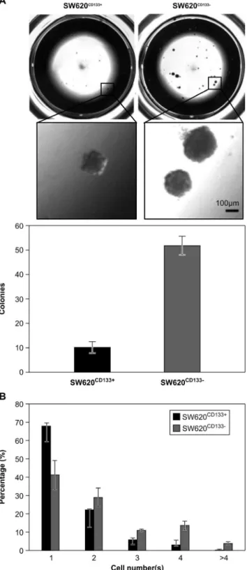
SW620CD1332cells exhibit higher colony formation capacity than SW620CD133+cells in a 3D Matrigel culture
The ability of extravasated tumor cells to grow despite immediate exposure to microenvironmental stressors at the metastatic site is essential for metastatic cells to progress to tumor colonization [26]. Matrigel, a soluble basement membrane protein extract derived from the Engelbreth-Holm-Swarm mouse sarco-ma, can serve as anex vivomicroenvironment in which cells are
exposed to 3D architecture, ECM interaction, and growth factor stimulation [27]. As such, a 3D Matrigel culture model has become widely used as an experimental model to measure tumorigenicity [28–30]. Using this model to compare the colony formation capacity of SW620CD133+
and SW620CD1332cells, 500 SW620CD133+and SW620CD1332 cells were seeded on top of a thick Matrigel culture. Quantification of visible colonies after 3 weeks of culture revealed that the SW620CD1332cells, which had formed a mean of 51.8 colonies (SD = 3.8), had a higher colony formation capability compared to the SW620CD133+
cells, which had formed a mean of 10.3 colonies (SD = 2.2; Figure 2A).
Cell proliferation in the early phase was further examined by quantification of the cell number in each clone. When approxi-mately 200 cells were seeded in a low-cell-density on 3D Matrigel culture and the cell number of each colony was counted one by one under microscope after 3 days incubation, the results revealed that the SW620CD133+
cells were less proliferative than SW620CD1332 cells (Figure 2B). Specifically, only a small proportion of SW620CD133+
cells (black bar) had been able to complete 2 (cell number = 4, 3.4%) or more (cell number .4, 0.4%) rounds of cell cycle, and most had experienced arrested growth, such that only 22.5% divided one time to become 2 cells and 67.7% did not undergo any cell division at all. The SW620CD1332cells (gray bar) were found to be more tolerant to proliferation inhibition due to exposure to the 3D Matrigel culture, with 4.7-fold more SW620CD1332 cells (18%) than SW620CD133+cells having been able to complete
$2 rounds of cell cycle. This 4.7-fold difference in large colony percentage (n$4) between the 2-cell types is similar to the 5-fold difference in
the number of visible colony numbers previously observed. This result indicates that different levels of tolerance, specifically the higher level of SW620CD1332cells, to proliferation inhibition from the microenvironment leads to different levels of tumorigenicity between SW620CD133+ and SW620CD1332 cells, providing SW620CD1332 cells with greater capacity to engage in tumor formation at early metastatic sites.
Figure 1. Purification of SW620CD133+and SW620CD1332cells. The CD133 expression of SW620 cells was analyzed and the cells were sorted by FACS Canto and FACS Aria, respectively. (A) Measurement of CD133 expression by flow cytometry revealed that 75% of the cells were SW620CD133+cells and 25% were SW620CD1332cells (white peak indicates staining control for mouse IgG). (B) The top 5% of the CD133+ subpopulation (CD133high) and bottom 5% of the CD133
2 subpopu-lation (CD133negative) were collected using FACS Aria. (C) Upper panel shows RT-PCR results for CD133 mRNA. Lower panel shows RT-PCR results for GAPDH mRNA used as a control to perform normalization. (D) Upper panel shows western blot results for CD133 protein in total cell lyse. Lower panel shows the results for alpha tubulin used as a loading control. (E) SW620CD133+
cell morphology under light microscopy (206).
(F) SW620CD1332cell morphology under light microscopy (206).
doi:10.1371/journal.pone.0061133.g001
Figure 2. Comparison of colony formation capacity of SW620CD133+ and SW620CD1332 cells on 3D Matrigel culture. Purified SW620CD133+
and SW620CD1332 cells were seeded on top of Matrigel for further analysis of cell proliferation and colonization. (A) Upper panel shows the entire and enlarged picture for colonies morphology; lower panel shows colonies count after 3 weeks of incubation. The SW620CD133+
cells formed a mean of 10 colonies (SD = 2.2) and the SW620CD1332cells a mean of 50 colonies (SD = 3.8). Bar = 100mm. (B) Cell count after 3 days of incubation.
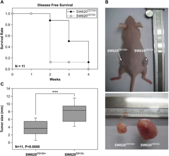
SW620CD1332 cells have a higher level of tumorigenicity than SW620CD133+in a nude mouse model
Thein vivotumorigenicity of SW620CD133+
and SW620CD1332 cells was examined in a nude (nu/nu) mouse model. After inoculation of 105cells in a subcutaneous region, tumor size was measured every 3 days for 4 weeks. The disease-free survival rate is shown in Figure 3A. Both cell types were able to generate tumor tissue after 4 weeks inoculation. Although hematoxylin and eosin (H&E) staining revealed the tumor morphology of the 2 cell types to be similar (Figure S3A and S3B), the growth rate was found to differ, with SW620CD1332 cells found able to generate larger tumor cells at an earlier stage. As shown in Figure 3C, by 2 weeks post inoculation, 78% of the mice inoculated with SW620CD1332 cells had developed tumors.2 mm in size (open circle) while only 11% of the mice inoculated with SW620CD133+
cells had done so (filled circle). Moreover, the average diameter of the tumors that had developed after SW620CD1332 inoculation was 1.9-fold greater than that after SW620CD133+
inoculation (Figure 3B and 3C). These results, which confirm that SW620CD1332cells have greater tumorigenicity than SW620CD133+cells and accord with the results obtained using theex vivoexperimental model, provide
the first cell-line evidence that the CD1332 subpopulation has greater potential to initiate tumor progression at metastatic sites [23].
Cell-type switching between SW620CD1332 and SW620CD133+subpopulations is induced by
microenvironmental stimulation
Although inducement of CD133 upregulation by exposure to certain stressors and forms of environmental stimulation was observed in several previous in vitro cell experiments [31–33],
switching of the CD1332subpopulation to the CD133+ subpop-ulation of brain tumor primary cultured cells was observed in a recentin vivoexperiment [34]. Similar switching was observed in
the NANK cell line, a primary colon cancer line. Although NANK is a CD1332 cell line isolated from ovarian metastatic colon cancer tissues, CD133+
cells were found in a NANK-induced tumor [21].
In accordance with these findings, switching between SW620CD1332and SW620CD133+
cells in an unbalanced manner was observed in the general culture condition in this study. Specifically, 16.7% of purified SW620CD1332 cells switched to SW620CD133+cells (Figure 4, white bar in right panel) while only 4.7% of SW620CD133+
cells switched to SW620CD1332cells (white bar in left panel, Figure 4). This 3.6-fold difference in switching capacity between SW620CD1332 and SW620CD133+
cells may explain the greater proportion of SW620CD133+
compared to SW620CD1332cells (3:1) in the general culture condition (Figure 1). This explanation is supported by the results of the MTT assay,
Figure 3. Comparison of tumorigenicity of SW620CD133+and SW620CD1332cells in nude mice.Purified SW620CD133+and SW620CD1332 cells were injected subcutaneously into nude mice. (A) Filled and unfilled circles refer to the disease-free survival rate of the SW620CD133+
and SW620CD1332cells, respectively, N = 11. (B) Upper photograph shows tumor formation due to SW620CD133+
cell inoculation on the left side and due to SW620CD1332cell inoculation on the right side. Lower photograph shows the sizes of the SW620CD133+
and SW620CD1332tumors. (C) Chart showing the mean diameters of the SW620CD133+
(3.8 mm, SD = 2.1) and SW620CD1332(8.1 mm, SD = 2.6) tumors, N = 11, P = 0.0006. doi:10.1371/journal.pone.0061133.g003
which revealed that the 2 populations have a similar proliferation rate, and thus excluded the possibility of growth competition.
Investigation of the tumor cells isolated during the previous xenograft experiments (Figure 3A) confirmed these findings. Whereas 90.3% of tumor cells that had proliferated from SW620CD1332 cells later switched to SW620CD133+
cells, only 4.8% of SW620CD133+
cells switched to SW620CD1332 cells (Figure 4, diagonal bar). These results suggest that cell-type switching might be modulated by stimulation from the in vivo
microenvironment. To test this hypothesis, the impact of exposure to hypoxia, a cell-adhesion-free environment, and ECM stimula-tion, 3 potential forms of stress in thein vivomicroenvironment was
examined. To establish a hypoxic condition, the oxygen concen-tration in the culture chamber was controlled at 1% using the PROOX 110 instrument and the temperature maintained at 37uC. Whereas exposure to hypoxia significantly increased switching of SW620CD1332cells to SW620CD133+
cells, specifically from 16.7% to 40% (p = 0.0064), it resulted in no significant change in the extent to which SW620CD133+
cells switched to SW620CD1332 cells (p = 0.13) after 4 weeks of culture in the hypoxia condition.
To test exposure to a cell-adhesion-free condition using a 3D culture system, cells were loaded onto Algimatrix, an artificial bio-scaffold material without an ECM component. Cells were cultured at low density (105per well) to prevent cell–cell contact. After 2 weeks of culture, cells were isolated from the gel and CD133 expression was measured by FACS. Whereas exposure to a cell-adhesion-free environment significantly increased the switching of SW620CD1332 cells to SW620CD133+, specifically from 16.7% to 64.5% (p,0.001), it resulted in no significant change in the extent to which SW620CD133+
cells switched to SW620CD1332 cells
(p = 0.57). To examine the impact of ECM stimulation, cell culturing was performed in a Matrigel-coated plate. Although exposure to ECM stimulation significantly inhibited CD133 expression in both cell types, it had a variable impact on the extent of cell-type switching. Whereas it decreased switching of SW620CD1332 cells to SW620CD133+ cells from 16.7% to 5% (black bar in right panel, p,0.001) in a Matrigel-coated plate and to 13% in a 3D Matrigel culture (Figure 4, right panel, horizontal filled bar, p = 0.19), it increased switching of SW620CD133+
cell group from 5% to 11% in a Matrigel-coated plate (p = 0.055) and to 27% in a 3D Matrigel culture (Figure 4, left panel, horizontal filled bar, p = 0.023).
Testing of exposure to the 3 forms of environmental stress revealed the existence of a significant regulation of CD133 switching in response to such exposures, with exposure to hypoxia and a cell-adhesion-free environment significantly promoting switching of SW620CD1332 cells to SW620CD133+
cells while exposure to ECM stimulation significantly promoting switching of SW620CD133+
cells to SW620CD1332 cells. These results suggest that regulation of SW620CD1332 and SW620CD133+
subpopula-tions in an in vivo environment is modulated by exposure to
multiple factors that act synergistically upon the cells, and that cell-type switching between subpopulations might be required for adaptation to the microenvironment in early tumor colonization.
Discussion
It has been widely discussed that CSCs play a key role in metastasis progression [7,35]. Of the several cell markers, including CD133, CD144, and CD166, that have been used to identify CSCs, a number of independent studies have demonstrated that the Figure 4. Modulation of CD133 expression in SW620 cells in reaction to exposure to stressors.Left panel shows SW620CD133+cells and
CD133+
subpopulation of colon cancer cells possess higher levels of stemness and tumorigenicity that the CD1332subpopulation. Two independent studies that isolated CD133+
and CD1332 subpopu-lations from colon tumor cells and transplanted them into NOD/ SCID mice found that only CD133+
cells can initiate tumor formation [15,17]. A study using a xenograft model found that the CD133+
subpopulation of colon tumor cells also display multi-lineage differentiation capacity and a high level of tumorigenicity [16].
However, 2 recent studies found that the CD1332 subpopula-tion of metastatic colon cancer cells in liver and ovarian cancer also initiate tumor formation, and ultimately induce greater tumor growth than the CD133+
subpopulation [21,23]. The conflicting nature of these various results may be due to the use of experimental models derived from different genetic backgrounds. The colon cancer cell line model provides a conventional and reproducible experimental model that allows for comparison of the 2 CD133 subpopulations. Of the 2 cell lines used as experimental models of colon cancer that can be divided into CD133+
and CD1332 subpopulations [32], the HCT116 cell line, which was established primarily from colon tumors, has a smaller (,5%) CD1332subpopulation than the SW620 cell line. In accordance with previous comparisons of cell viability and tumorigenicity between HCT116CD1332and HCT116CD133+cells by MTT assay and xenograft experiments, HCT116CD1332and HCT116CD133+ cells have been found to have similar levels of viability and tumorigenicity [22].
In the present study, the SW620CD1332 and SW620CD133+ subpopulations were found to have a similar morphology, proliferation rate, and drug response in a general culture condition. However, the SW620CD1332cells displayed significant-ly greater growth in a 3D Matrigel culture and in mouse xenograft experiments. These results provide the first pieces of evidence that CD1332 colon cells have greater tumorigenicity than CD133+ colon cells in experimental models with the same genetic background, suggesting that certain environmental factors could induce different rates of growth. Unfortunately, exposure to one form of stress, such as hypoxia or ECM stimulation, alone, was not found to affect the proliferation rate (Figure S4A), indicating that inhibition might be induced by a complex mechanism of regulation and/or a number of factors that act synergistically in the microenvironment. The existence of such a complex mechanism remains to be investigated.
Switching between CD133+
and CD1332 subpopulations has been observed in many types of tumor cells [36–38]. It is well known that CD133+CSCs will switch to CD1332 cells during differentiation [16], and that such switching is unidirectional and irreversible. In the chemically induced differentiation model, sodium butyrate treatment of the human colon cancer cell lines HT-29 and HCT116 has been found to induce CD133 downregulation and epithelial differentiation [39]. Several tumor studies have found that switching of CD1332cells to CD133+
cells can be induced by exposure to growth factor and environmental stressors. Specifically, exposure to TGF-beta1 has been found to trigger CD133 upregulation in the Huh7 and A549 hepatocellular carcinoma and lung cancer cell lines and increase cell tumorige-nicity in a nude mice model [33,36]. While exposure to hypoxia has been found to induce CD133 expression in lung and pancreatic cancer cells, these transformed CD133+ tumor cells gain more stemness and malignancy [31,40]. Hypoxia induced CD133 expression in brain tumor also correlates with higher tumor aggressiveness [41].
In a xenograft model of metastatic colon tumor, CD133+ cells were detected in a tumor generated from purified CD1332cells
[21], suggesting that cell-type switching might also occur in colon cancer cells. The results of this study clearly demonstrate the existence of bi-directional switching between SW620CD133+
and SW620CD1332 subpopulations, and that such switching can be modulated by exposure to various stressors. Specifically, exposure to either a Matrigel-coated plate or a 3D Matrigel culture was found to significantly induce switching of SW620CD133+
to SW620CD1332cells, the latter of which has been found to exhibit greater growth in ex vivo and in vivo models. In pilot studies,
exposure to Type I and IV collagen and laminin, the 3 major ECM components, was observed to induce inhibition of CD133 expression to a similar extent (data not shown). In contrast, exposure to hypoxia or a cell-adhesion-free condition was found to promote switching of SW620CD1332 cells in the present study. This result was also observed using the hand-drop culture system, which provides exposure to a cell-adhesion-free environment, in which SW620CD133+
cells were found in SW620CD1332-forming spheres, suggesting that they play a role in driving tumorigenesis [32].
In the conventional model of metastasis progression, both CD133+
and CD1332 cancer cells are released into the bloodstream when metastasis is initiated [42], but only CD133+ cells are able to survive and initialize tumor colonization (Figure 5, upper panel). However, several studies of colon cancer cell lines have reported that both CD133+
and CD1332cells can initialize tumor formation [21–23]. The present study, which observed this phenomenon in the SW620 cell line, also found that colon cancer cells may engage in cell-type switching between the SW620CD1332 and SW620CD133+
subpopulations in response to microenviron-mental stress. Based on these observations, an alternative hypothetical model of colon cancer colonization at the metastasis site, which is illustrated in Figure 5, is proposed here. In the early metastatic phase, cancer cells migrate into the metastatic site and begin colonizing. During this phase, the cells are directly exposed to many forms of environmental stress and stimulation, among which ECM stimulation promotes the switching of CD133+
to CD1332cells, the latter of which have a growth advantage due to their greater resistance to proliferation inhibition from the microenvironment. After tumor colonization has been established and the tumor cells have been organized into a solid tumor, other forms of environment stress, including exposure to hypoxia and a cell-adhesion-free environment, promote the switching of CD1332 to CD133+cells, which, because the latter type has been found to have greater stemness and malignancy in colon and other types of cancer [36,43], may increase later tumor growth.
This hypothetical model explains the conflicting results obtained from previous studies of CD133+
and CD1332colon cancer cells. Although CD133 is known as a stem-cell marker, the results of previous studies, as well as the present study, indicate that both CD133+
and CD1332cells can generate tumor growth, suggesting that CD133 might not be an absolute biomarker for cancer stem cells in colon cancer. To help cancer cells adapt to different environmental stressors during tumor regression, the 2 subpopu-lations often engage in cell-type switching, which may explain why CD133+
cells are frequently found at metastatic sites. This model provides a means of explaining the many observations regarding colon cancer metastasis that cannot be explained by the conventional CSC model.
Materials and Methods
Cell culture and growth conditions
The SW620 human colorectal cancer cell line used in this study was obtained from the Bioresource Collection and Research
Center (Hsinchu, Taiwan). SW620 cells were grown in Leibovitz’s L-15 Medium supplemented with 10% fetal bovine serum and 1% penicillin/streptomycin (Invitrogen) at 37uC. Cells were grown in a humidified environment to 80% confluence before further experiments and passage. A hypoxic condition consisting of 1% O2and 99% N2was controlled using the PROOX 110 instrument (BioSpherix, Lacona, NY, USA). Western blot for HIF-1alpha was performed to confirm achievement of a hypoxic condition. Matrigel was diluted with a serum-free medium (1:20) and coated on a Petri dish to conduct the experiments that followed.
FACS
After removal from the cell culture medium and washing in phosphate-buffered saline (PBS), cells were dissociated from the Petri dish using TE buffer (Invitrogen) and labeled with mouse anti-human CD133-PE antibody (Miltenyi Biotec) following the standard protocol on ice for 30 min. The labeled cells were then analyzed using FACS Canto (BD, San Jose, CA, USA) and sorted using FACS Aria (BD). The top 5% of the CD133+
subpopulation and bottom 5% of the CD1332subpopulation were collected for further experiments.
CFSE staining
For this process, 16106 cell/ml were labeled with 5mM of CFSE (Invitrogen) at 37uC for 10 min, and 56105 cells were seeded onto a Petri dish for further culturing. Staining with CD133-APC antibody was performed on days 0 and 3 to analyze CD133 expression and cell division by FACS Canto.
RT-PCR
The CD133 protein has been found to have various splice forms that can lead to loss of the AC133 epitope on the cell surface during cell differentiation but not of the CD133 protein [24]. Based on this finding, a specific primer was designed to amplify the
conserved region for all CD133 alternative splicing forms on exons 17 to 24 in the CD133 gene using RT-PCR. Purified SW620CD133+
and SW620CD1332cells were treated with TRIzol (Gibco-BRL; Gaithersburg, MD, USA) following the standard protocol to extract total RNA and reverse-transcribed into first-strand cDNA. Subsequently, cDNA was added to the PCR reaction with CD133 forward (59- ACGGCACTCTTCACCTG-CAG-39) and reverse (59-CGATGCCACTTTCTCACTGA-39) primers (94uC for 30 s, 55uC for 30 s, and 72uC for 30 s for 27 cycles). Glyceraldehyde 3-phosphate dehydrogenase (GAPDH), forward (59- AGCGAGATCCCTCC -39) and reverse (59 -GCAGGAGGCATTGC -39) was used as a control. The PCR product was separated on 2% agarose gel and the signal measured at 592 bp and 221 bp respectively.
In vivotumorigenicity assay
Six-to-eight week old immunodeficient nu/nu mice were obtained from BioLasco Taiwan Co. Ltd. and maintained within facilities operated by the National Yang-Ming University Labo-ratory Animal Center. Approval for all mouse experiments was obtained from the Institutional Animal Care and Use Committee of the National Yang-Ming University. Purified SW620CD133+and SW620CD1332cells (16105) were suspended in PBS and injected subcutaneously into the dorsal region near the thigh, which was then observed twice a week for 4 weeks. Mice were sacrificed 4 weeks after inoculation for excision of tumors and preparation of paraffin-embedded sections to H&E staining. Excised tumors were incubated with 10 U of collagenase I (Sigma-Aldrich) for 2 h at 37uC, washed twice with PBS, and filtered with a mesh to obtain single cells for CD133 expression analysis by FACS.
Western blot analysis
Cells were washed twice with cold PBS and lysed by RIPA buffer (Cell Signaling Technology) with protease inhibitors (Roche) on ice. Equal amounts of proteins were separated by SDS-PAGE and transferred to polyvinylidene difluoride (PVDF) membranes. Membranes were blotted with anti-CD133 monoclo-nal antibody (Cell Sigmonoclo-naling Technology), HIF1alpha anti-body (Cell Signaling Technology), anti-alpha tubulin antianti-body (Abcam), and secondary antibodies conjugated with horseradish peroxidase (HRP). The proteins were detected using an enhanced chemiluminescence kit (Pierce, Rockford, IL) in 133 kDa for CD133, 120 kDa for HIF1-alpha and 50 kDa for alpha tubulin.
MTT assay
Purified SW620CD133+ and SW620CD1332 cells (1
6103) were seeded into 96-well plates and treated with different anti-tumor drugs, including taxol Aldrich, 1 uM), cisplatin (Sigma-Aldrich, 10 uM), actinomycin D (Millipore, 10 nM), and campto-thecin (Millipore, 2 nM) to perform drug-resistance assay. After 24, 48, and 72 h of incubation, MTT reagent was added and the cells incubated at 37uC for 3 h. The solution was then replaced with DMSO to determine the absorbance at OD590 using a 620-nm reference filter.
3D Algimatrix culture
Purified SW620CD133+
and SW620CD1332 cells (16105) were seeded onto Algimatrix and cultured in a humidified environment at 37uC. After 2 weeks of incubation during which the medium was replaced every 3 days, the cells were dissociated from the Algimatrix using Algimatrix-dissolving buffer (Invitrogen), following the standard protocol to label with mouse anti-human CD133-PE Figure 5. Hypothetical model of early tumor colonization in
metastatic lesions.Upper panel shows conventional CSC model, in which only CD133+
colon cancer cells colonize at the metastatic site before differentiating into CD1332cells during tumor growth. Lower panel shows the hypothetical model, in which both CD133+ and
CD1332cells colonize at the metastatic site and in which CD1332cells have greater colonization capacity. While ECM stimulation promotes switching of CD133+
cells to CD1332 cells in the early phase of colonization, exposure to hypoxia and 3D architecture after coloniza-tion induces switching of CD1332cells to CD133+
antibody on ice for 30 min and analysis of CD133 expression by FACS.
3D Matrigel culture
In accordance with Lee et al.’s protocol [30], 400ml of Matrigel was added into 24-well plates and allowed to stand at 37uC for 30 minutes to gelatinize. Purified SW620CD133+
and SW620CD1332 cells (500 cells) were seeded on top of each well in 800ml medium that was changed every 3 days. After 3 weeks, the number of colonies was counted and the colonies were photographed (2.56objective, NA 0.007). The cells were then recovered from the Matrigel using Cell Recovery Solution (BD, San Jose, CA) for further experiments. The early phase cell proliferation assay used 200ml of Matrigel in 48-well plates. Cells were seeded in very low cell density (,200 cells/well), the
dispersion of each cell were validated under microscope (DM-IRBE, Leica). After 3 days incubation, the cell numbers of each colony were counted one by one under microscope.
Immunohistochemical staining
Paraffin-embedded, formalin-fixed tissue sections (5mm in thickness) were deparaffinized and rehydrated with xylene (Sigma-Aldrich) and gradient alcohol (Sigma-Aldrich, 100%, 90%, 80%). Following antigen retrieval were use Trilogy (Cell Marque, CA) and boiled for 30 min in a pressure cooker. Endogenous peroxidase activity was blocked by incubation with peroxidase block (Dako, EnVision system) for 10 min. Non-specific binding was blocked by incubation with antibody diluent (Dako, CA) for 10 min. Further block endogenous mouse immunoglobulin on mouse tissue with Rodent Block M (Biocare Medical, CA) for 30 min. The slides were incubated with antibodies against human CD133 (Miltenyi Biotec) antibody with antibody diluent at 4uC overnight in a moist chamber. Using diaminobenzidine tetrahydrochloride (DAB) as the substrate. Nuclear counterstaining with hematoxylin. The stained slides were scanned using a Scanscope CS (Aperio).
Immunofluorescence staining
Cells were fixed with 4% paraformaldehyde (in PBS) for 15 min, blocked with 1% BSA in PBS for 30 min. Incubated with human CD133 (Miltenyi Biotec) antibody in 1% BSA/PBS at 4uC overnight. After PBS washes, secondary antibodies (30 mg/ml goat anti-mouse IgG conjugated with FITC, Jackson ImmunoR-esearch) were applied at room temperature for 1 h. After HBSS washes, WGA (Invitrogen, alexa fluor 594 conjugate, 1:200) and hoechst33342 (Invitrogen, 1:100000) in HBSS were applied at room temperature for 10 min. Following HBSS washes, cells were mounted in mounting medium (Vector Laboratories, Inc., CA) and observed by confocal microscopy (Leica SP5).
Statistical Analysis
Student’s t-test was used to evaluate the statistical significance of the difference in the present study, * indicate that p,0.05, ** indicate that p,0.01, *** indicate that p,0.001.
Supporting Information
Figure S1 Comparison of proliferation capacity of SW620CD133+
and SW620CD1332cells.CD133 Expression of
SW620 were shown by IF staining for CD133 (green). Nuclear (hoechst33342, blue) and plasma membrane (WGA, red) of cells were co-stained. The arrow indicates spherical type of SW620, while bipolar type was pointed by arrow-head (Figure S1A and S1B). SW620CD133+
and SW620CD1332 cells were observed to have similar morphologies. MTT assay and CFSE staining revealed no significant difference in cell proliferation ability between the 2 cell subpopulations after 3 and 6 days of culture (Figure S1C). In the CFSE staining results, the x-axis (CD133-APC) shows the CD133+
(upper) and CD1332 (lower) cell populations, the y-axis (CFSE-FITC) indicates the timing of cell division, the left panel shows the day 0 results, and the right panel shows the day 3 results. The CFSE staining results demonstrate that SW620CD133+
and SW620CD1332 cells have similar cell-doubling rates (Figure S1D).
(TIF)
Figure S2 Comparison of drug-resistance capacity of SW620CD133+
and SW620CD1332 cells. The results of MTT assay after Taxol (Figure S2A), Cisplatin (Figure S2B), Actinomy-cin D (Figure S2C), and CamptotheActinomy-cin (Figure S2D) treatment revealed that all treatments inhibit cell proliferation to a similar extent in both subpopulations.
(TIF)
Figure S3 Histology staining on SW620CD133+ and SW620CD1332 forming tumor. H&E staining revealed the tumor morphology were similar between SW620CD133+
and SW620CD1332(Figure S3A and S3B). CD133 expressions of two groups were also similar in immunohistochemical staining (Figure S3C and S3D).
(TIF)
Figure S4 Effect of exposure to hypoxia and ECM coating on viability of SW620CD133+
and SW620CD1332 cells.While SW620CD133+
and SW620CD1332cells show a similar level of proliferation capacity in a conventional culture system, they show differing levels in a 3D Matrigel culture andin vivo in
tumors. These differences were further examined by comparing the effect of exposure to hypoxia or ECM coating on cell proliferation by MTT assay. In a less than 1% O2concentration culture chamber (filled diamond in Figure S4A), the proliferation capacity of both subpopulations are significantly lower than in the control (filled circle) after 3 days in culture (SW620CD133+
: p = 0.007 and SW620CD1332: p = 0.003). This hypoxic condition was validated by HIF1-alpha expression (Figure S4B). On an ECM-coated Petri dish (filled triangle), both subpopulations displayed more rapid proliferation than the control (p = 0.024 and 0.022 in SW620CD133+
and SW620CD1332 respectively). However, no significant differences were observed between the subpopulations regarding their reaction to exposure to hypoxia or ECM coating.
(TIF)
Author Contributions
Conceived and designed the experiments: CSH CYT. Performed the experiments: CSH. Analyzed the data: CSH. Contributed reagents/ materials/analysis tools: CYT CYY CHL. Wrote the paper: CSH CYT.
References
1. Davies JM, Goldberg RM (2011) Treatment of metastatic colorectal cancer. Semin Oncol 38: 552–560.
2. Sanoff HK, Sargent DJ, Campbell ME, Morton RF, Fuchs CS, et al. (2008) Five-year data and prognostic factor analysis of oxaliplatin and irinotecan
combinations for advanced colorectal cancer: N9741. J Clin Oncol 26: 5721– 5727.
3. Shibue T, Weinberg RA (2011) Metastatic colonization: settlement, adaptation and propagation of tumor cells in a foreign tissue environment. Semin Cancer Biol 21: 99–106.
4. Sampieri K, Fodde R (2012) Cancer stem cells and metastasis. Semin Cancer Biol 22: 187–193.
5. Soltanian S, Matin MM (2011) Cancer stem cells and cancer therapy. Tumour Biol 32: 425–440.
6. Malanchi I, Santamaria-Martinez A, Susanto E, Peng H, Lehr HA, et al. (2012) Interactions between cancer stem cells and their niche govern metastatic colonization. Nature 481: 85–89.
7. Vaiopoulos AG, Kostakis ID, Koutsilieris M, Papavassiliou AG (2012) Colorectal cancer stem cells. Stem Cells 30: 363–371.
8. Pang R, Law WL, Chu AC, Poon JT, Lam CS, et al. (2010) A subpopulation of CD26+cancer stem cells with metastatic capacity in human colorectal cancer. Cell Stem Cell 6: 603–615.
9. Miraglia S, Godfrey W, Yin AH, Atkins K, Warnke R, et al. (1997) A novel five-transmembrane hematopoietic stem cell antigen: isolation, characterization, and molecular cloning. Blood 90: 5013–5021.
10. Wang Q, Chen ZG, Du CZ, Wang HW, Yan L, et al. (2009) Cancer stem cell marker CD133+ tumour cells and clinical outcome in rectal cancer. Histopathology 55: 284–293.
11. Li CY, Li BX, Liang Y, Peng RQ, Ding Y, et al. (2009) Higher percentage of CD133+cells is associated with poor prognosis in colon carcinoma patients with stage IIIB. J Transl Med 7: 56.
12. Horst D, Kriegl L, Engel J, Kirchner T, Jung A (2008) CD133 expression is an independent prognostic marker for low survival in colorectal cancer. Br J Cancer 99: 1285–1289.
13. Horst D, Scheel SK, Liebmann S, Neumann J, Maatz S, et al. (2009) The cancer stem cell marker CD133 has high prognostic impact but unknown functional relevance for the metastasis of human colon cancer. J Pathol 219: 427–434. 14. Puglisi MA, Sgambato A, Saulnier N, Rafanelli F, Barba M, et al. (2009)
Isolation and characterization of CD133+cell population within human primary and metastatic colon cancer. Eur Rev Med Pharmacol Sci 13 Suppl 1: 55–62. 15. Ricci-Vitiani L, Lombardi DG, Pilozzi E, Biffoni M, Todaro M, et al. (2007) Identification and expansion of human colon-cancer-initiating cells. Nature 445: 111–115.
16. Vermeulen L, Todaro M, de Sousa Mello F, Sprick MR, Kemper K, et al. (2008) Single-cell cloning of colon cancer stem cells reveals a multi-lineage differentiation capacity. Proc Natl Acad Sci U S A 105: 13427–13432. 17. O’Brien CA, Pollett A, Gallinger S, Dick JE (2007) A human colon cancer cell
capable of initiating tumour growth in immunodeficient mice. Nature 445: 106– 110.
18. Haraguchi N, Ohkuma M, Sakashita H, Matsuzaki S, Tanaka F, et al. (2008) CD133+CD44+population efficiently enriches colon cancer initiating cells. Ann Surg Oncol 15: 2927–2933.
19. Wu Y, Wu PY (2009) CD133 as a marker for cancer stem cells: progresses and concerns. Stem Cells Dev 18: 1127–1134.
20. Hongo K, Tanaka J, Tsuno NH, Kawai K, Nishikawa T, et al. (2011) CD133(2) Cells, Derived From a Single Human Colon Cancer Cell Line, Are More Resistant to 5-Fluorouracil (FU) Than CD133(+) Cells, Dependent on the beta1-Integrin Signaling. J Surg Res.
21. Navarro-Alvarez N, Kondo E, Kawamoto H, Hassan W, Yuasa T, et al. (2010) Isolation and propagation of a human CD133(2) colon tumor-derived cell line with tumorigenic and angiogenic properties. Cell Transplant 19: 865–877. 22. Dittfeld C, Dietrich A, Peickert S, Hering S, Baumann M, et al. (2009) CD133
expression is not selective for tumor-initiating or radioresistant cell populations in the CRC cell lines HCT-116. Radiother Oncol 92: 353–361.
23. Shmelkov SV, Butler JM, Hooper AT, Hormigo A, Kushner J, et al. (2008) CD133 expression is not restricted to stem cells, and both CD133+ and
CD1332metastatic colon cancer cells initiate tumors. J Clin Invest 118: 2111– 2120.
24. Kemper K, Sprick MR, de Bree M, Scopelliti A, Vermeulen L, et al. (2010) The AC133 epitope, but not the CD133 protein, is lost upon cancer stem cell differentiation. Cancer Res 70: 719–729.
25. Leibovitz A, Stinson JC, McCombs WB, 3rd, McCoy CE, Mazur KC, et al. (1976) Classification of human colorectal adenocarcinoma cell lines. Cancer Res 36: 4562–4569.
26. Chiang AC, Massague J (2008) Molecular basis of metastasis. N Engl J Med 359: 2814–2823.
27. Kleinman HK, Martin GR (2005) Matrigel: basement membrane matrix with biological activity. Semin Cancer Biol 15: 378–386.
28. Harma V, Virtanen J, Makela R, Happonen A, Mpindi JP, et al. (2010) A comprehensive panel of three-dimensional models for studies of prostate cancer growth, invasion and drug responses. PLoS One 5: e10431.
29. Benton G, George J, Kleinman HK, Arnaoutova IP (2009) Advancing science and technology via 3D culture on basement membrane matrix. J Cell Physiol 221: 18–25.
30. Lee GY, Kenny PA, Lee EH, Bissell MJ (2007) Three-dimensional culture models of normal and malignant breast epithelial cells. Nat Methods 4: 359–365. 31. Iida H, Suzuki M, Goitsuka R, Ueno H (2012) Hypoxia induces CD133 expression in human lung cancer cells by up-regulation of OCT3/4 and SOX2. Int J Oncol 40: 71–79.
32. Yang ZL, Zheng Q, Yan J, Pan Y, Wang ZG (2011) Upregulated CD133 expression in tumorigenesis of colon cancer cells. World J Gastroenterol 17: 932–937.
33. Pirozzi G, Tirino V, Camerlingo R, Franco R, La Rocca A, et al. (2011) Epithelial to mesenchymal transition by TGFbeta-1 induction increases stemness characteristics in primary non small cell lung cancer cell line. PLoS One 6: e21548.
34. Wang J, Sakariassen PO, Tsinkalovsky O, Immervoll H, Boe SO, et al. (2008) CD133 negative glioma cells form tumors in nude rats and give rise to CD133 positive cells. Int J Cancer 122: 761–768.
35. Vermeulen L, de Sousa e Melo F, Richel DJ, Medema JP (2012) The developing cancer stem-cell model: clinical challenges and opportunities. Lancet Oncol 13: e83–89.
36. You H, Ding W, Rountree CB (2010) Epigenetic regulation of cancer stem cell marker CD133 by transforming growth factor-beta. Hepatology 51: 1635–1644. 37. Sun Y, Kong W, Falk A, Hu J, Zhou L, et al. (2009) CD133 (Prominin) negative
human neural stem cells are clonogenic and tripotent. PLoS One 4: e5498. 38. Tabu K, Sasai K, Kimura T, Wang L, Aoyanagi E, et al. (2008) Promoter
hypomethylation regulates CD133 expression in human gliomas. Cell Res 18: 1037–1046.
39. Feng HL, Liu YQ, Yang LJ, Bian XC, Yang ZL, et al. (2010) Expression of CD133 correlates with differentiation of human colon cancer cells. Cancer Biol Ther 9.
40. Kolenda J, Jensen SS, Aaberg-Jessen C, Christensen K, Andersen C, et al. (2011) Effects of hypoxia on expression of a panel of stem cell and chemoresistance markers in glioblastoma-derived spheroids. J Neurooncol 103: 43–58. 41. Hashimoto O, Shimizu K, Semba S, Chiba S, Ku Y, et al. (2011) Hypoxia
Induces Tumor Aggressiveness and the Expansion of CD133-Positive Cells in a Hypoxia-Inducible Factor-1alpha-Dependent Manner in Pancreatic Cancer Cells. Pathobiology 78: 181–192.
42. Iinuma H, Watanabe T, Mimori K, Adachi M, Hayashi N, et al. (2011) Clinical significance of circulating tumor cells, including cancer stem-like cells, in peripheral blood for recurrence and prognosis in patients with Dukes’ stage B and C colorectal cancer. J Clin Oncol 29: 1547–1555.