Research Article
Bone-Implant Contact around Crestal and Subcrestal Dental
Implants Submitted to Immediate and Conventional Loading
Ana Emília Farias Pontes,
1Fernando Salimon Ribeiro,
1Giovanna Iezzi,
2Juliana Rico Pires,
1Elizangela Partata Zuza,
1Adriano Piattelli,
2and Elcio Marcantonio Junior
31Master of Science Program, UNIFEB Educational Foundation of Barretos, Rua Prof. Roberto Frade Monte 389,
Aeroporto, Barretos, SP, Brazil
2Department of Oral Health Care Sciences, Dental School, University of Chieti-Pescara (UNICH), Chieti, AB, Italy 3Department of Periodontology, Araraquara Dental School, S˜ao Paulo State University (UNESP), Araraquara, SP, Brazil
Correspondence should be addressed to Ana Em´ılia Farias Pontes; [email protected]
Received 4 July 2014; Accepted 12 August 2014; Published 14 October 2014
Academic Editor: Stefano Corbella
Copyright © 2014 Ana Em´ılia Farias Pontes et al. his is an open access article distributed under the Creative Commons Attribution License, which permits unrestricted use, distribution, and reproduction in any medium, provided the original work is properly cited.
he present study aims to evaluate the inluence of apicocoronal position and immediate and conventional loading in the percentage of bone-implant contact (BIC). hus, 36 implants were inserted in the edentulous mandible from six dogs. hree implants were installed in each hemimandible, in diferent positions in relation to the ridge: Bone Level (at crestal bone level), Minus 1 (one millimeter apical to crestal bone), and Minus 2 (two millimeters apical to crestal bone). In addition, each hemimandible was submitted to a loading protocol: immediate (prosthesis installed 24 hours ater implantation) or conventional (prosthesis installed 120 days ater implantation). Ninety days ater, animals were killed, and implant and adjacent tissues were prepared for histometric analysis. BIC values from immediate loaded implants were 58.7%, 57.7%, and 51.1%, respectively, while conventional loaded implants were 61.8%, 53.8%, and 68.4%. Diferences statistically signiicant were not observed among groups (� = 0.10, ANOVA test). hese indings suggest that diferent apicocoronal positioning and loading protocols evaluated did not interfere in the percentage of bone-implant contact, suggesting that these procedures did not jeopardize osseointegration.
1. Introduction
Over the years, relevant studies have been conducted to inves-tigate the inluence of tridimensional implant positioning on esthetic outcomes. Tarnow et al. [1] observed in humans that a remodeling occurs around implant platform, and bone loss occurs in the vertical and horizontal directions, resulting in a saucer or cup shape hard tissue defect. Ater that, Hermann et al. [2], Todescan et al. [3], and Piattelli et al. [4], based on preclinical studies, concluded that implants inserted apically to crestal bone may present signiicantly more bone absorption than those inserted more coronally.
Based on these indings, it would seem logical to assume that the optimal treatment plan should include positioning of
the top of the implant coronal to bone crest and thus prevent further absorption. However, clinically it could represent a risk for esthetics, as long as the metal prosthetic component or implant platform could become apparent, not to mention that it can lead a poor emergence proile [5]. Garber et al. [6] reinforce that implant placement deeper than usual could be beneicial for aesthetic improvement.
It is interesting to consider that apical positioning could beneit not only esthetics but also bone-implant contact (BIC). Negri et al. [7], Boquete-Castro et al. [8], and Calvo-Guirado et al. [9, 10] inserted implants at crestal level or 2 mm subcrestal, in dogs, under immediate implantation conditions. hen, comparisons were performed with regards to the use of diferent implant-prosthesis connections, and
to diferent healing periods. Hence, those authors found that around subcrestal group, not only bone remodeling was greater, but also boneimplant contact tended to be higher.
Considering that mechanical loading is another factor that afects bone maintenance [11], Pontes et al. [12, 13] investigated whether biologic width was inluenced by apico-coronal position of implants (crestal, 1 and 2 mm apical to the crest) submitting them to immediate or conventional loading. he results suggested that apical positioning of the top of the implant may not jeopardize the position of sot peri-implant tissues and that immediate loading can be beneicial to minimize lateral bone loss. However, the evaluation of BIC was not investigated.
he present study aims to evaluate the inluence of apico-coronal position and immediate and conventional restoration in the percentage of BIC.
2. Material and Methods
he present study was approved by the Ethical Committee in Animal Research from the State University of S˜ao Paulo (protocol number 242003). Six mongrel dogs, featuring good health, weighing23.0 ± 6.30kg were included in the present study. Previous to the irst surgical intervention, the dogs were submitted to coronal scaling and were molded with condensation silicon.
hirty-six dental implants (Conect, Conex˜ao Sistema de Pr´otese Ltda, S˜ao Paulo, Brazil) were used in this study (4.3×10 mm, sandblasted with titanium oxide, root-form, internal hexagon). In each dog, six dental implants were inserted, three per hemimandible, each one representing an experimental group. he experimental groups were designed according to the distance between the implant abutment junction and the crestal bone: Bone Level group (inserted at crestal bone level), Minus 1 group (one millimeter below crestal bone), and Minus 2 group (two millimeters below crestal bone). Each hemimandible was submitted to a dif-ferent loading protocol: conventional loading (prostheses installation occurred 120 days ater implant placement) or immediate loading (prostheses installation occurred 24 hours ater implant placement). hus, six sets of arrangement were designed, so that an implant representing each group was inserted one time in any site.
In order to carry out surgical procedures, 1% acepro-mazine (0.02 mg/kg, 0.1 mL/kg, intramuscular) was admin-istered, followed by thiopental (10 mg/kg, 0.5 mL/kg, intra-venous). he oral cavity was disinfected with gauzes soaked in 0.12% chlorhexidine solution, and local anesthesia was performed with 2% mepivacaine HCl with norepinephrine 1 : 100.000. An intrasulcular incision was performed, and ater the mucoperiosteal lap was relected, bicuspids were sectioned with high-speed bur under saline irrigation. All mandibular premolars were extracted with forceps, and laps were closed with 4.0 nylon suture. Ater the surgical procedures, antibiotic association (penicillin and strepto-mycin, 24.000 UI/kg, 0.1 mL/kg, intramuscularly) and anal-gesic ketoprofen (2 mg/kg, 0.4 mL/kg, intramuscularly) were administered. In the following 2 days, the dogs received addi-tional doses of analgesic. During the irst week ater surgery,
the animals were fed a sot diet. Ten days ater surgical procedures, sutures were removed. During the experimental period, animals were submitted to a rigorous plaque control with tooth brushing using 0.12% chlorhexidine gel, 3 times a week. hese preoperative and postoperative cares were repeated in the following surgical procedures.
Ater a 90-day period of healing, a crestal incision was performed on the hemimandible designed to be submitted to conventional loading, maintaining similar quantities of keratinized tissue on each side of the incision, and a mucope-riosteal lap was relected. Dental implants representing each group were inserted, using the mesial crestal bone as reference point. Horizontal distances were determined as follows: 6 mm between the surfaces of adjacent implants and 4 mm between the mesial surface of the irst molar and the implant. In sequence, laps were sutured.
Ninety days aterwards, on the same side, a crestal incision was performed, the cover screws were removed, and healing caps were screwed. he heights of healing caps were selected according to commercial availability, 3 mm, 4 mm, and 5.5 mm, and were used, respectively, in Bone Level, Minus 1 and Minus 2 sites. hen, laps were closed.
hirty days ater, on the conventional loading side, the healing caps were removed, abutments were placed, and impression was taken using custom-made trays with con-densation silicone. On the other side, a crestal incision was performed, the dental implants were inserted, abutments were placed, impression was taken, and laps were closed. he abutments heights corresponded to those from healing caps.
Twenty-four hours later, metallic ixed partial prostheses were passively screwed. Special attention was taken to avoid occlusal contact. he animals were followed up for 90 days ater prostheses installation.
Ater the animals were killed, mandible and maxilla were dissected, and the specimens were prepared according to a method previously described by Piattelli et al. [14]. he ixation process was accomplished by using 10% neutral formalin for 48 hours. he specimens were dehydrated by using increasing alcohol concentrations, from 60 to 100%. hen, plastic iniltration was processed, with combinations of alcohol and resin (Technovit 7200 VLC. Kulzer, Wehrheim, Germany).
he specimens were polymerized, sectioned at about 150�m using a speciic system (Precise 1 Automated System, Assing, Rome, Italy), and ground down to about 100�m. Slides were stained with toluidine blue and acid fuchsine and were analyzed using a microscope connected to a video cam-era interfaced to a computer, where speciic processing sot-ware was used for measurements (ImageJ 1.34, National Insti-tutes of Health, Bethesda, MA, USA). Images were measured with regard to the percentage of bone-implant contact all around implant body.
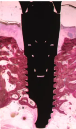
Figure 1: Histometric analysis in the percentage of bone-implant contact was evaluated. In this case, the calculated percentage was 57.6%.
3. Results
Healing was uneventful in all animals, no loss of either implants or prostheses was observed during the experimental period, and a direct contact was observed between living bone and all implants without interposed sot tissues at the light microscope level (Figure 1).
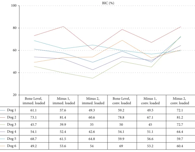
Mean (±standard deviation) BIC values from immediate loaded implants were58.7 ± 10.9%,57.7 ± 13.7%, and51.1 ±
11.1%, respectively, for Bone Level, Minus 1, and Minus 2 groups, while conventional loaded implants were 61.8 ±
10.5%,53.8 ± 7.6%, and68.4 ± 8.4% (Figure 2). Diferences
statistically signiicant were not observed among groups (� =
0.10, ANOVA test).
In all groups, the presence of compact bone was observed and evenly distributed around the entire implant surface, from the coronal portion to the apex of the implant; and there was no interposition of ibrous tissue interface bone-implant. In Bone Level group submitted to immediate loading, at the apex of the implant, where bone usually tends to be more trabecular, compact bone was present. Some threads were surrounded by marrow spaces where there was an intense osteoblastic activity.
In Minus 1 group submitted to immediate loading, the presence of compact bone was observed especially at the coronal and medial portion of the implants. New bone formation with osteoblastic activity was detected in the coronal portion of the implant.
In Minus 2 group submitted to immediate loading, bone remodeling occurred at the coronal portion of the implants; in a general manner, bone had a solid and mature aspect.
In Bone Level group submitted to conventional loading, the threads of the implants were surrounded by newly formed bone tissue. Implants were surrounded by trabecular bone with wide marrow spaces, and near the surface of the implant, bone was more compact and marrow spaces were smaller.
In Minus 1 group submitted to conventional loading, the presence of bone tissue was observed, especially at the coronal third and medium. A compact bone was present, with small marrow spaces and secondary osteons. Adjacent to the threads, there were numerous areas of bone remodeling. Trabecular bone with wide marrow spaces was observed especially at the middle and apical third of the plant.
In Minus 2 group submitted to conventional loading, the presence of new bone formation was detected in the coronal portion of the implant. he implants were surrounded by trabecular bone with wide marrow spaces, especially in the apical portion.
4. Discussion
his study evaluated bone-implant contact of implants inserted under diferent clinical conditions. he implication is that, at least under the conditions studied, submerging two-piece implants and submitting to immediate or conventional loading do not necessarily jeopardize osseointegration.
he methodology of this study was designed to clarify whether the osseointegration of the implants would be inluenced by the absorption of crestal bone that occurred adjacent to the platform of the implant or by the low bone density observed mainly in the most apical portion of those implants inserted apically to the crestal bone.
Diferent loading protocols were evaluated, because mechanical loading inluences bone remodeling [11], and this variable was not considered in the studies from Hermann et al. [2], Todescan et al. [3], Negri et al. [7], Boquete-Castro et al. [8], and Calvo-Guirado et al. [9,10], in which prostheses were not installed.
BIC values ranged from 51.1% to 69.4%, and these values seem to be enough to warrant stability, since values were higher than 50% [15]. In the study from Todescan et al. [3], mean BIC values were the following: 46.8% for implants inserted 1 mm coronal to the crest, 53.7% at crestal position, and 49.0% 1 mm apical to the crest. Such values, lower than those from present study, may be justiied by lower mechani-cal stimulus.
Similarly, Boquete-Castro et al. [8] developed a study in which BIC of 53.85% was reported for 2 mm subcrestal group
and39.50 ± 9.25% for crestal group. hen, Calvo-Guirado et
al. [9] reported that BIC values tended to increase in 2 mm subcrestal group (47.33% at 8 weeks and 53.85% at 12 weeks) and to decrease in crestal group (44.52% at 8 weeks and 39.50% at 12 weeks).
BIC (%) 100
80
60
40
20
Bone Level, immed. loaded
Bone Level, conv. loaded Minus 1,
immed. loaded
Minus 2, immed. loaded
Minus 1, conv. loaded
Minus 2, conv. loaded
Dog1
Dog2
Dog3
Dog4
Dog5
Dog6
61.1
73.1
45.7
54.1
68.7
49.2
57.6
81.4
39.9
52.4
61.5
53.6
49.3
60.6
35
42.6
64.8
54
59.2
78.8
50
54.1
59.9
69
49.5
67.1
45
51.1
56.6
53.2
72.1
81.2
72.7
64.4
59.7
60.4
Figure 2: BIC values (%) 90 days ater loading for each dog. Immed. loaded = immediately loaded; conv. loaded = conventionally loaded. Statistically signiicant diferences were not observed among groups (� = 0.10, ANOVA test).
54.88±11.73.%, while those inserted at crestal level resulted in
42.52±8.67%,35.19±18.12%, and47.46±11.50%, respectively.
Diferences between each subgroup in the test and the control groups were statistically signiicant.
Negri et al. [7] compared bone-implant contact from diferent implant designs. Tapered and cylindrical implants resulted in33.85 ± 5.21% and45.87 ± 2.02% in subcrestal group and29.50 ± 9.25% and42.52 ± 8.78% for crestal group, respectively. hus, there was less bone resorption in the subcrestal group than crestal group. Additionally, cylindrical implants, as those used in present study, led to higher BIC values.
Present study and those from Boquete-Castro et al. [8], Calvo-Guirado et al. [9,10], and Negri et al. [7] corroborate the arguments from Garber et al. [6], who suggested that two-piece implants may be successfully indicated.
In the present study, developed in posterior mandible, large mongrel dogs were used, because it was necessary to install commercially available implants even 2 mm apical to crestal bone and screw their abutments and prosthesis. A split-mouth design and random selection of groups were performed in order to reduce bias; moreover, histologic evaluation, which is considered as a gold standard, was carried to clarify precisely newly formed tissues.
However, main limitations of this study were that im-plants were installed neither in esthetic area nor in humans.
Implants were not installed in esthetic zone because it was important to insert implants in an area long enough to support six implants, in a lat alveolar bone, which was achieved ater surgical extraction of the premolars and regu-larization of the posterior border of the mandible. Secondly, a preclinical model was preferred to limit the variable involved and to allow histologic evaluation.
Future studies with longer healing periods and human clinical and radiographic trials should be conducted to provide data concerning the stability of the implants to support these indings and evaluate clinical outcome ater the insertion of implants in the esthetic zone, where bone tends to assume a scalloped shape.
5. Conclusions
Within the sample studied, it could be concluded that diferent apicocoronal positioning and the restoration pro-tocols evaluated did not interfere in the percentage of BIC, suggesting that these procedures did not jeopardize its osseointegration.
Conflict of Interests
Acknowledgments
he authors would like to express their gratitude to Conex˜ao Sistema de Pr´otese Ltda for providing the implants and related supplies used in the present study. his research pro-ject was supported by CAPES (Government Agency for the Development of Higher Education, scholarship no. PDEE 0989/05-3) and FAPESP (S˜ao Paulo Foundation for the Support of Research, Grant no. 04/08141-3).
References
[1] D. P. Tarnow, S. C. Cho, and S. S. Wallace, “he efect of inter-implant distance on the height of inter-inter-implant bone crest,” Journal of Periodontology, vol. 71, no. 4, pp. 546–549, 2000. [2] J. S. Hermann, D. Buser, R. K. Schenk, J. D. Schoolield, and D. L.
Cochran, “Biologic Width around one- and two-piece titanium implants—a histometric evaluation of unloaded nonsubmerged and submerged implants in the canine mandible,”Clinical Oral Implants Research, vol. 12, no. 6, pp. 559–571, 2001.
[3] F. F. Todescan, F. E. Pustiglioni, A. V. Imbronito, T. Albrektsson, and M. Gioso, “Inluence of the microgap in the peri-implant hard and sot tissues: a histomorphometric study in dogs,”he International Journal of Oral & Maxillofacial Implants, vol. 17, no. 4, pp. 467–472, 2002.
[4] A. Piattelli, G. Vrespa, G. Petrone, G. Iezzi, S. Annibali, and A. Scarano, “Role of the microgap between implant and abutment: a retrospective histologic evaluation in monkeys,”Journal of Periodontology, vol. 74, no. 3, pp. 346–352, 2003.
[5] D. Buser, W. Martin, and U. C. Belser, “Optimizing esthetics for implant restorations in the anterior maxilla: anatomic and surgical considerations,”International Journal of Oral and Maxillofacial Implants, vol. 19, pp. 43–61, 2004.
[6] D. A. Garber, H. Salama, and M. A. Salama, “Two-stage versus one-stage—is there really a controversy?”Journal of Periodon-tology, vol. 72, no. 3, pp. 417–421, 2001.
[7] B. Negri, J. L. Calvo-Guirado, M. P. Ram´ırez-Fern´andez, J. Mat´e S´anchez-de Val, J. Guardia, and F. Mu˜noz-Guz´on, “Peri-implant bone reactions to immediate implants placed at diferent levels in relation to crestal bone. Part II: a pilot study in dogs,”Clinical Oral Implants Research, vol. 23, no. 2, pp. 236–244, 2012. [8] A. Boquete -Castro, G. G´omez-Moreno, A. Aguilar-Salvatierra,
R. A. Delgado-Ruiz, G. E. Romanos, and J. L. Calvo-Guirado, “Inluence of the implant design on osseointegration and crestal bone resorption of immediate implants: a histomorphometric study in dogs,”Clinical Oral Implants Research, 2014.
[9] J. L. Calvo-Guirado, A. Boquete-Castro, B. Negri, R. Delgado Ruiz, G. G´omez-Moreno, and G. Iezzi, “Crestal bone reactions to immediate implants placed at diferent levels in relation to crestal bone. A pilot study in Foxhound dogs,”Clinical Oral Implants Research, vol. 25, no. 3, pp. 344–351, 2014.
[10] J. L. Calvo-Guirado, G. Gomez Moreno, A. Aguilar-Salvatierra, J. E. Mate Sanchez de Val, M. Abboud, and C. E. Nemcovsky, “Bone remodeling at implants with diferent conigurations and placed immediately at diferent depth into extraction sockets,” Clinical Oral Implants Research, 2014.
[11] H. M. Frost, “he role of changes in mechanical usage set points in the pathogenesis of osteoporosis,”Journal of Bone and Mineral Research, vol. 7, no. 3, pp. 253–261, 1992.
[12] A. E. F. Pontes, F. S. Ribeiro, G. Iezzi, A. Piattelli, J. A. Cirelli, and E. Marcantonio Jr., “Biologic width changes around
loaded implants inserted in diferent levels in relation to crestal bone: histometric evaluation in canine mandible,”Clinical Oral Implants Research, vol. 19, no. 5, pp. 483–490, 2008.
[13] A. E. F. Pontes, F. S. Ribeiro, V. C. Da Silvas et al., “Clinical and radiographic changes around dental implants inserted in diferent levels in relation to the crestal bone, under diferent restoration protocols, in the dog model,”Journal of Periodontol-ogy, vol. 79, no. 3, pp. 486–494, 2008.
[14] A. Piattelli, A. Scarano, and M. Quaranta, “High-precision, cost-efective cutting system for producing thin sections of oral tissues containing dental implants,”Biomaterials, vol. 18, no. 7, pp. 577–579, 1997.
Submit your manuscripts at
http://www.hindawi.com
Hindawi Publishing Corporation
http://www.hindawi.com Volume 2014
Oral Oncology
Journal ofDentistry
International Journal ofHindawi Publishing Corporation
http://www.hindawi.com Volume 2014
Hindawi Publishing Corporation
http://www.hindawi.com Volume 2014
International Journal of
Biomaterials
Hindawi Publishing Corporation
http://www.hindawi.com Volume 2014
BioMed
Research International
Hindawi Publishing Corporation
http://www.hindawi.com Volume 2014
Case Reports in
Dentistry
Hindawi Publishing Corporation
http://www.hindawi.com Volume 2014
Oral Implants
Journal ofHindawi Publishing Corporation
http://www.hindawi.com Volume 2014
Anesthesiology Research and Practice
Hindawi Publishing Corporation
http://www.hindawi.com Volume 2014
Radiology
Research and Practice
Environmental and Public Health
Journal of
Hindawi Publishing Corporation
http://www.hindawi.com Volume 2014
The Scientiic
World Journal
Hindawi Publishing Corporation
http://www.hindawi.com Volume 2014
Hindawi Publishing Corporation
http://www.hindawi.com Volume 2014
Dental Surgery
Journal ofDrug Delivery
Journal of Hindawi Publishing Corporationhttp://www.hindawi.com Volume 2014
Hindawi Publishing Corporation
http://www.hindawi.com Volume 2014
Oral Diseases
Journal ofHindawi Publishing Corporation
http://www.hindawi.com Volume 2014
Computational and Mathematical Methods in Medicine
Scientifica
Hindawi Publishing Corporationhttp://www.hindawi.com Volume 2014
Pain
Research and Treatment
Hindawi Publishing Corporation
http://www.hindawi.com Volume 2014
Hindawi Publishing Corporation
http://www.hindawi.com Volume 2014
Endocrinology
International Journal ofHindawi Publishing Corporation
http://www.hindawi.com Volume 2014
Hindawi Publishing Corporation
http://www.hindawi.com Volume 2014