Implications in Cancer and HIV Pathogenesis
Robert L. Furler1,3, Christel H. Uittenbogaart1,2,3,4*
1Department of Microbiology, Immunology, and Molecular Genetics, David E. Geffen School of Medicine, University of California Los Angeles, Los Angeles, California, United States of America,2Department of Pediatrics, David E. Geffen School of Medicine, University of California Los Angeles, Los Angeles, California, United States of America,3UCLA AIDS Institute, University of California Los Angeles, Los Angeles, California, United States of America,4Jonsson Comprehensive Cancer Center, David E. Geffen School of Medicine, University of California Los Angeles, Los Angeles, California, United States of America
Abstract
Elevated levels of the immunoregulatory cytokine TGF-b1 in cancer and HIV infection have been linked to the suppression of protective immune responses. The transcriptional regulation of TGF-b1 is complex and still not completely understood. We report here for the first time that the transcription factor GLI2 regulates the expression of TGF-b1 in human CD4+T cells. In silicoscreening revealed five novel putative GLI binding sites in the humanTGF-b1promoter. At least two of these sites within the humanTGF-b1promoter are regulated by the GLI2 activator as knockdown ofGLI2in regulatory CD4+CD25hiT
cells, high producers of TGF-b1, significantly decreasedTGF-b1transcription. Additionally, naı¨ve CD4+T cells, low producers of TGF-b1, increased their basal level ofTGF-b1mRNA following lentiviral infection with GLI2. The transcriptional regulation ofTGF-b1by GLI2 is a new extension to Sonic Hedgehog (SHH) andTGF-b1cross-regulation and may provide insight into the detrimental elevation of TGF-b1 leading to pathogenesis in cancer and HIV infection.
Citation:Furler RL, Uittenbogaart CH (2012) GLI2 Regulates TGF-b1 in Human CD4+T Cells: Implications in Cancer and HIV Pathogenesis. PLoS ONE 7(7): e40874. doi:10.1371/journal.pone.0040874
Editor:Derya Unutmaz, New York University, United States of America
ReceivedJuly 26, 2011;AcceptedJune 18, 2012;PublishedJuly 31, 2012
Copyright:ß2012 Furler, Uittenbogaart. This is an open-access article distributed under the terms of the Creative Commons Attribution License, which permits unrestricted use, distribution, and reproduction in any medium, provided the original author and source are credited.
Funding:This work was supported by grants from the National Institutes of Health AI-081636 and AI-028697 (University of California Los Angeles, Center for AIDS Research), a Jonsson Comprehensive Cancer Center Seed Grant, and a Stein Oppenheimer Endowment Award. The funders had no role in study design, data collection and analysis, decision to publish, or preparation of the manuscript.
Competing Interests:The authors have declared that no competing interests exist. * E-mail: [email protected]
Introduction
Transforming Growth Factor-b1 (TGF-b1) is a pleiotropic cytokine that regulates the immune response in many diseases including cancer and HIV infection and plays an important role in preventing pathogenesis[1–5]. It is produced by several cell types including CD4+
CD25hiregulatory T cells, macrophages, as well as by fibroblasts. TGF-b1 decreases proliferation of T and B lymphocytes, effector functions of many cells of the immune system and induces peripheral regulatory T cells [6]. Although TGF-b1 is critical throughout mammalian development and to prevent aberrant activation of the host’s immune system, elevated expression of TGF-b1 can dampen protective immune responses to tumors and infections.
Aberrant high production of TGF-b1 by malignant cells was shown to increase metastasis as well as to prevent a protective host immune response to tumors [7]. In chronic HIV infection,
TGF-b1 is elevated in several compartments including lymphoid tissues, blood, and cerebrospinal fluid[8–10]. Increased levels of TGF-b1 can induce the development of induced (i)Treg observed in chronic HIV infection [11,12], and may contribute to dysfunction of the immune system during late-stage disease. The HIV-1 Tat protein induces TGF-b1 in human leukocytes [13–15], chondro-cytes [16], and also in murine CD2+T cells of a Tat-transgenic
mouse model [17]. The mechanisms of tumor-induced and Tat-mediated TGF-b1 induction are unknown. Cancer and AIDS are only two of the many diseases in which TGF-b1 plays a role in pathogenesis.
Expression of active TGF-b1 is tightly regulated at the transcriptional, translational, and post-translational levels. Al-though TGF-b1 expression has been a focus of intense investiga-tion, we are still far from understanding this cytokine’s complex regulation. In silico analysis of the TGF-b1 promoter region indicates several putative binding sites of known transcription factors. Some of these transcription factors, like Sp-1 and Zf9, have previously been found to mediateTGF-b1transcription, but these factors alone are not sufficient to induce mRNA expression [18,19]. Therefore we investigated which other transcription factors induce TGF-b1 mRNA.
We found six putative binding sites (five previously unreported) for the human GLI transcription factors byin silicoanalysis of the humanTGF-b1promoter. The GLI2 transcription factor is one of the main downstream effectors of the Sonic Hedgehog (SHH) pathway, which was shown to play a role in the function of the adaptive immune system [20,21]. The SHH pathway modulates activation [22], proliferation [23,24], and cytokine regulation [25] in CD4+
T cells. Since several interactions between the TGF-b1 and SHH pathways exist (Table 1), we focused on regulation of TGF-b1 by SHH pathway family members, i.e. GLI proteins, in the immune system.
We provide evidence that at least two of the six putative GLI-binding sites are required for GLI2-mediated transcriptional regulation of the humanTGF-b1promoter. At least one of these two sites must be functional to observe TGF-b1 regulation by GLI2. Furthermore, by knocking down GLI2 in human CD4+
expression. The transcriptional regulation ofTGF-b1by GLI2 has not yet been described and increases our understanding of the complexities ofTGF-b1regulation.
Materials and Methods
Ethics Statement
The use of anonymous peripheral blood mononuclear cells to obtain T cells and the epithelial 293T cell line was reviewed by the UCLA Institutional Review Board (IRB), which concluded that this activity did not involve human subjects, and therefore did not require IRB review or certification.
In Silico Screening
Screening for putative GLI binding sites in the humanTGF-b1
promoters was done byin silicoanalysis using the software program MatInspector (http://www.genomatix.de). A range of twenty kilobases surrounding the first human TGF-b1 promoter were used to screen for putative transcription factor binding sites. All six putative GLI binding sites within the first kilobase of TGF-b1
promoter#1 (five potential sites) or promoter#2 (one potential site) had a matrix similarity score equal to or greater than 0.8. Binding sites found within the first proximal promoter region are novel and are the focus of this study.
Luciferase & Promoter Studies
The human TGF-b1 promoter-luciferase reporter constructs, phTG5 and phTG1, were kindly provided by S.J. Kim [18]. The wild-type phTG5 construct was mutated at the putatitve GLI binding sites (site A, B, or A and B) using site-directed mutagenesis (SITE A: 59-CAGCCCCCCCATGCC-39 to 59 -CAGCaa-CaaaATGC-39, SITE B: 59-GAGCCCGCCCACGCGA-39 to 59-GAGCaaGaaaACGCGA-39, mutated bases in lower case). The wild-type phTG1 construct was mutated at putative GLI binding Site E using site-directed mutagenesis (SITE E: 59 -CCCAC-CACCCACGAA-39 to 59CCaAaaAaaaACGAA-39, mutated ba-ses in lower case). The wild-type promoter, the mutated promoters, or a pGL3 empty-vector control were individually co-transfected with the constitutive activator vector pGLI2DN (kindly provided by E. Roessler [26]) or the control pcDNA3 with Lipofectamine 2000 using standard procedures into 293T cells, a human epithelial cell line containing Adeno and SV-40 viral DNA sequences, obtained from the American Type Culture Collection (ATCC). The pGLI2DN plasmid contains a constitutively active GLI2 gene due to an amino-terminal truncation. The amino terminal domain of the human GLI2 gene has a repressive function, and it’s removal has been shown to increase transcription of downstream target genes such as GLI1 [26]. The 293T cell line was chosen because of its transfection efficiency and low levels of
GLI1 expression, which is our readout for GLI2 activation. Luciferase expression was measured at 48 hours post-transfection.
Cell Isolation, Culture, & Stimulation
Naı¨ve CD4+
T cells, expressing CD4, CD31, CD45RA, and CD62L, were purified and isolated from human peripheral blood mononuclear cells (PBMC) using negative selection. CD4+CD25hi regulatory T cells were enriched from human blood using RosetteSep and Robosep kits from Stem Cell Technologies. Primary cells were cultured in serum-free medium consisting of Iscove’s Modified Dulbecco’s Medium supplemented with delipi-dated BSA (Sigma-Aldrich) at 1100mg/mL, human transferrin (Sigma-Aldrich) at 85.5mg/mL, 2 mM glutamine, and penicillin/ streptomycin at 25 U/25mg/mL. All stimulation conditions of
primary T cells were done with aCD3/aCD28-coated M-450 beads from Invitrogen. Cell surface and intracellular immuno-phenotypes were validated by flow cytometry as described previously [27]. For detection of intracellular FoxP3, cells were first stained for cell surface markers, fixed and permeabilized with eBioscience recommended buffers following manufacturer instruc-tions, then incubated with FITC or eFluor 450 conjugated monoclonal antibodies against FoxP3 (eBioscience, clone PCH101).
siRNA Knockdown
Knockdown ofGLI2in primary human regulatory T cells was done using ACCELL siRNA designed and validated by Thermo-Fisher/Dharmacon. A non-targeting siRNA was used as a negative control. A positive control siRNA for GAPDH was used to verify knockdown. Knockdown of GLI2 was done using a commercially available and validated siRNA from Dharmacon (A-006468-14 - Target sequence - 5’ GGUUUGAAUCUGAAUG-CUA 3’). A separate independent negative control using a scrambled GLI2 siRNA sequence (5’- AAGGUAUGCG-CUUAAUUU-3’) was used to address off-target effects. GLI2
knockdown was verified by assessing GLI1 mRNA expression following stimulation. GLI1 has been previously shown to be transciptionally regulated by GLI2 and is the positive control for
GLI2knockdown. Purified CD4+
CD25hiFoxP3+
regulatory T cells were incubated with the siRNA and serum-free ACCELL medium (Dharmacon) for 72 hours prior to stimulation with aCD3/aCD28 coated beads. Twenty-four hours post-stimulation, cells were collected to measureTGF-b1mRNA levels by RT-PCR. TGF-b1 levels were normalized to18S rRNAand compared to unstimulated cells.
RT-PCR
Taqman Gene Expression primer/probe sets forTGF-b1, GLI1, 18S rRNA, andGAPDHfrom Applied Biosystems Inc. were used
Table 1.The TGF-band SHH Pathways Exhibit Extensive Cross-Regulation.
ASSOCIATION REFERENCE
Constitutive SHH pathway activation in transgenic mouse have elevated TGF-b1 transcription.
(Eichenmulleret al., 2007)
GLI3 binds to SMAD proteins. (Liuet al., 1998)
BMP-2, 4, & 7 (TGFbfamily members) all have GLI-responsive elements in their promoters.
(Miyashitaet al., 2008; Suguira T., 1999; Kawai, S. and T. Sugiura, 2001)
TGF-b2 promoter contains a SHH-responsive element. (Linet al.,2006)
TGF-b1 and SMAD3 upregulate GLI2 and GLI1 (Dennleret al., 2007; Dennleret al.,2009)
to measure transcripts levels in various conditions. The expression of TGF-b1, GLI1, and GAPDH were normalized to 18S rRNA levels and relative expression between conditions was calculated to assess upregulation/downregulation of transcription.
Lentiviral Infections
TheGLI2DNtransgene was placed into a third-generation HIV-1 vector at the UCLA Vector Core. The majority of the HIV viral coding sequence was removed and replaced by the transgene. The LTRs were modified to be "self-inactivating,’’ and the transcrip-tion of the vector genome in the packaging cells was driven by a non-HIV promoter. Virus was made using 293T cells that were co-transfected with the vector plasmid and three other plasmids encoding A) the viral enzymes and structural proteins, except Env, B) Rev, which is involved in HIV RNA transport, and C) VSV glycoprotein, which is a pan-tropic envelope that allows virus entry into almost any cell type. Naı¨ve CD4+T cells were infected with
100 ng p24 per million cells in serum-free medium for 96 hours. RNA was isolated for determination of changes in gene expression using RT-PCR. A GFP-containing lentivirus was used as a negative control and also to measure infection efficiency.
Chromatin Immunoprecipitation (ChIP)
ChIP analysis was done to determine whether GLI2 binds to the
TGF-b1 promoter. Regulatory CD4+
CD25hi T cells were negatively isolated as described above. The cells were stimulated withaCD3/aCD28-coated beads overnight. The GLI2 Exacta-ChIP kit (R&D Systems) was used for chromatin immunoprecip-itation. Cells were fixed, lysed, and sonicated prior to incubation with eitheraGLI2-coated or controlaIgG-coated agarose beads. PCR was done following ChIP to assess GLI binding to the TGF-b1 promoter. Putative GLI2 binding site ‘E’ (Figure 1) was detected using designed primers (59 -CTGGGGTCAGCTCTGA-CAGT-39 and 59-CAGTTGGCGAGAACAGTTGG-39; 290 bp fragment). The Bcl2 promoter region was detected as a positive GLI-binding control using primers from the ExactaChIP kit. A 238 bp region of theIL-10promoter which contained no putative GLI2 binding site was measured as a negative control (59 -TGATTTCCTGGGGAGAACAG-39 and 59 -CCCACCCCCT-CATTTTTACT-39).
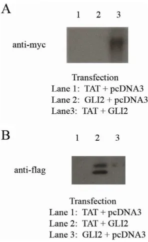
Co-Immunoprecipitation
Co-immunopreciptation was performed to assess the binding of human GLI2 to HIV-1 Tat. Dual transfections were done in 293T cells using three different sets of plasmids: (GLI2-6xMYC+HIV-1 Tat-FLAG), (GLI2-6xMYC + PCDNA3 control), (HIV-1 Tat-FLAG + PCDNA3 control). Forty-eight hours post-transfection, the cells were lysed and the released proteins were collected for co-IP (Pierce Co-co-IP Kit from Thermo Scientific). The agarose resin was coated with one of three antibodies: Sigma F1804 anti-FLAG M2 antibody (to bind Tat to bead), CST 71D10 anti-MYC antibody (to bind GLI2 to bead), or an isotype control antibody. Following co-IP, the elutions were run on an SDS-PAGE gel followed by Western Blotting.
Statistics
All analyses were conducted by the UCLA AIDS Institute Biostatistics Core or with GraphPad Prism 5 Software. Variables were expressed as means with standard error of the mean. ANOVA and two-tailed Wilcoxon Rank sum test, paired when appropriate, were used to analyze differences between popula-tions. A p value lower or equal to 0.05 was considered significant.
Results
Six Putative GLI Binding Sites Lie Within the Human TGF-b1Promoters
Through in silico analysis using MatInspector (http://www. genomatix.de) we found six putative GLI binding sites within the two reported human TGF-b1 promoters, including a previously reported site within the second promoter [28] in addition to five sites which were previously unidentified. Both Genomatix software and UCSC genome (www.genome.ucsc.edu) programs indicate a high degree of sequence conservation surrounding the TGF-b1
promoter region in mammals which suggests the functional relevance of the human TGF-b1 promoters. The consensus binding sequence for the GLI transcription factors has been identified as 59-TGGGTGGTC-39[29,30]. The six putative GLI binding sites reported in this study have a high-degree of matrix similarity43and are listed in Figure 1. The abundance of possible GLI binding sites within the first thousand base pairs of the transcription start site of theTGF-b1promoter suggests that this proximal promoter region may be regulated by the GLI transcription factors.
GLI2 Activates the First HumanTGF-b1Promoter Through At Least Two of the Putative GLI-binding Sites
The putative GLI binding site in the second TGFb-1 promoter has been reported to be non-responsive to SHH stimulation by Eichenmulleret al.[28]. However, the five putative sites in the first promoter have not previously been reported to be regulated by GLI proteins. We used a reporter construct containing the first 500 base pairs of the human TGF-b1 promoter which was placed upstream of the firefly luciferase gene by Kimet al.(phTG5) [18]. This expression vector was co-transfected into 293T cells along with a constitutive GLI activator construct pGLI2DN [26] or a pcDNA3 plasmid control to assess GLI2-mediated regulation of the promoter of the human TGF-b1 gene.
Following co-transfection of GLI2DN and phTG5 into 293T cells, we observed an approximately 4-fold increase of luciferase
Figure 1. Five Novel Putative GLI Binding Sites Lie Within the First Human TGF-b1 Promoter.There are two reported promoters in the humanTGF-b1gene.In silicoanalysis of the humanTGF-b1proximal promoter regions using the Genomatix software program revealed five previously unidentified putative GLI-binding sites (A through E) in a highly conserved region of the human genome. Site F in the second promoter has previously been identified and studied [28]. Sites A and B (which overlaps with C), all lie within the luciferase reporter construct phTG5 used in the promoter mutagenesis studies. Site E is present in the phTG1 construct. Approximate base pair positions of potential GLI binding sites are shown relative to the two reported transcription start sites for the humanTGF-b1gene.
expression over the controls (Figure 2). As the promoter region of phTG5 contains three of the six putative GLI binding sites (A, B, & C) we used site-directed mutagenesis to mutate these putative GLI binding sites individually or in combination (denoted phTG5A, phTG5B, and phTG5AB respectively). Although mutation of a single site only modestly reduced luciferase expression, mutation of both sites reduced reporter gene expression down to control levels. These findings indicate that GLI2 can activate transcription at the humanTGF-b1promoter
and that binding to at least one of these sites is required for this to occur. Mutation at Site E, which lies further upstream of the transcription start site, was done to assess its role in GLI2-induced TGF-b1 expression. We used a luciferase reporter construct phTG1 containing21362 to+11 bases near the transcription start site of the human TGF-b1 promoter [18]. Following mutation of the possible GLI-binding site, the luciferase expression was decreased approximately two-fold following cotransfection with the constitutive GLI2 activator. Further analysis will be needed to
Figure 2. GLI Activators Induce Transcription at the HumanTGF-b1Promoter.The humanTGF-b1promoter region was placed into a pGL3-basic luciferase vector. (A) The three GLI binding sites within this region (Sites A, B, and C) were mutated separately or together (WT = wild-type
TGF-b1promoter phTG5, MUT-A = site A mutation, MUT-B = sites B/C mutation, MUT-A/B = sites A/B/C mutation, pGL3 = control vector). TheseTGF-b1 promoter:luciferase constructs were co-transfected into 293T cells with the constitutive GLI activator (GLI2dN) and normalized to the negative control (pcDNA3). The GLI activator induced a four-fold increase of luciferase activity in phTG5 over the control. Mutation of both GLI binding sites (phTG5AB) abrogated this induction (n = 10, *p = 0.0011, ** p = 0.0033). (B) The putative GLI binding Site E was mutated in the reporter plasmid phTG1 (WT = wild-type TGF-1 promoter phTG1, MUT-E = site E mutation, pGL3 = control vector). The GLI activator induced a six-fold increase of luciferase activity in phTG1 over the control. Mutation of GLI binding Site E decreased this induction nearly two-fold (n = 12, *** p = 0.0025).
examine whether putative GLI binding sites other than sites A, B, and E are also functional.
Activated GLI2 Can EnhanceTGF-b1Transcription in Naı¨ve CD4+T cells
In order to investigate the effect of the GLI transcription factors in primary cells, we infected primary naı¨ve CD4+
T cells with lentiviral vectors containing the constitutive activator GLI2DN or a GFP control. Expression of GLI2DN was confirmed by Western blot (see insert in Figure 3). Naı¨ve CD4+
T cells do not constitutively express high levels of TGF-b1. We measured the level of TGF-b1mRNA by RT-PCR before and after infection
with the GLI constructs. As shown in Figure 3A, overexpression of the constitutively activated GLI2DN in naı¨ve CD4+
T cells significantly increased TGF-b1 mRNA expression (n = 6, p = 0.0313) following stimulation with aCD3/aCD28-coated beads. Upregulation of GLI1 mRNA, a known GLI2-mediated gene, indicates that the GLI2DN protein is activating transcription in these lentivirally infected cells (data not shown). The infection efficiency was measured by infecting naı¨ve CD4+
T cells with a control lentivirus expressing GFP (69% infection efficiency, Figure 3B). The phenotype of the naı¨ve cells was CD4+
CD45RA+
CD45RO-(Figures 3C & 3D).
Figure 3. Lentiviral Infection of Naı¨ve CD4+T cells with GLI2 Activator IncreasesTGF-b1transcription.Naı¨ve human CD4+T cells were
isolated by negative selection and infected with a lentivirus containing a GLI2DN activator construct or a GFP control. 72 hours post-infection, cellular RNA was collected to measureTGF-b1transcripts. (A) RelativeTGF-b1mRNA was measured by RT-PCR and normalized to 18S rRNA.TGF-b1mRNA was significantly increased (n = 6, * p = 0.0313) in cells infected with GLI2DN as compared to cells infected with GFP control. (B) The infection efficiency was measured at 69% using a GFP expressing control lentivirus. (C & D) The phenotype of the naı¨ve CD4+
T cells was greater than 95% of the cells at Day 0 were CD4+CD45RA+CD45RO-naı¨ve T cells. Expression of GLI2DN was confirmed by Western Blot (insert).
GLI2 RegulatesTGF-b1Transcription in Primary Human CD4+ CD25hiTreg
Although overexpression of transgenic GLI proteins can modulateTGF-b1transcription in naı¨ve CD4+
T cells, we wanted to assess the physiological role of GLI2 activation in primary cells that express high levels of TGF-b1. Previous studies have used siRNA to knockdown specific genes in primary Treg [31,32]. Knockdown of GLI2 was done using commercially available Dharmacon ACCELL siRNA to assess its function in TGF-b1 transcription in primary human Treg.GLI2siRNA knockdown in
primary human CD4+
CD25hi Treg stimulated with aCD3/
aCD28 coated beads decreased the expression ofTGF-b1mRNA by approximately 5-fold (Figure 4A, p = 0.0189). To assess the knockdown of GLI2 activity, GLI1 mRNA expression was measured by RT-PCR. GLI1 gene transcription is a known downstream target of the activated GLI2 transcription factor. The decreased expression of GLI1 mRNA supports the siRNA-mediated knockdown ofGLI2(Figure 4B, p = 0.0278). A separate independent negative control using a scrambled GLI2 siRNA sequence (5’- AAGGUAUGCGCUUAAUUU-3’) was used to
Figure 4. GLI2 Knockdown in CD4+CD25+FoxP3+Treg Decreases Levels ofTGF-b1mRNA.CD4+CD25+FoxP3+Treg were enriched from
PBMC and then incubated for 72 hours with non-targeting (NT) orGLI2siRNA. The cells were then stimulated (1aCD3/aCD28 bead: 1 cell) for an additional 24 hours. RT-PCR was used to measureTGF-b1 mRNA levels. RelativeTGF-b1or GLI1mRNA was normalized to 18S rRNA. (A) GLI2 knockdown in Treg significantly decreasedTGF-b1transcription (n = 7, * p = 0.0189). (B)GLI1mRNA was also measured as a control GLI2-regulated gene; showing that knockdown ofGLI2in Treg also diminishedGLI1mRNA (n = 5, ** p = 0.0278). (C) The sequence of theGLI2siRNA was scrambled (SCRAM) as an additionalGLI2siRNA negative control and was not significantly different from the non-targeting control.
address off-target effects (Figure 4C). These results indicate that GLI2 is important in regulatingTGF-b1transcription in primary human Treg. The sorted Treg were CD3+CD8
-CD4+CD25hi -FoxP3+
T cells. The purity of the Treg was more than 85% (Figure 4D).
GLI2 Binds to the HumanTGF-b1 Promoter
Chromatin immunoprecipitation (ChIP) was done to assess the binding of GLI2 to the humanTGF-b1promoter. Primary human regulatory CD4+CD25hi
T cells were negatively isolated and stimulated with aCD3/aCD28-coated beads overnight. The stimulated Treg were incubated with agarose beads coated with
aGLI2 oraIgG control antibodies. As shown in Figure 5, GLI2 bound to theTGF-b1promoter. GLI2 binding was shown to be at ‘Site E’ (Figure 5) in the humanTGF-b1promoter. However GLI2 did not bind to a non-targeting region in the human IL-10
promoter (Lane 8).
HIV-1 Tat Binds to GLI2
Increased plasma and cerebral spinal fluid (CSF) levels of immunosuppressive cytokines like TGF-b1 are found during HIV-1 disease progression[8–HIV-10] leading to an increase in iTreg [11,12]. The HIV-1 transactivator and viral protein Tat has been implicated as the inducer of TGF-b1 in bothin vitroas well asin vivo models [13–17]. Although Tat has been shown to increase TGF-b1, the underlying transcriptional mechanism has not yet been clearly defined.
Since we found that GLI2 regulates TGF-b1 at the transcrip-tional level, we tested whether HIV-1 Tat binds to human GLI2 and can thereby increase or stabilize TGF-b1 transcription using co-immunopreciptation (co-IP) to assess the binding of human GLI2 to HIV-1 Tat. Dual transfections were done in 293T cells with three different sets of plasmids: (GLI2-6xMYC+HIV-1 Tat-FLAG), (GLI2-6xMYC+PCDNA3 control), (HIV-1 Tat-FLAG+ PCDNA3 control). Forty-eight hours post-transfection, the cells were lysed and the released proteins were collected for co-IP (Pierce Co-IP Kit from Thermo Scientific). The agarose resin was
coated with one of three antibodies: Sigma F1804 anti-FLAG M2 antibody (to bind Tat to bead), CST 71D10 anti-MYC antibody (to bind GLI2 to bead), or an isotype control antibody. Following co-IP, the elutions were run on an SDS-PAGE gel followed by Western Blotting. As shown in Figure 6, HIV-1 Tat bound specifically to GLI2 which was bound to a bead. The reverse reaction also showed specificity, indicating that these two proteins can indeed interact. The binding of HIV-1 Tat to GLI2 suggests a potential mechanism for TGF-b1 induction in HIV-1 infection. Further experiments will need to be done to test the functional synergy of HIV-1 Tat and GLI2 to affect transcription at the TGF-b1 promoter.
Discussion
Our results show that the transcription factor GLI2 plays a previously unknown, but important role in the complex transcrip-tional regulation ofTGF-b1.Throughin silicoscreening analysis we revealed six potential GLI-binding sites in the human TGF-b1 promoter, five of which were previously unknown. Using mutational analysis of the human TGF-b1promoter, we found that GLI2 enhancesTGF-b1transcription in at least three of the five putative GLI-binding sites (Sites A, B, & E). In addition our results show that infection of primary naı¨ve CD4+
T cells (low TGF-b1 expressing cells) with a constitutively active GLI2 lentivirus enhances TGF-b1 transcription. Furthermore, siRNA knockdown ofGLI2in primary human CD4+
CD25hiFoxP3+
Treg (high TGF-b1 expressing cells) significantly decreased TGF-b1
transcription. We also show by ChIP that GLI2 directly binds to ‘‘Site E’’ in the TGF-b1 promoter. This newly identified mechanism GLI2-mediated TGF-b1 regulation may have impli-cations in diseases such as cancer and AIDS, where high levels of TGF-b1 contribute to pathogenesis.
Although the role of GLI-mediated regulation of TGF-b1
transcription has not previously been shown, there is indirect evidence for this mechanism. Eichenmulleret al.discovered that
Ptch1 knockout mice, which have constitutive activation of the SHH pathway, have more than 400-fold increased levels of TGF-Figure 5. GLI2 binds to the humanTGF-b1promoter.Chromatin immunoprecipitation (ChIP) was done to assess the binding of GLI2 to the humanTGF-b1promoter. Primary human regulatory CD4+
CD25hiT cells were negatively isolated and stimulated withaCD3/aCD28-coated beads overnight. PCR was done to assess GLI2 binding. Whole DNA was used as a positive control for the PCR. GLI2 bound to theTGF-b1promoter at ‘Site E’ in the humanTGF-b1promoter. GLI2 did not bind to a non-targeting region in the humanIL-10promoter (Lane 8).
b1mRNA transcripts [28]. Although the investigators found that the putative GLI binding site in the second promoter was not responsive to GLI1, they did not investigate the role of GLI2 in regulating the five putative GLI binding sites that we discovered in the first TGF-b1 promoter.
Additionally, several reports emphasize the close relationship between the TGF-b1 and SHH/GLI families [33,34]. Liu et al.
reported the interaction between the SHH pathway molecule GLI3 and the SMAD proteins (TGF-b1 pathway transcription factors) [35]. Additionally Dennleret al. reported that GLI2 and GLI1 could be induced through a SMAD-mediated mechanism [36]. The TGF-bsuperfamily genesBMP-2, 4, &7all have GLI-responsive elements in their promoters[37–39]. Linet al.found a SHH-responsive element in the 59-upstream promoter region of
TGF-b2 [40]. TGF-b1 and its effector molecule SMAD3 have been shown to upregulate GLI2 and GLI1 [36,41]. Despite the
extensive cross-regulatory network of TGF-b and SHH, the regulation ofTGF-b1by SHH family members, such as the GLI proteins, has not been previously reported.
Several types of tumors (basal cell carcinomas, gliomas, medullablastomas, and prostate cancer) express abnormally high levels of GLI proteins or increased SHH pathway activation, which may be targets for therapy [42]. Although aberrant activation of SHH/GLI signaling leads to elevated expression of immunosuppressive factors like TGF-b1[43–49], a clear mecha-nism of cancer-induced TGF-b1 induction is still lacking. One possible mechanism is through activation of GLI2 by the ERK and AKT pathways in tumor cells. The ERK and AKT pathways are hyperactivated in several tumors and are critical for GLI-regulation in both tumor cells [50,51] as well as primary mammalian cells [52,53]. Our data indicate that the ERK and AKT pathways are also critical for GLI activation in CD4+T cells
(data not shown). The hyperactivation of the ERK and AKT pathways in tumor cells may provide an environment for GLI2-mediated transcription of the immunosuppressive TGF-b1.
We also show that HIV-1 Tat binds to GLI2. The interaction between HIV-1 Tat and GLI2 may be a contributing factor to the elevated levels of TGF-b1 in HIV infection and contributes to immune dysregulation, a key characteristic of chronic HIV-1 disease. Increased plasma and cerebrospinal fluid (CSF) levels of immunosuppressive cytokines like TGF-b1 are found during HIV-1 disease progression[8–HIV-10] leading to an increase in iTreg [11,12]. This increase of immunoregulatory CD4+CD25+FoxP3+
T-cells (Treg) during chronic HIV infection disables the immune system’s control over viral replication as well as opportunistic infections [12]. The HIV-1 transactivator Tat has been implicated as the inducer of TGF-b1 in bothin vitroas well asin vivomodels [13–17], although the underlying transcriptional mechanism has not been clearly defined.
Our current findings support those of Browning et al., who found that human GLI2 bound to HIV-1 Tat [54]. The role of the GLI proteins in retroviral replication have previously been reported by several groups[54–57]. Specifically, GLI-2 has been previously shown to interact with retroviral transactivators to increase viral transcription[54–57]. We suggest that the reported induction of TGF-b1 transcription by HIV-1 Tat[13–17] may be in part mediated by the binding of Tat to GLI2 at theTGF-b1
promoter. Although further studies will need to be done to validate this mechanism, the novel role of GLI2 inTGF-b1transcription may provide an interesting link between HIV-1 replication and TGF-b1 induction.
The role of TGF-b1 in pathogenesis of cancer, AIDS, and several other diseases warrants interest in the regulation of this immunoregulatory cytokine. The regulatory role of GLI2 in TGF-b1transcription extends our understanding of how the SHH and TGF pathways interact in mammalian biology. How GLI2 is regulated by the ERK and AKT pathways and by HIV-1 Tat may lead to insight into TGF-b1 induction during cancer progression and HIV infection.
Acknowledgments
We wish to thank Ruth Getachew and Josh Craft for their excellent technical assistance throughout this project and Stephen Smale for his critical review of the manuscript. Additionally, we are grateful for the plasmids and materials provided by Seong-Jin Kim, Erich Roessler, David Harrich and recognize the important contributions of the University of California Los Angeles, (UCLA) Vector, CFAR Virology, and CFAR Flow Cytometry Core Facilities.
Author Contributions
Conceived and designed the experiments: RLF. Performed the experi-ments: RLF. Analyzed the data: RLF CHU. Contributed reagents/
materials/analysis tools: RLF CHU. Wrote the paper: RLF CHU. Obtained permission for plasmids, vectors and other materials: RLF.
References
1. Taylor AW (2009) Review of the activation of TGF-bin immunity. J Leukoc Biol 85: 29–33.
2. Luo X, Tarbell KV, Yang H, Pothoven K, Bailey SL, et al. (2007) Dendritic cells with TGF-b1 differentiate naive CD4+CD25- T cells into islet-protective Foxp3+regulatory T cells. Proc Natl Acad Sci U S A 104: 2821–2826. 3. de St Groth BF, Landay AL (2008) Regulatory T cells in HIV infection:
pathogenic or protective participants in the immune response? AIDS 22: 671– 683.
4. Chang H, Brown CW, Matzuk MM (2002) Genetic analysis of the mammalian transforming growth factor-bsuperfamily. Endocr Rev 23: 787–823. 5. Flavell RA, Sanjabi S, Wrzesinski SH, Licona-Limon P (2010) The polarization
of immune cells in the tumour environment by TGFb. Nat Rev Immunol 10: 554–567.
6. Li MO, Flavell RA (2008) TGF-b: a master of all T cell trades. Cell 134: 392– 404.
7. Prud’homme GJ (2007) Pathobiology of transforming growth factorbin cancer, fibrosis and immunologic disease, and therapeutic considerations. Lab Invest. 8. Garba ML, Pilcher CD, Bingham AL, Eron J, Frelinger JA (2002) HIV antigens
can induce TGF-b(1)-producing immunoregulatory CD8+T cells. J Immunol 168: 2247–2254.
9. Wiercinska-Drapalo A, Flisiak R, Jaroszewicz J, Prokopowicz D (2004) Increased plasma transforming growth factor-b1 is associated with disease progression in HIV-1-infected patients. Viral Immunol 17: 109–113.
10. Johnson MD, Kim P, Tourtellotte W, Federspiel CF (2004) Transforming growth factor b and monocyte chemotactic protein-1 are elevated in cerebrospinal fluid of immunocompromised patients with HIV-1 infection. J NeuroAIDS 2: 33–43.
11. Andersson J, Boasso A, Nilsson J, Zhang R, Shire NJ, et al. (2005) The prevalence of regulatory T cells in lymphoid tissue is correlated with viral load in HIV-infected patients. J Immunol 174: 3143–3147.
12. Amarnath S, Dong L, Li J, Wu Y, Chen W (2007) Endogenous TGF-b
activation by reactive oxygen species is key to Foxp3 induction in TCR-stimulated and HIV-1-infected human CD4+CD25- T cells. Retrovirology 4: 57.
13. Reinhold D, Wrenger S, Kahne T, Ansorge S (1999) HIV-1 Tat: immunosup-pression via TGF-b1 induction. Immunol Today 20: 384–385.
14. Rasty S, Thatikunta P, Gordon J, Khalili K, Amini S, et al. (1996) Human immunodeficiency virus tat gene transfer to the murine central nervous system using a replication-defective herpes simplex virus vector stimulates transforming growth factorb1 gene expression. Proc Natl Acad Sci U S A 93: 6073–6078. 15. Zauli G, Davis BR, Re MC, Visani G, Furlini G, et al. (1992) tat protein stimulates production of transforming growth factor-b1 by marrow macro-phages: a potential mechanism for human immunodeficiency virus-1-induced hematopoietic suppression. Blood 80: 3036–3043.
16. Lotz M, Clark-Lewis I, Ganu V (1994) HIV-1 transactivator protein Tat induces proliferation and TGFbexpression in human articular chondrocytes. J Cell Biol 124: 365–371.
17. Brady HJ, Abraham DJ, Pennington DJ, Miles CG, Jenkins S, et al. (1995) Altered cytokine expression in T lymphocytes from human immunodeficiency virus Tat transgenic mice. J Virol 69: 7622–7629.
18. Kim SJ, Glick A, Sporn MB, Roberts AB (1989) Characterization of the promoter region of the human transforming growth factor-b1 gene. J Biol Chem 264: 402–408.
19. Kim Y, Ratziu V, Choi SG, Lalazar A, Theiss G, et al. (1998) Transcriptional activation of transforming growth factorb1 and its receptors by the Kruppel-like factor Zf9/core promoter-binding protein and Sp1. Potential mechanisms for autocrine fibrogenesis in response to injury. J Biol Chem 273: 33750–33758. 20. Benson RA, Lowrey JA, Lamb JR, Howie SE (2004) The Notch and Sonic
hedgehog signalling pathways in immunity. Mol Immunol 41: 715–725. 21. Rowbotham NJ, Hager-Theodorides AL, Cebecauer M, Shah DK,
Drakopou-lou E, et al. (2007) Activation of the Hedgehog signaling pathway in T-lineage cells inhibits TCR repertoire selection in the thymus and peripheral T-cell activation. Blood 109: 3757–3766.
22. Crompton T, Outram SV, Hager-Theodorides AL (2007) Sonic hedgehog signalling in T-cell development and activation. Nat Rev Immunol 7: 726–735. 23. Chan VS, Chau SY, Tian L, Chen Y, Kwong SK, et al. (2006) Sonic hedgehog promotes CD4+T lymphocyte proliferation and modulates the expression of a subset of CD28-targeted genes. Int Immunol 18: 1627–1636.
24. Lowrey JA, Stewart GA, Lindey S, Hoyne GF, Dallman MJ, et al. (2002) Sonic hedgehog promotes cell cycle progression in activated peripheral CD4(+) T lymphocytes. J Immunol 169: 1869–1875.
25. Stewart GA, Lowrey JA, Wakelin SJ, Fitch PM, Lindey S, et al. (2002) Sonic hedgehog signaling modulates activation of and cytokine production by human peripheral CD4+T cells. J Immunol 169: 5451–5457.
26. Roessler E, Ermilov AN, Grange DK, Wang A, Grachtchouk M, et al. (2005) A previously unidentified amino-terminal domain regulates transcriptional activity
of wild-type and disease-associated human GLI2. Hum Mol Genet 14: 2181– 2188.
27. Schmid I, Uittenbogaart CH, Giorgi JV (1991) A gentle fixation and permeabilization method for combined cell surface and intracellular staining with improved precision in DNA quantification. Cytometry 12: 279–285. 28. Eichenmuller M, Bauer R, Von Schweinitz D, Hahn H, Kappler R (2007)
Hedgehog-independent overexpression of transforming growth factor-b1 in rhabdomyosarcoma of Patched1 mutant mice. Int J Oncol 31: 405–412. 29. Kinzler KW, Vogelstein B (1990) The GLI gene encodes a nuclear protein
which binds specific sequences in the human genome. Mol Cell Biol 10: 634– 642.
30. Hallikas O, Palin K, Sinjushina N, Rautiainen R, Partanen J, et al. (2006) Genome-wide prediction of mammalian enhancers based on analysis of transcription-factor binding affinity. Cell 124: 47–59.
31. Lee SM, Gao B, Fang D (2008) FoxP3 maintains Treg unresponsiveness by selectively inhibiting the promoter DNA-binding activity of AP-1. Blood 111: 3599–3606.
32. Tran DQ, Andersson J, Hardwick D, Bebris L, Illei GG, et al. (2009) Selective expression of latency-associated peptide (LAP) and IL-1 receptor type I/II (CD121a/CD121b) on activated human FOXP3+regulatory T cells allows for their purification from expansion cultures. Blood 113: 5125–5133.
33. Li M, Li C, Liu YH, Xing Y, Hu L, et al. (2008) Mesodermal deletion of transforming growth factor-breceptor II disrupts lung epithelial morphogenesis: cross-talk between TGF-band Sonic hedgehog pathways. J Biol Chem 283: 36257–36264.
34. Fernandez-Zapico ME (2008) Primers on molecular pathways GLI: more than just Hedgehog? Pancreatology 8: 227–229.
35. Liu F, Massague J, Ruiz i Altaba A (1998) Carboxy-terminally truncated Gli3 proteins associate with Smads. Nat Genet 20: 325–326.
36. Dennler S, Andre J, Alexaki I, Li A, Magnaldo T, et al. (2007) Induction of sonic hedgehog mediators by transforming growth factor-b: Smad3-dependent activation of Gli2 and Gli1 expression in vitro and in vivo. Cancer Res 67: 6981–6986.
37. Miyashita T, Hanashita T, Toriyama M, Takagi R, Akashika T, et al. (2008) Gene cloning and biochemical characterization of the BMP-2 of Pinctada fucata. Biosci Biotechnol Biochem 72: 37–47.
38. Sugiura T (1999) Cloning and functional characterization of the 5’-flanking region of the human bone morphogenetic protein-2 gene. Biochem J 338 (Pt 2): 433–440.
39. Kawai S, Sugiura T (2001) Characterization of human bone morphogenetic protein (BMP)-4 and -7 gene promoters: activation of BMP promoters by Gli, a sonic hedgehog mediator. Bone 29: 54–61.
40. Lin SL, Chang SJ, Ying SY (2006) Transcriptional control of Shh/Ptc1 signaling in embryonic development. Gene 367: 56–65.
41. Dennler S, Andre J, Verrecchia F, Mauviel A (2009) Cloning of the human GLI2 Promoter: transcriptional activation by transforming growth factor-bvia SMAD3/b-catenin cooperation. J Biol Chem 284: 31523–31531.
42. Merchant AA, Matsui W (2010) Targeting Hedgehog–a cancer stem cell pathway. Clin Cancer Res 16: 3130–3140.
43. Hishii M, Nitta T, Ishida H, Ebato M, Kurosu A, et al. (1995) Human glioma-derived interleukin-10 inhibits antitumor immune responses in vitro. Neurosur-gery 37: 1160–1166; discussion 1166–1167.
44. Elliott LH, Brooks WH, Roszman TL (1992) Suppression of high affinity IL-2 receptors on mitogen activated lymphocytes by glioma-derived suppressor factor. J Neurooncol 14: 1–7.
45. Platten M, Wick W, Weller M (2001) Malignant glioma biology: role for TGF-b
in growth, motility, angiogenesis, and immune escape. Microsc Res Tech 52: 401–410.
46. Hao C, Parney IF, Roa WH, Turner J, Petruk KC, et al. (2002) Cytokine and cytokine receptor mRNA expression in human glioblastomas: evidence of Th1, Th2 and Th3 cytokine dysregulation. Acta Neuropathol (Berl) 103: 171–178. 47. Parney IF, Farr-Jones MA, Chang LJ, Petruk KC (2000) Human glioma
immunobiology in vitro: implications for immunogene therapy. Neurosurgery 46: 1169–1177; discussion 1177–1168.
48. Daya-Grosjean L, Couve-Privat S (2005) Sonic hedgehog signaling in basal cell carcinomas. Cancer Lett 225: 181–192.
49. Taipale J, Beachy PA (2001) The Hedgehog and Wnt signalling pathways in cancer. Nature 411: 349–354.
50. Riobo NA, Haines GM, Emerson CP Jr (2006) Protein kinase C-delta and mitogen-activated protein/extracellular signal-regulated kinase-1 control GLI activation in hedgehog signaling. Cancer Res 66: 839–845.
51. Riobo NA, Lu K, Ai X, Haines GM, Emerson CP Jr (2006) Phosphoinositide 3-kinase and Akt are essential for Sonic Hedgehog signaling. Proc Natl Acad Sci U S A 103: 4505–4510.
53. Riobo NA, Manning DR (2007) Pathways of signal transduction employed by vertebrate Hedgehogs. Biochem J 403: 369–379.
54. Browning CM, Smith MJ, Clark NM, Lane BR, Parada C, et al. (2001) Human GLI-2 is a tat activation response element-independent Tat cofactor. J Virol 75: 2314–2323.
55. Smith MJ, Gitlin SD, Browning CM, Lane BR, Clark NM, et al. (2001) GLI-2 modulates retroviral gene expression. J Virol 75: 2301–2313.
56. Tanimura A, Dan S, Yoshida M (1998) Cloning of novel isoforms of the human Gli2 oncogene and their activities to enhance tax-dependent transcription of the human T-cell leukemia virus type 1 genome. J Virol 72: 3958–3964. 57. Dan S, Tanimura A, Yoshida M (1999) Interaction of Gli2 with CREB protein