cholerae
Vera H. I. Fengler, Eva C. Boritsch, Sarah Tutz, Andrea Seper, Hanna Ebner, Sandro Roier, Stefan Schild, Joachim Reidl*
Institute of Molecular Biosciences, University of Graz, Humboldtstrasse, Graz, Austria
Abstract
Virulence factor production inVibrio choleraeis complex, with ToxRS being an important part of the regulatory cascade. Additionally, ToxR is the transcriptional regulator for the genes encoding the major outer membrane porins OmpU and OmpT. ToxR is a transmembrane protein and contains two cysteine residues in the periplasmic domain. This study addresses the influence of the thiol-disulfide oxidoreductase system DsbAB, ToxR cysteine residues and ToxR/ToxS interaction on ToxR activity. The results show that porin production correlates with ToxR intrachain disulfide bond formation, which depends on DsbAB. In contrast, formation of ToxR intrachain or interchain disulfide bonds is dispensable for virulence factor production and in vivo colonization. This study further reveals that in the absence of ToxS, ToxR interchain disulfide bond formation is facilitated, whereat cysteinyl dependent homo- and oligomerization of ToxR is suppressed if ToxS is coexpressed. In summary, new insights into gene regulation by ToxR are presented, demonstrating a mechanism by which ToxR activity is linked to a DsbAB dependent intrachain disulfide bond formation.
Citation:Fengler VHI, Boritsch EC, Tutz S, Seper A, Ebner H, et al. (2012) Disulfide Bond Formation and ToxR Activity inVibrio cholerae. PLoS ONE 7(10): e47756. doi:10.1371/journal.pone.0047756
Editor:Michael Hensel, University of Osnabrueck, Germany
ReceivedAugust 2, 2012;AcceptedSeptember 20, 2012;PublishedOctober 29, 2012
Copyright:ß2012 Fengler et al. This is an open-access article distributed under the terms of the Creative Commons Attribution License, which permits unrestricted use, distribution, and reproduction in any medium, provided the original author and source are credited.
Funding:This work was supported by the Austrian Science Fund (FWF) W901 (DK Molecular Enzymology) to VHIF, AS, SR, SS and JR and by the German Research Foundation (DFG) Re1561/4-1. The funders had no role in study design, data collection and analysis, decision to publish, or preparation of the manuscript.
Competing Interests:The authors have declared that no competing interests exist.
* E-mail: [email protected]
Introduction
Vibrio choleraeis a Gram-negative, facultative anaerobic bacte-rium. It is the causative agent of cholera, which is endemic in India, Bangladesh, Southeast Asia, Africa and South America [1]. Infection starts with the oral ingestion ofV. choleraebacteria from the environment through contaminated food or water supplies [2,3]. V. cholerae bacteria pass through the gastric acid compart-ment of the stomach, penetrate the mucus lining of the intestinal epithelia and start colonizing the small intestine. This compart-ment contains growth inhibitory substances, such as bile salts and organic acids and also factors of the innate immune system, e.g., complement secreted by intestinal epithelial cells [4] and defensins produced by Paneth cells [5]. Therefore,V. choleraehas developed the ability to survive, colonize and produce virulence factors [6] in spite of harsh stress conditions [7,8].
Extensive studies of cholera pathogenesis revealed that produc-tion of the main virulence factors, namely cholera toxin (CT) and toxin-coregulated pili (TCP), is coordinated by a regulatory network [9]. This system is directly controlled by four transcrip-tional activator complexes identified so far, which act in a regulatory cascade and include AphAB, TcpPH, ToxRS and ToxT [10–14]. AphAB act at the beginning of the cascade and regulate transcription of the inner membrane located transcrip-tional regulator components TcpPH [14] and ToxRS [15]. ToxR is critical for regulation of virulence genes and together with TcpP, it activates transcription of toxT [12,16–21]. Subsequently, the AraC-like transcriptional activator ToxT directly activates tran-scription ofctxandtcploci, as well as additional genes [7,19,22].V.
choleraestrains lacking ToxT or ToxR do not produce CT or TCP and are avirulent [23]. Moreover, ToxR regulates the transcrip-tion of more than 150 additranscrip-tional genes [16], includingompTand ompU, which encode the outer membrane proteins OmpT and OmpU. Both porin genes are inversely regulated [24,25], i.e., ompU transcription is activated, whereas ompT is repressed by ToxR as determined by OMP analysis and in vivo colonization [25,26]. For thetoxT promoter, ToxR and TcpP binding occurs such that ToxR binds on the distal end and acts as a ‘‘scaffold’’ protein by facilitating TcpP binding adjacent to the RNA polymerase binding site [27,28]. Recently, it was shown that point mutations in the cytoplasmic domain of ToxR differentially activateompUandtoxTtranscription. Based on this observation it was proposed that the orientation of ToxR on its corresponding operators differs for theompUandtoxTpromoter regions [29].
two cysteine residues at amino acid position 236 and 293, which either can form homodimer or intrachain disulfide bonds [34]. ThetoxSgene is cotranscribed downstream oftoxR[21]. ToxS is an inner membrane protein [7] and evidence suggests that ToxR and ToxS are interacting partners in the periplasm [38] and can form a heterodimer [34]. Additionally, knockout mutants intoxS negatively influence the transcriptional activity of ToxR [21], suggesting that ToxS facilitates the activity of ToxR or has implications for ToxR protein stability [38,39]. However, major questions remain to be answered. What defines an active ToxR conformation? Is ToxR activity dependent on reduced or oxidized disulfide bonds? Is ToxR/ToxS interaction necessary to yield active ToxR complexes, e.g., homodimer, oligomer or heterodi-mer? And finally, does ToxR activity respond to environmental stimuli, cellular growth physiology and other input signals?
In this report, the molecular mechanisms that control activity of the membrane bound transcription factor ToxR were addressed using epidemic O1 El Tor and O395 classical strains. The study includes the interplay between ToxR and ToxS and the formation of ToxRS heterodimer. Furthermore, the redox state of ToxR cysteines were characterized in strains encoding knockout mutations indsbABand cysteine to serine substitutions in ToxR and ToxR activities were determined for virulence factor and porin expression.
Materials and Methods
Ethics statement
Mice were used for competition colonization experiments in strict accordance to the Guide for the Care and Use of Laboratory Animals of the National Institutes of Health, the national ‘‘Bundesgesetzblatt fuer die Republik Oesterreich’’. Animal protocol (39/158 ex 2000/10), has been approved by the Austrian Federal Ministry of Science and Research Ref. II/10b and the Committee on the Ethics of Animal Experiments of the University of Graz. Housing of mice was conducted with food and water ad libitum and monitored in accordance with the rules of the Institute of Molecular Biosciences at the University of Graz.
Bacterial strains, plasmids and culture conditions Strains and plasmids used in this study are listed in Table 1. For construction of deletion and chromosomally encoded amino acid substitution mutations intoxR, the suicide vectors pCVD442 and pKEK229 were used. If not stated otherwise,E. coliandV. cholerae strains were transformed by electroporation. E. coli strain SM10lpir was used to introduce plasmids into V. cholerae by conjugation. V. cholerae P27459-S, a spontaneous streptomycin resistant mutant ofV. cholerae O1 El Tor clinical isolate P27459 [40] or O395 [41], a spontaneous streptomycin resistant mutant of V. choleraeO1 classical clinical isolate were used as WT strains in all experiments.E. coli strains were grown using LB broth at 37uC. Unless stated otherwise, V. choleraestrains were grown using LB broth or minimal medium M9 supplemented with glycerol (0.4%) as a carbon source at 37uC. For optimal induction of virulence genes,V. choleraeO1 El Tor was grown using AKI conditions [42], whereas theV. choleraeO1 classical strain was grown in LB broth (pH 6.5) at 30uC [25]. If required, streptomycin, ampicillin and kanamycin were used at final concentrations of 100, 100, and 50mg/ml, respectively. Other supplements were used in the following concentrations: IPTG (0.05 or 0.005 mM), glucose (0.2%), arabinose (0.2%) and sucrose (10%). Strains were stock frozen using LB medium containing glycerol (20%) at280uC. If not noted otherwise,E. colirefers to XL1-Blue,V. choleraeto O1 El Tor isolate P27459-S and classical to classical O1 isolate O395.
Recombinant DNA techniques and construction of deletion mutants, point mutants and expression vectors
Oligonucleotide primers used in this study are listed in Table 2 and were purchased from Life Technologies (Life Technologies, Lofer). Chromosomal DNA was prepared as described previously by using ethanol salt precipitation [43]. Purification of DNA fragments from PCR samples and plasmid DNA preparations were performed using QIAgen QIAquick gel extraction, QIAquick PCR purification and QIAprep Spin Miniprep Kits according to the manufacturer’s instructions (Quiagen, Hilden). Phusion high-fidelity polymerase (Finnzyme, Espoo) was used in PCR for DNA fragment generation for further subcloning and sequencing. For all other reactions, Taq DNA polymerase, restriction endonucleases and T4 DNA ligase were obtained from New England Biolabs (NEB, Ipswich). DNA sequencing was performed by the dideox-ynucleotide chain termination method of Sanger et al. [44] with an automated DNA sequencer, performed at LGC Genomics, Berlin. Deletion mutations were generated as described by Donnenberg and Kaper [45] and represent start to stop codon deletions. DNA fragments of approximately 800 bp upstream and downstream of genes of interest were amplified by PCR (oligonucleotides labeled in the format X_Y, in which X stands for the restriction enzyme and Y for the respective gene) and digested with the corresponding restriction endonuclease. After ligation into suicide vector pCVD442 or pKEK229, derivatives were transformed intoE. coli SM10lpir and were subsequently conjugated intoV. cholerae[45]. Homologous integration of the plasmid into the chromosome, followed by negative selection for loss of plasmid in the presence of sucrose, allowed mutant strains to be constructed that have a deletion in the gene of interest. To improve selection of dsb deletion mutants, a kmrcassette was derived from pKanp, Table 1, as anEcoRI fragment and ligated in between thedsbup and down flanking DNA fragments. The resultingdsbmutant strains carried chromosomal replacements of thedsbgenes by kmrcassettes. The correct deletion for all mutants was confirmed by PCR (BioRad, Wien) (data not shown). Insertion mutants where constructed by using suicide plasmid pGP704 [25]. Therefore an approximately 500 bp internal fragment of the respective gene was amplified by using oligonucleotides labeled in the format X_Y, in which X stands for the restriction enzyme and Y for the respective gene, restricted with the corresponding endonucleases and ligated into similarly digested pGP704. Derivatives were transformed intoE. coli SM10lpir and further conjugated into V. cholerae. Correct homologous integration of the plasmid into the chromosome was confirmed by PCR (BioRad, Wien) (data not shown) and maintenance of the plasmid was ensured by culturing the respective strains on media containing ampicillin.
For construction of expression plasmids, using pFLAG-MACTM
Table 1.Bacteria strains and plasmids used in this study.
Strain or plasmid Relevant characteristic Reference
E. colistrains
XL1-Blue F9::Tn10proA+
B+
lacq
D(lacZ)M151recA1 endA1 gyrA46(Nalr)thi hsdR17(r
K2mK+)supE44 relA1 lac NEB
DH5alpir F2W80dlacZDM15D(argF lac)U169 deoR recA1 endA1 hsdR17(r
K2mK+)supE44 thi-1 gyrA69 relA1
lrecA::RPA-2-Te::MulpirR6K, kmr
[69]
SM10lpir thi thr leu tonA lacY supE recA::RPA-2-Te::MulpirR6K, kmr [25]
V. choleraestrains
O395 O1 Ogawa, classical, clinical isolate, India 1964, spontaneous smr [41]
P27459-S O1 Inaba, El Tor, clinical isolate, Bangladesh 1976, spontaneous smr [40]
O395DdsbA::km dsbAreplaced by km cassette, smr, kmr This study
O395DdsbB::km dsbBreplaced by km cassette, smr, kmr This study
O395DtoxR Deletion intoxR, smr This study
O395DtoxRS Deletion intoxRandtoxS, smr This study
P27459-SDdsbA::km dsbAreplaced by km cassette, smr, kmr This study
P27459-SDdsbA::kmDtoxRS dsbAreplaced by km cassette, deletion oftoxRandtoxS, smr, kmr This study
P27459-SDdsbB::km dsbBreplaced by km cassette, smr, kmr This study
P27459-SdsbC::pGP dsbCinserted by pGP704, amr This study
P27459-SDtoxR Deletion intoxR, smr This study
P27459-SDtoxRS Deletion intoxRandtoxS, smr This study
O395DtoxR::FLAGtoxR toxRreplaced by FLAGtoxR, smr This study
O395DtoxR::FLAGtoxRCC
toxRreplaced by FLAGtoxRC236SC293S
, smr This study
O395DtoxR::FLAGtoxRDtoxS toxRreplaced by FLAGtoxR, deletion intoxS, smr This study
P27459-SDtoxR::FLAGtoxR toxRreplaced by FLAGtoxR, smr This study
P27459-SDtoxR::FLAGtoxRCC
toxRreplaced by FLAGtoxRC236SC293S
, smr This study
P27459-SDtoxR::FLAGtoxRDlacZ toxRreplaced by FLAGtoxR, deletion inlacZ, smr This study
P27459-SDtoxR::FLAGtoxRDtoxS toxRreplaced by FLAGtoxR, deletion intoxS, smr This study
Plasmids
pKEK229 OriR6KmobRP4 sacB, apr [70]
pGP704 OriR6KmobRP4, apr [25]
pCVD442 OriR6KmobRP4 sacB, apr [45]
pKanp kmr [71]
pBAD18 Expression vector, arabinose inducible, apr [72]
pFLAG-MACTM Expression vector with N-terminal FLAG-Tag, IPTG inducible, apr Sigma-Aldrich
pGPdsbC pGP704 carrying internat fragment ofdsbC9apr This study
pKEK229dsbA::km pCVD442 carrying up and down fragment ofdsbAflanking a km cassette, apr This study
pKEK229dsbB::km pCVD442 carrying up and down fragment ofdsbBflanking a km cassette, apr This study
pCVD442lacZ pCVD442 carrying up and down fragments oflacZ, apr This study
pCVD442toxR pCVD442 carrying up and down fragment oftoxR, apr This study
pCVD442toxRS pCVD442 carrying up fragment oftoxRand down fragments oftoxS, apr This study
pdsbA dsbAof P27459-S in pBAD18, apr This study
pdsbB dsbBof P27459-S in pBAD18, apr This study
pFLAGtoxR toxRof P27459-S in pFLAG-MACTM, apr This study
pFLAGtoxRCC toxRC236SC293S
point mutant of P27459-S in pFLAG-MACTM, apr This study
pFLAGtoxRS toxRandtoxSof P27459-P in pFLAG-MACTM, apr This study
pFLAGtoxRCCS
toxRC236SC293Spoint mutant andtoxSof P27459-P in pFLAG-MACTM, apr This study
pFLAGtoxRS(D264) pFLAGtoxRS carrying a 264 bp deletion intoxSgenerated by two internalAccI sites, apr This study
pFLAGtoxRS_ompU toxR,toxSand operator region ofompUof P27459-P in pFLAG-MACTM, apr This study
pFLAGtoxRS_toxT toxR,toxSand operator region oftoxTof P27459-P in pFLAG-MACTM, apr This study
pFLAGtoxRS_ompU(D264) pFLAGtoxRS_ompU carrying a 264 bp deletion intoxSgenerated by two internalAccI sites, apr This study
the resulting PCR fragments were digested withHindIII andKpnI and ligated into similarly digested pFLAG-MACTM. pFLAG-toxRS and pFLAGtoxRCCS were constructed by using P27459-S
DtoxR::FLAGtoxRor P27459-S DtoxR::FLAGtoxRCC as templates and oligonucleotides KpnI_toxRS_59_FLAG and BglII_ toxRS_39_FLAG. PCR fragments were digested with KpnI and BglII and ligated into similar digested pFLAG-MACTM. The resulting plasmids were digested withBamHI to integrateompUor toxT operator sites. ompU and toxT operator fragments were amplified with oligonucleotides BamHI_ompU_59 and Bam-HI_ompU_39or BamHI_toxT_59and BamHI_toxT_39, respec-tively, and also digested with BamHI. Similar constructs were digested withAccI to generate a 264 bp deletion oftoxS. Constructs were confirmed by PCR and DNA sequencing (data not shown). Chromosomal FLAG-taggedtoxR and amino acid substitution mutants were constructed by using SOE PCR. For amplification of PCR fragments, pFLAGtoxR and pFLAGtoxRCCwere used as templates. Oligonucleotides c_FLAGtoxR_59_F2 and c_FLAG-toxR_39_F2 respectively c_FLAGtoxRC293S_39_F2 were used for generation for P27459-S DtoxR::FLAGtoxR and P27459-S
DtoxR::FLAGtoxRCC. For construction of P27459-SDtoxR ::FLAG-toxR DtoxS oligonucleotides c_FLAGtoxR_59_F2 and c_FLAG-toxRtoxS_39_F2 were used. Fragments with about 800 bp each of flanking DNA regions oftoxRwith one end complementary to the first PCR fragment (see above) were amplified by PCR using oligonucleotides SacI_toxRS_1 and c_FLAGtoxR_39_F1 and c_FLAGtoxR_59_F3 or c_FLAGtoxRC293S_59_F3 and XbaI_ toxRS_4 for P27459-S DtoxR::FLAGtoxR and P27459-S DtoxR:: FLAGtoxRCC, respectively. For construction of P27459-S DtoxR:: FLAGtoxR DtoxSoligonucleotides c_FLAGtoxR toxS_59_F3 and XbaI_toxRS_4 were used. The three PCR products were used as templates in the second PCR with SacI_toxRS_1 and XbaI_ toxRS_4 and the resulting PCR fragments were digested withSacI andXbaI and ligated into pCVD442 that had been digested with same restriction enzymes. Resulting ligation products were transformed intoE. coli SM10lpir and were further transferred intoV. choleraeDtoxRorDtoxRSby conjugation.V. choleraecells in which integration of the plasmid occurred by homologous recombination via one of the two fragments and a second homologous recombination step via the other fragment resulted in mutant strains harboring an integration of either FLAG-tagged toxRor FLAG-taggedtoxRcysteine to serine substitution mutant in the toxR gene locus. The correct integration of all mutants was confirmed by PCR and DNA sequencing (data not shown).
Membrane protein preparation
Proteins of the membrane and outer membrane ofV. cholerae strains were prepared from cells either grown in LB medium or minimal medium M9 glycerol (0.4%). Cells were harvested by centrifugation, washed with HEPES pH 7.5 (10 mM) and lysed by sonification on ice according to standard protocols (Branson
Sonifier 250A, Branson Ultrasonics Corp., Danbury). OMP preparations were performed as described previously [47]. For preparation of membrane proteins, lysed cells were centrifuged (13,000 g, 8 min, RT). The supernatants were then transferred to a new tube and centrifuged again (20,000 g, 30 min, RT). The membrane pellets were resuspended in HEPES pH 7.5 (10 mM) supplemented with protease inhibitor (CompleteTM, Boehringer Mannheim) to obtain proteins of the inner and outer membrane. In order to separate outer membrane proteins, the pellets were resuspended in HEPES pH 7.5 (10 mM) with sarcosyl (1%) and incubated for 30 min at RT. The suspensions were centrifuged (20,000 g, 30 min, RT) and the pellets, containing outer membrane proteins, were first washed and then resuspended in HEPES pH 7.5 (10 mM) supplemented with protease inhibitor. Protein amounts were determined by using the UV absorption 260/280 nm protocol according to Warburg and Christian [48].
SDS-PAGE and immunoblot analyses
For whole cell extracts,E. coliorV. choleraecultures were either grown in LB, induced with IPTG (0.005 to 0.05 mM) for one to two hours or in M9 glycerol (0.4%) minimal media and induced with IPTG (0.005 to 0.05 mM) for 6.5 h. Equal amounts of cells were harvested by centrifugation in an Eppendorf centrifuge. For immunoblot analyses of whole cell extracts the overall protein contents were assessed to contain similar protein levels by SDS-PAGE coomassie blue staining. Cell pellets were washed with media, resuspended in sample buffer either with or without the reducing agentb-mercaptoethanol and boiled for 10 min. OMP preparations and whole cell extracts were then separated by SDS-PAGE in polyacrylamide (15%) gels, using Mini-PROTEAN Tetra cell (Bio-Rad, Vienna). For detection of membrane and outer membrane proteins, equal protein amounts (60mg and 4mg, respectively) were loaded. After SDS-PAGE, proteins were either stained with Coomassie brilliant blue as previously described [49] or transferred for immunoblot analysis to a nitrocellulose membrane (Amersham-Bioscience, Freiburg). Immunoblot analy-ses were performed as described previously [50]. After transfer and blocking, the membrane was incubated for 2 h at RT with the primary antibody, mouse monoclonal anti-FLAG M2 antibody (Sigma-Aldrich, Taufkirchen) or anti-DDK monoclonal antibody (OriGene Technologies, Inc., Rockville) diluted 1:2,000 in skim milk (10%) in TBS. The membrane was washed twice in TBS-TT (Tris-HCl pH 7.5, 20 mM, NaCl, 500 mM, Tween 20, 0.05%, Triton X-100, 0.2%) and once in TBS for 10 min each. The membrane was incubated with secondary antibody (horseradish peroxidase-conjugated goat anti-mouse, Dianova GmbH, Ham-burg), diluted 1:10,000 in skim milk (10%) in TBS, for 1 h at RT. Subsequently, the nitrocellulose membrane was washed three times in TBS-TT and once in TBS for 10 min each. Chemilu-minescent detection was performed using the Immun-StarTM WesternCTMKit (Bio-Rad, Vienna) and the result visualized using
Table 1.Cont.
Strain or plasmid Relevant characteristic Reference
pFLAGtoxRCCS_ompU toxRC236SC293S
point mutant,toxSand operator region ofompUof P27459-P in pFLAG-MACTM, apr This study
pFLAGtoxRCCS_toxT toxRC236SC293S
point mutant,toxSand operator region oftoxTof P27459-P in pFLAG-MACTM, apr
pCVD442FLAGtoxR pCVD442 carrying FLAGtoxR, apr
pCVD442FLAGtoxRCC pCVD442 carrying FLAGtoxRC236SC293S , apr
pCVD442FLAGtoxRS pCVD442 carrying FLAGtoxRandtoxS, apr
Table 2.Oligonucleotide primers.
Oligonucleotides Sequence (59- 39)a
SacI_dsbA_1 TTTGAGCTCCAAGAAGAGATCCCGATCGTCC
EcoRI_dsbA_2 TTTGAATTCCATGACTTTCTCCATTGGATTTATT
EcoRI_dsbA_3 AATGAATTCTAATCTCAACCCATGATTCGGTAT
XbaI_dsbA_4 TTTTCTAGAGATTAAACTGTTGCTGCCGTCAG
SacI_dsbB_1 TTTGAGCTCGTCTTCCTGCCAATGTT
EcoRI_dsbB_2 TTTGAATTCCACAGATAGATCCTTGTTAAAAAGA
EcoRI_dsbB_3 TTTGAATTCTAAGCCAATCGCATCGCTCAAT
XbaI_dsabB_4 TTTTCTAGATAGCATGGAGAGTGAGCCGCCACT
EcoRI_dsbC_1 ATTGAATTCGTGCAAACGTCTGGTGGT
XbaI_dsbC_2 ATTTCTAGAGAGCTCGTGACCCAGCAT
SacI_toxRS_1 TTTGAGCTCATTTGGAAATCACATCGCGCAAAC
BamHI_toxRS_2 TTTGGATCCTCCTAATCCGAACATCTAATGTCC
BamHI_toxR_3 TTTGGATCCAACCCTAACGATGCCATCAAAGT
BamHI_toxRS_3 TTTGGATCCTCCGATGACAATAGTGCAGAAAG
XbaI_toxRS_4 TTTTCTAGAATGACGTTTCCCCGCGGTGAG
XbaI_lacZ_1 TAATCTAGAACACATAACCCTGCAGTA
XhoI_lacZ_2 TTTCTCGAGCTCTACGGCGTACATCCCT
XhoI_lacZ_3 TTTCTCGAGTGCGTGTGGAATGTGACGAT
SacI_lacZ_4 ATGAGCTCTTATTGTGGGGATGACGCTTT
SacI_dsbA_59 AATGAGCTCGCCACTTTACAAGAACCCCCG
XbaI_dsbA_39 ATTTCTAGAGATTTACAAAGCCGATTAGCACTG
SacI_dsbB_59 AATGAGCTCCAATTGAAACTGAAACTAATCCAAG
XbaI_dsbB_39 AATTCTAGACTTTAAGCGCCTTTTTTATCAACC HindIII_toxR_59_FLAG AATAAGCTTATGTTCGGATTAGGACACAACTCA KpnI_toxR_39_FLAG AATGGTACCCTACTCACACACTTTGATGGCAT KpnI_toxRC293S_39_FLAG AATGGTACCCTACTCAGACACTTTGATGGCATCGTTAb
toxRC236S_59 GGCTACCGTCAATCGAACTGAGCGTTAAAAAATACAATGAb
toxRC236S_39 TCATTGTATTTTTTAACGCTCAGTTCGATTGACGGTAGCCb
KpnI_toxRS_59_FLAG AATGGTACCCATGTTCGGATTAGGACACAACTCA BglII_toxRS_39_FLAG TTAAGATCTTTAAGAATTACTGAACAGTACGGT
BamHI_ompU_59 ATTGGATCCTCCTAAATCGGGTCGGGTT
BamHI_ompU_39 AATGGATCCGGCTCAGCCATTTTCGTGGC
BamHI_toxT_59 TTAGGATCCGTATAGCAAAGCATATTCAGAGA
BamHI_toxT_39 ATTGTCGACTAAATAAACGCAGAGAGCCATC
c_FLAGtoxR_39_F1 TGTCATCGTCGTCCTTGTAGTCCATCTAATGTCCCAGTATCTCCCTGT c_FLAGtoxR_59_F2 GGGACAGGGAGATACTGGGACATTAGATGGACTACAAGGACGACGATGA c_FLAGtoxR_39_F2 CTACTCACACACTTTGATGGCAT
c_FLAGtoxRC293S_39_F2 CTACTCAGACACTTTGATGGCATb
c_FLAGtoxRtoxS_39_F2 CTTTCTGCACTATTGTCATCGGTCTACTCACACACTTTGATGGCAT c_FLAGtoxR_59_F3 AACCAGTTAACGCTGAATTACATTC
c_FLAGtoxRC293S_59_F3 GTTGCTAACCCTAACGATGCCATCAAAGTGTCTGAGb
c_FLAGtoxRtoxS_59_F3 ATGCCATCAAAGTGTGTGAGTAGTCCGATGACAATAG
rpoB_fwc CTGTCTCAAGCCGGTTACAA
rpoB_rvc TTTCTACCAGTGCAGAGATGC
VC0633_fw CTCGCGTACGTCTAAACTTCTTGG
VC0633_rv CGGTTGTCTAGGCTGTTGTTAGAC
VC0984_fw CCTTCATCAGCCACTGTAGTGAAC
VC0984_rv GACCGCTATCAGAATAAGCAGTCG
VC1854_fw ACCCACTAGTGATCGATGAAGACG
a Molecular Imager ChemiDocTM XRS System (Bio-Rad, Vienna).
qRT-PCR analyses
Primers used for quantitative reverse transcriptase PCR (qRT-PCR) are listed in Table 2 (labeled x-fw and x-rv, in which x stands for the respective gene and fw and rv for forward and reverse primers). The efficiencies of fluorescence signaling of the primer pairs amplifying the target and reference genes used in this study are at least 80% compared to a complete duplication per cycle and qRT-PCR was performed as described previously [51]. Briefly, RNA was isolated from six independent cultures grown in M9 glycerol (0.4%) medium at defined time points by using peqGOLD Bacterial RNA Kit (Peqlab, Erlangen). To remove chromosomal DNA, RNA was treated with RQ1 RNase-Free DNase (Promega, Mannheim). By using an iScriptTM Select cDNA Synthesis Kit (Bio-Rad, Vienna) cDNA was synthesized from 200 ng RNA, including controls lacking reverse transcriptase. Quantification of cDNA was performed with SYBR GreenERTMqPCR SuperMix for ABI PRISMHinstrument (Invitrogen, Lofer), utilizing a Rotor-GeneTM 600 and Rotor-Rotor-GeneTM 600 Series Software 1.7 (GenXpress, Wiener-Neudorf). Each reaction mixture contained primers (400 nM) and template (10 ng). Each independent sample was tested in triplicate. For each sample, the mean cycle threshold of the test transcript was normalized to that of reference generpoB [51] or 16 s rRNA and to one randomly selected rpoB or 16 s rRNA reference sample of the WT. Values above or below 1 indicate that the transcript is present in higher or lower numbers, respectively, in the mutant compared to the WT strain.
CTX-kmWtransduction
TCP production was determined by phage transduction frequency, utilizing phage CTX-kmWand TCP producing cells [52]. In short, lysogenic strain O395 CTX-kmWwas grown over night in LB broth. Cells were centrifuged and the supernatant, containing CTX-kmW, was sterilized by filtration through a Whatman Klari-Flex filter unit with pore size of 0.22mm (GE Healthcare, Vienna). Aliquots were stored at 4uC. For induction of TCP production,V. choleraeO1 El Tor was grown in AKI andV. cholerae O1 classical in LB broth (pH 6.5) at 30uC. Cells were incubated with CTX-kmW lysate for 30 min and subsequently plated in parallel on LB plates with sm and sm/km to determine transduction frequency.
CT ELISA
CT production in culture supernatants was determined by the ganglioside GM1ELISA [53].V. choleraeO1 El Tor was grown in
AKI andV. choleraeO1 classical in LB broth (pH 6.5) at 30uC to induce CT production. Cells were removed from CT containing supernatants by centrifugation and supernatants were stored at 220uC. ELISA plates (BD Falcon, Heidelberg) were coated with
GM1ganglioside (10mg/ml) in Na2CO3(10 mM) (Sigma-Aldrich,
Taufkirchen) for 4 h at 37uC and washed four times with PBS-T pH 7.4 consisting of NaCl (137 mM), KCl (2.7 mM), Na2HPO462 H2O (8.1 mM), KH2PO4 (1.76 mM), Tween-20
(0.05%). Free binding sites were blocked with BSA (4 mg/ml) for 1 h at RT. After washing as described above, CT containing supernatants were diluted in PBS and added to the plate. Additionally, purified CT in PBS (Sigma-Aldrich, Taufkirchen) was inoculated in separate wells to generate a standard curve. ELISA plates were incubated with supernatants and purified CT for 1 h at RT and were washed again as described above. After incubation with the primary antibody (anti-CT antibody produced in rabbit, Sigma Aldrich, Taufkirchen), diluted 1:2,000 in PBS containing BSA (4 mg/ml) for 1 h at RT, ELISA plates were washed four times with PBS-T. After incubation with the secondary antibody (goat anti-rabbit IgG - horseradish peroxidase, Amersham-Biosiences, Freiburg), diluted 1:5,000 in PBS contain-ing BSA (4 mg/ml) for 1 h at RT and subsequent washcontain-ing, the ELISA plates were incubated with TMB Substrate Reagent Set (BioLegend, Vienna) for detection of CT. The reaction was stopped, by adding H3PO4 (1 M) and ELISA plates were
measured at OD450 by using a microplate reader (FLUOstar
Omega, BMG LABtech, Vienna).
In vivo colonization studies
Competition assays for intestinal colonization in infant C57Bl/6 mice (Harlan Laboratories, Inc., Udine) (in vivo) and for growth in LB broth (in vitro) were performed as previously described [54], with a mixture of mutant (LacZ2) and isogenic WT (LacZ+
) strains. The competitive index (CI) is the ratio calculated of CFU of mutant to WT recovered at 24 h, normalized to the input ratio.
Statistical analyses
For data analyses Mann-Whitney U test, Kruskal-Wallis followed by Dunns test of selected pairs of columns or unpaired t test were used. Differences were considered significant for P values of,0.05.
Results
dsbABmutants and porin production
ToxR contains two cysteine residues in its periplasmic domain at amino acid position 236 and 293 [30]. Inter- as well as intrachain disulfide bond formation was found to exist within ToxR, which may alter ToxR activity [34]. To address whether ToxR activity depends on disulfide bond formation via the Dsb system [55], porin production was monitored by comparing aV. choleraeWT strain and correspondingdsbA,dsbBanddsbCdeletion mutants. WT and thedsbAmutant showed similar growth kinetics under the conditions tested (Fig. S1). It is essential to monitordsb defects during growth of cells in minimal media, because in rich LB broth small organic molecules are present that act as oxidizing
Table 2.Cont.
Oligonucleotides Sequence (59- 39)a
VCr001_fw AGGGAGGAAGGTGGTTAAGT
VCr001_rv CGCTACACCTGAAATTCTACCC
aRestriction sites are underlined.
bBold letters indicate codons changed to obtain desired amino acid mutations. cOligonucleotides for
agents and hence replace Dsb function [56]. Also for monitoring Dsb activity it might be of advantage to use C-sources, which are used for respiration rather than fermentation to establish ubiquinone depending e-transfer activating DsbB. Therefore, cultures were grown in M9 glycerol medium with high respiration to either stationary (24 h) or late growth phase (72 h) and then subjected to OMP preparation. As shown for O1 El Tor (Fig. 1A, B) and classical strain O395 (Fig. S2A), OmpU production was significantly decreased in samples derived fromdsbAanddsbB, but not in samples derived from dsbC mutants (Fig. 1A). Comple-menting activity was observed by expressing dsbA ordsbB from pBAD18 in correspondingdsb knockout mutant strains (Fig. 1A, B). Importantly, if cells were grown in LB broth or M9 supplemented with glucose, no significant change in porin
production was observed, neither indsbAnordsbBmutant strains (data not shown).
Furthermore, qRT-PCR was performed for toxR, ompU and ompTtranscription in WT anddsbAmutant strains derived from late stationary grown cells (72 h) in M9 glycerol medium (Fig. 1B). In accordance with the OMP profile, the qRT-PCR data of WT compared with a dsbA mutant showed a 8-fold reduced ompU transcription, whereasompTtranscription was 3-fold upregulated. Notably, no alteration was observed for toxR transcription, indicating that adsbAdeletion has no effect ontoxRtranscription under the conditions tested.
ToxR disulfide bond formation depends on ToxS and DsbA
In order to monitor ToxR protein in V. cholerae, toxRS was amplified using chromosomal DNA as template and toxRS encoding reporter plasmids were constructed. They additionally contained a DNA sequence encoding a FLAG tag peptide (N-DYKDDDDK-C) fused in frame to the toxR59 end, termed as FLAG-taggedtoxRS. Cysteine residue 236 in ToxR was shown to contribute to ToxR intrachain disulfide bond formation [34]. Hence, the two periplasmic cysteine residues 236 and 293 were replaced with serine residues to construct a toxRCCS mutant. Furthermore, to address ToxS function on ToxR disulfide bond formation a control plasmid pFLAGtoxRS(D264) was constructed, harboring a 264 bp internal deletion in toxS, yielding an incomplete ToxS protein. Immunoblot analysis was performed using specific FLAG-antibodies to detect ToxR proteins. The
Figure 1.dsbknockout mutations and porin production inV. choleraeP27459-S.Panel A, B shown are OMP profiles on SDS-PAGE of WT,DtoxR,DdsbA,DdsbA(pBAD18),DdsbA(pdsbA),DdsbB, DdsbB
(pBAD18), DdsbB (pdsbB) and dsbC::pGP704 (only panel B) strains derived from cells grown for 24 h and 72 h in M9 glycerol, respectively. Arrows mark OmpU and OmpT. As a negative control,DtoxRmutant strain showed no production of OmpU and derepressed OmpT protein level. The arrowhead on the right indicates a ToxR independent protein band used as loading control. Panel C, shown are qRT-PCR analyses of WT andDdsbAstrain forompU,ompTandtoxRtranscripts. Fold change ratios were calculated by comparing cDNA levels of genes of interest and the reference generpoB, derived from cells grown in M9 glycerol for 72 h. Data are presented as median fold change and the error bars indicate the interquartile range of each data set. Experiments were performed with at least six independent samples, utilizing the Mann-Whitney U test,P,0.05.
doi:10.1371/journal.pone.0047756.g001
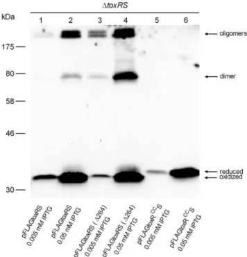
Figure 2. toxRS coexpression inV. cholerae P27459-S DtoxRS
mutant strain acts negatively on ToxR disulfide bond homo-dimer and oligomers.Shown is an immunoblot analysis derived from SDS-PAGE analysis performed under non-reducing conditions, utilizing anti-FLAG antibodies andV. choleraecells harboring various pFLAG-toxRS expressing plasmids, grown in LB medium to mid-log phase and induced with IPTG. Molecular markers are indicated on the left side. Two different IPTG concentrations are indicated, showing different ToxR levels. Immunoblot analysis was performed at least three times, and results were reproducible.
latter were produced by plasmid encoded FLAG-tagged toxRS, toxRS(D264) andtoxRCCSin aV. choleraeDtoxRSstrain grown in LB medium. Cell extracts were sampled in Laemmli buffer, both with and without the reducing agent ß-mercaptoethanol. In the presence of ß-mercaptoethanol, only a single reduced ToxR or ToxRCC protein band with 35 kDa was observed (data not shown). If cell extracts were not treated with reducing agent, then additional ToxR protein bands became visible. As shown (Fig. 2, lane 4, from bottom to top), ToxR protein bands were visible as an oxidized form at,33 kDa and a minor reduced monomer band
at 35 kDa, followed by a 70 kDa homodimer and oligomeric forms .170 kDa. In contrast, FLAG-tagged toxRCCS expression (Fig. 2, lane 5 and 6) only yielded one single monomeric ToxR form migrating at 35 kDa, indicating no existing disulfide bond formation. Interestingly, it was observed that by expressing FLAG-tagged toxRS(D264), the formation of ToxR disulfide bond dependent homodimer and oligomers was enhanced (Fig. 2, compare lane 1, 2 with lane 3, 4). This observation was also shown in E. coli (see below) and demonstrates that toxRS coexpression negatively influences the formation of cysteinyl dependent ToxR homodimers and oligomers.
ToxR disulfide bond formation of plasmid encoded FLAG-tagged toxRS was monitored in V. cholerae DtoxRS and dsbA+/2
strain backgrounds. Cells were grown under two different conditions, M9 glycerol and LB broth. As shown (Fig. 3), if FLAG-taggedtoxRSwas expressed in adsbA+
background, mainly oxidized ToxR and oligomeric forms were visible, regardless of the growth medium. In contrast, expression of FLAG-taggedtoxRSin a dsbA strain in M9 glycerol medium resulted in both ToxR monomer forms and no observable oligomers, while in LB broth, the oxidized monomer was the dominant expressed ToxR form followed by very minor expressed oligomer forms. Thus, these data demonstrate that disulfide bond formation of ToxR differs in
adsbAdependent manner. Importantly, ToxR intrachain disulfide bond formation existed independently of DsbA under LB broth growth conditions, but responded to DsbA function under M9 glycerol growth conditions (Fig. 3). These results indicate a correlation between the activity of DsbA, ToxR intrachain disulfide bond formation and ToxR activity.
toxRCCmutant affects porin production
As shown above, a dsbA knockout mutation interfered with disulfide bond formation of ToxR and furthermore resulted in a loss of ToxR activity. Therefore,toxRCCexpression should cause a similar defect in ToxR activity. ToxR regulation is considered non-physiological if plasmid encoded toxR is used, because elevated ToxR protein levels counteract ToxR regulation sensi-tivity [21]. Accordingly, no difference in OmpU and OmpT production was observed by analyzing the OMP profile ofDtoxRS strains expressing plasmid encoded FLAG-taggedtoxRSortoxRCCS (data not shown). Therefore, single copy gene number and chromosomally expressed toxR and toxRCC were tested under different growth conditions. FLAG-tagged toxRand toxRCC gene alleles were transferred into the chromosomal toxR expression locus of a V. cholerae DtoxR strain (see Material and Methods). Subsequently, chromosomally produced FLAG-tagged ToxR proteins were collected from membrane extracts derived from cells grown to stationary phase in LB broth and monitored by immunoblot analysis. It is important to note that ToxR proteins could be detected under these conditions, however, the signal intensity was weak such that monitoring ToxR was only possible under high magnification sensitivity (Fig. S3). No signals were detected for samples treated without ß-mercapthoethanol (data not shown). As shown recently, the production of the porins OmpU and OmpT was significantly changed inV. choleraecells if they were grown in a complex broth medium, such as LB, compared to minimal T medium [57]. It was further shown that the pattern of OMP production observed for cells grown in LB broth could be mimicked by the addition of the amino acids L-asparagine, -arginine, -glutamate and –serine (termed NRES) to minimal growth medium [57]. In order to monitor the effect of thetoxRCC mutant on OMP production, standard growth conditions were tested that generally affect porin [57] and virulence factor production, i.e., AKI growth medium [42]. When cells were grown in AKI, M9 glycerol or M9 glycerol NRES medium, the pattern of OmpU and OmpT production in a FLAG-taggedtoxR strain was similar to that observed for WT (Fig. 4A, B, C, lane 1 and 2, respectively). In contrast and shown by others [25], no OmpU and derepressed OmpT was observed for the DtoxR mutant strain (Fig. 4A, B, C, lane 3). Interestingly, the toxRCC mutant cells, grown in M9 glycerol medium (Fig. 4A, lane 4), showed no OmpU and derepressed OmpT, similarly as observed for thedsbABmutations (Fig. 1A). Such an effect was also observed in the classical strain O395 (Fig. S2B). Furthermore, production of OmpU was only partially restored and did not reach WT levels in toxRCCmutant cells grown in M9 NRES medium (Fig. 4B, lane 4). Interestingly, if thedsbAmutant strain was grown in M9 glycerol with NRES, OmpU production was similar to that observed for NRES activated WT cells (data not shown). Considering the recently published effects of NRES [57], these data indicate that dsbAmutation does not disrupt NRES activated OmpU produc-tion, buttoxRCCmutation does. To quantify the effects oftoxRCCon transcription,ompU,ompTandtoxRmRNA levels were determined by qRT-PCR analysis of cells grown in M9 glycerol medium for 24 h. The results showed thattoxRgene transcript levels of FLAG-taggedtoxRcompared totoxRCCwere similar (Fig. 5). In contrast, ompUtranscription showed a 15-fold reduction in a FLAG-tagged
Figure 3.dsbAknockout mutant and ToxR forms.Immunoblot analyses are shown using anti-FLAG antibodies to detect FLAG-tagged ToxR produced in V. cholerae P27459-S DtoxRS and DtoxRS DdsbA
mutant strain (as indicated in the figure). Bacterial cultures harboring pFLAGtoxRS were grown to mid-log phase in M9 glycerol and in LB broth and induced with IPTG. ToxR mobility in the different samples was monitored and differences for intrachain disulfide bond formation were detected. Immunoblot analysis was performed at least three times, and results were reproducible.
toxRCCmutant compared to the FLAG-taggedtoxRstrain, whereas ompTtranscription was 6-fold increased. Thereby, the transcrip-tional pattern corresponds to the observed porin production profile and is related to a similar transcriptional pattern seen in a dsbAmutant (Fig. 1B). It is important to note that in AKI grown WT, FLAG-tagged toxR or FLAG-tagged toxRCC cells, OmpU levels were strongly upregulated, as shown in Fig. 4C. To quantify this observation we monitoredtoxRandompUtranscription in WT, using qRT-PCR, by comparing cells grown in M9 glycerol and AKI medium. The obtained results showed a 4-fold upregulation forompUtranscription, but no difference intoxRtranscription (Fig. S4). Therefore, the increase in OmpU production did not correlate with elevatedtoxRtranscription.
ToxR operators negatively influence ToxRS heterodimerization
If pFLAGtoxRS was expressed inE. colicells,grown in LB broth (mid-log phase OD600of 0.5 and subsequently induced with IPTG
for 1 h), a novel and stable SDS-resistant FLAG related protein band of about 55 kDa was observed (Fig. 6A, B, lane 4). This size corresponded well with a reported ToxRS heterodimer [34]. This heterodimer is disulfide bond independent, since ToxS does not have any cysteine residues and the heterodimer was also present in SDS-PAGE analysis using samples treated with ß-mercaptoetha-nol (Fig. 6A, lane 4). To confirm that this protein band represents a ToxRS heterodimer, again pFLAGtoxRS(D264) served as negative control, resulting in the loss of the 55 kDa protein band (Fig. 6A, B, lane 1). Thus, these data indicate that this protein band indeed represented a ToxRS heterodimer. Furthermore, as observed in Fig. 6A, B, lane 7,toxRCCSexpression also yielded a heterodimer, which was less pronounced and indicated a decreased interaction of both proteins. Prompted by the observa-tion that ToxRS heterodimer formaobserva-tion only occured inE. coli (Fig. 6, lane 4), but not in V. cholerae (Fig. 2, lane 1, 2), pFLAGtoxRS plasmids were constructed to additionally contain ToxR operator sites, either ofompU[58] ortoxT[19] (see Material and Methods). Such plasmids were expressed again in E. coli, showing that in the presence of ToxR binding sites, ToxRS heterodimer formation appeared strongly diminished (Fig. 6B, lane 4–6) and mainly ToxR monomers with the intrachain disulfide bond were detectable. Furthermore, also a slight decrease in ToxRCCS heterodimerization was observed in the presence of ToxR operators (Fig. 6A, B, lane 8, 9). Notably, the presence of ToxR operators had no effect on the appearance of cysteinyl dependent ToxR homodimer or oligomer formation if expressed as toxRS(D264) (Fig. 6B, lane 1–3). Thus, it seems that ToxR binding sites negatively influenced ToxRS heterodimerization.
Figure 4. Chromosomal expression of FLAGtoxR and
FLAG-toxRCC and porin regulation. Panel A, B and C, shown are OMP profiles derived from OM preparations, representing WT,DtoxR
::FLAG-toxR,DtoxRandDtoxR::FLAGtoxRCC
strains, grown to stationary phase in M9 glycerol (A), M9 glycerol NRES (B) and AKI (C) during the anaerobic growth phase, respectively. Arrows mark OmpU and OmpT. Arrowheads on the right indicate a ToxR independent protein band used as loading control.
doi:10.1371/journal.pone.0047756.g004
Figure 5. Transcriptional analysis oftoxRand porin genesompU
and ompT in V. cholerae P27459-S. Using qRT-PCR analysis, transcriptional activity of chromosomal encoding FLAG-tagged toxR
andtoxRCC
strains was monitored for the porin genesompUandompT
and also fortoxRandtoxRCC. mRNA levels ofrpoB(used as a reference gene) were determined and correlated with the mRNA level of the genes of interest. Data are presented as median fold change and the error bars indicate the interquartile range of each data set. Experiments were performed with six independent samples, the Mann-Whitney U test was used,P,0.05.
Figure 6.toxRScoexpression inE. coliXL1-Blue strain.Shown are immunoblot analyses utilizing anti-FLAG antibodies to monitor FLAG-tagged ToxR production of pFLAGtoxRS constructs, performed under reducing (panel A) and non-reducing conditions (panel B). pFLAGtoxRS was expressed in E. colicells grown in LB broth to mid-log phase (OD600of 0.5) and subsequently induced with IPTG for 1 h. From left to right, shown are pFLAGtoxRS(D264), pFLAGtoxRS and pFLAGtoxRCCS, either containing no ToxR operator sequence orompUortoxToperator sequences, respectively. A 55 kDa ToxR cross-reacting protein band, associated with pFLAGtoxRS and pFLAGtoxRCCS, is indicated by an arrow. To note, cysteinyl dependent homodimer and oligomer ToxR bands occurred diminished as observed for pFLAGtoxRS in comparison to pFLAGtoxRS(D264). Molecular size markers are indicated on the left. Immunoblot analysis was performed at least three times, and results were reproducible.
These data provide a rational explanation for the failure to observe a heterodimer in V. cholerae, because numerous ToxR regulated genes [16] and therefore multiple ToxR operators are present in V. cholerae.
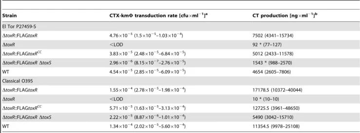
Virulence factor production oftoxRCC mutants in El Tor and classical strains and in vivo colonization of mice
In order to characterize atoxRCC mutant strain for virulence factor production, the strain encoding chromosomally FLAG-taggedtoxRCCwas compared with a chromosomally FLAG-tagged toxR strain. The levels of CT and TCP were assessed in both biotypes, O1V. choleraeEl Tor and O395 classical strain, Table 3. As shown, no significant differences were detectable between FLAG-tagged toxRCC and toxR, while slightly enhanced CT production was observed by comparing FLAG-tagged toxR and WT strains. In contrast, atoxRdeletion strain was about 82-fold reduced for CT production compared to a FLAG-taggedtoxRO1 El Tor strain and .1,700-fold reduced in the classical strain, Table 3. AtoxSknockout was about 5- and 3-fold below the CT level compared to the parental strains with FLAG-taggedtoxRO1 El Tor and classical, respectively see Table 3. Additionally, the CTX-kmW transduction frequency was determined for both biotypes, Table 3. This assay relies on the production level of the type IV bundle forming pili TCP and subsequent CTX-kmW
transduction utilizing a kanamycin encoding CTX-kmW [52]. CTX-kmWtransduction frequencies of WT, FLAG-tagged toxR and toxRCC strains did not show significant differences and for
DtoxR mutants, no detectable CTX-kmW transductants were observed. DtoxS mutants of O1 El Tor and classical strains compared to a corresponding FLAG-tagged toxR strain showed 16- and 7-fold lower frequencies, respectively. In addition, in vivo and in vitro competition assays were performed to further elucidate a putative loss of function for thetoxRCCmutant. A 1:1 mixture of chromosomally encoded FLAG-tagged toxR (LacZ2) and FLAG-taggedtoxRCC(LacZ+
) strains was administered orally to infant mice (in vivo). As a control, LB broth was inoculated with this mixture and incubated for 24 h at 37uC (in vitro). Competitive indices from at least four independent competition experiments were obtained, Table 4, demonstrating no significant difference in colonization fitness between both strains tested. Finally, and as shown above, OMP analysis of the FLAG-taggedtoxRCCmutant strain grown in AKI medium showed a similar porin pattern as obtained for a WT strain (Fig. 4C). Thus, under AKI growth conditions, FLAG-tagged toxRCC did not disrupt the porin production pattern. In summary, expression of toxRCC has no effect on virulence gene transcription under the conditions tested. Consequently, these data suggest that other ToxR activation mechanisms exist that do not require ToxR disulfide bond formation.
Discussion
A previous study [34] reported that ToxR formed a heterodi-mer with ToxS and also existed as a homodiheterodi-mer and monoheterodi-mer based on inter- and intrachain disulfide bonds of cysteine 236 and 293. Moreover, in vitro analysis using the purified periplasmic domain of ToxR showed that ToxR homodimers exist and rely on cysteine 293 by forming an intermolecular disulfide bond [59]. We revisited these earlier characterizations with our work, because we observed that dsbAB mutants affect porin production of OmpU and OmpT. Therefore, we focused our studies on the influence of cysteinyl dependent ToxR forms, ToxR activity, ToxRS interac-tion and response to the Dsb system. As identified in this study, dsbA,dsbBdeletions andtoxRCCmutants showed altered activity for porin regulation. InV. cholerae,dsbABanddsbCDhomologues exist, encoding functions for disulfide bond formation [60] and
Table 3.Virulence factor production of chromosomal encoded FLAG-taggedtoxRand FLAG-taggedtoxRCCmutants.
Strain CTX-kmWtransduction rate [cfu6ml21]a CT production [ng6ml21]b
El Tor P27459-S
DtoxR::FLAGtoxR 4.7661025(1.5
61025–1.03
61024) 7502 (4341–15734)
DtoxR ,LOD 92 * (77–127)
DtoxR::FLAGtoxRCC
3.8361025(2.4861025–6.8461025) 5012 (2433–11578)
DtoxR::FLAGtoxRDtoxS 2.9661026(8.15
61027–2.76
61025) 1543 * (988–2570)
WT 4.5461025(2.8561025–6.0961025) 4654 (2605–7806)
Classical O395
DtoxR::FLAGtoxR 1.5561024(2.78
61025–1.98
61024) 17178.5 (10372–40044)
DtoxR ,LOD 10 * (10–10)
DtoxR::FLAGtoxRCC 5.7161025(1.6361025–3.1361024) 12725.5 (3961–48650)
DtoxR::FLAGtoxRDtoxS 2.2261025(8.87
61026–1.01
61024) 5490 (3042–15710)
WT 1.3461024(2.0261025–5.6061024) 11354.5 (9978–25108)
amedian and interquartile range of at least 7 independent experiments. bmedian and interquartile range of 9 independent experiments.
*significant by Kruskal-Wallis test followed by Dunn’s test of selected pairs of columns withP,0.05.
,LOD below limit of detection of 561028.
doi:10.1371/journal.pone.0047756.t003
Table 4.In vitro and in vivo competition of chromosomal encoded FLAG-taggedtoxRversus FLAG-taggedtoxRCC mutant in El Tor P27459-S.
in vitroa in vivob
1.04 (0.76–1.22) 2.94 (1.23–3.52)
amedian and interquartile range of 12 independent experiments. bmedian and interquartile range of 5 independent experiments.
correction of protein folding [61] that are located in the periplasm. InE. coli, it is known that DsbAB activity is important for disulfide bond formation if cells are grown in minimal medium. This is because in full broth media, such as LB, small organic molecules are present, which act as oxidizing agents on cysteine residues and will therefore also lead to disulfide bond formation [56]. As observed in this study,dsbAordsbB, but notdsbCdeletion mutants affected ToxR dependent porin regulation. As shown fordsbAor dsbBmutant strains grown under M9 glycerol growth conditions, low OmpU and derepressed OmpT levels were observed, which corresponded to alteredompUandompTtranscription if compared to the WT strain. Interestingly, in a dsbA mutant a statistic significant increase ofompT transcription was observed, also the OMP profile exposed a slightly higher expressed OmpT band. However, the latter observation also indicated a higher OmpT expression indsbmutants if compared withtoxRknockout mutant, thereby it can be excluded that dsbA deficient disulfide bond formation of ToxR is the cause of increased OmpT expression. Further investigation is necessary to provide an explanation for this observation. No alteration in porin production was observed in the dsbCmutant, which suggests that the DsbCD system does not participate in regulating ToxR activity. However, its participation cannot be entirely excluded since DsbCD activity may have an influence on porin production under conditions not tested in this study. Since thedsbABas well astoxRCCphenotypes were also detectable in classical strain O395, we conclude that the herein characterized ToxR cysteine requirement is significant for V. choleraestrains in general.
To identify whether DsbA disulfide oxidoreductase activity per se is influencing ToxR forms, FLAG-taggedtoxRSexpression was analyzed in a dsbA toxRS knockout strain. Interestingly, it was demonstrated that by expression oftoxRS in the absence ofdsbA ToxR showed intrachain disufide bond and reduced monomer forms in M9 glycerol, but only the intrachain disulfide bond form in LB broth. The latter is explainable because small organic molecules are present in LB and can catalyze intrachain disulfide bond formation supporting ToxR activity. Taken together, these data imply that ToxR activity is stimulated by DsbA or alternative oxidizing mechanisms, producing thiol-dependent intrachain disulfide bond formation.
To confirmdsbAdependent ToxR phenotypes, atoxRCCmutant was characterized, which is defined by amino acid substitutions of cysteines to serines. The appearance and electrophoretic mobility of ToxRCCtested without reducing agents in SDS-PAGE was as expected. ToxRCC showed no intrachain disulfide bonded monomer, homodimer or oligomeric forms. Instead, ToxRCC only produceed a single protein band corresponding to the size of a reduced ToxR monomer. Therefore, the activity of ToxRCCwas associated with the reduced monomeric form. By using a chromosomally encodedtoxRCCmutant, it was demonstrated that porin production is influenced profoundly, quite similar to that observed for thedsbA ordsbB deletion mutants. Additionally, we tested a double mutant comprisingdsbAandtoxRCCand observed no differences in the OMP profile compared to atoxRCCmutant (data not shown). Furthermore, no difference was observed betweentoxRortoxRCCtranscription levels, hence we argue that toxRCCrepresents a mutation affecting ToxR activity. Therefore, we provide evidence that DsbA activity is targeting cysteine residues of ToxR and this influences ToxR activation.
As shown recently [57], when an O1 El TorV. choleraestrain was grown in minimal medium OmpT was expressed as a major porin and no OmpU protein was observed. In LB broth, the porin production pattern was reversed. If NRES amino acids were added to the minimal medium, the porin production profile
appeared similar to that observed for cells cultured in LB broth. Mey et al. further showed that NRES amino acids added to the minimal medium led to elevatedtoxR transcription, which they concluded, is the cause for the switched porin production. As shown in here, we also confirmed the NRES effect in showing that addition of NRES to M9 glycerol medium enhances OmpU production for WT and FLAG-taggedtoxR strains. Also FLAG-taggedtoxRCC
mutant cells responded to NRES but to a lower extend. However, toxRCC mutant cells grown in AKI showed maximum OmpU production, similar as observed for FLAG-taggedtoxRor WT strains. Hence, we assumed that under AKI growth conditions, elevated toxRand toxRCC transcription would occur, explaining the increase in OmpU production. This assumption was not confirmed, since qRT-PCR analysis of WT cells cultured in M9 glycerol compared with AKI cultures showed no significant difference intoxRtranscription. These data indicate, that other mechanisms exist, which influence ToxR activity. For example, we cannot rule out, that DsbAB activity influencesompU post-transcriptional, -translational or secretion pathways, neither can we exclude thatdsbAortoxRCCmutants are solely responsible for the observed decreasedompUtranscription. Therefore, other yet unknown factors may contribute or facilitate OmpU expres-sion, especially under growth conditions such as AKI or LB broth media. Also to mention is that iftoxRCCis expressed from multi-copy plasmid, then we observed that cells were producing high OmpU levels. Similar behavior of ToxR activity was observed earlier [21], however such conditions were regarded as non-physiological, hence we will not address this for further discussions. In summary, our data consistently show that under minimal growth conditions, porin regulation solely depends on ToxR intrachain disulfide bond formation iftoxR is expressed from its chromosomal loci.
ToxR operator sites. Interestingly, in another report [39] it was shown that ToxS was not necessary for ToxR binding to DNA in vitro. Therefore, it seems questionable whether ToxRS heterodi-mers can be found in a DNA bound state. So far it can only be speculated whether disaggregation of heterodimer is a conse-quence of ToxR binding and this needs further characterization.
Finally, we tested virulence factor production in O1 El Tor or classical strains and neither the level of CT, nor CTX-kmW
transduction frequencies showed any significant differences between FLAG-tagged toxRCC and toxR strains. This indicated that under virulence factor inducing conditions, ToxRCC can participate in the regulation cascade of the virulence factor production system. Moreover, thetoxRCCmutant in O1 El TorV. cholerae strain did also not display a phenotype for in vivo colonization. In summary, this suggests that cysteine associated ToxR forms are dispensable for ToxR activity under virulence factor inducing conditions. Thereby, we argue that multiple ToxRS activation conditions may exist, which do not rely on thiol-dependent disulfide bond forms. So far, we cannot explain why porin gene regulation seems to respond sensitive to ToxR disulfide bond formation, whereas virulence gene expression is not. Recently, a possible hint was provided by Morgan and colleagues [29]. They published data addressing the isolation oftoxR point mutations, which differentially targettoxTandompUtranscription. All of theirtoxRmutations were found in the cytoplasmic part of ToxR. For example, for amino acid residues V71, F69 and E39 it was proposed that they interfere with RNAP engagement on the ompUpromoter, rather then with DNA binding. Interestingly, the same point mutants had much less effect on toxT activation. In contrast, amino acids R65 and D73 affected more severelyompU activation thantoxT. Based on these results, the authors concluded that the facing of ToxR upon operator binding seems differently oriented for the promoter regions ofompUandtoxT. Thereby, it can only be speculated that ToxRCC protein configuration is
sufficient to serve fortoxTactivation by correctly facing to thetoxT promoter, but becomes conditionally insufficient for ompU activation for yet unknown reasons.
Finally, the question arises whether ToxR may represent a thiol-based redox switch regulator. Although, several periplasmic proteins depend on DsbAB folding activity to obtain function, e.g. PhoA [65], while no examples to our knowledge exist for DsbAB depending thiol-based redox switches for periplasmic proteins. However, the latter does not exclude the possibility that defined environmental conditions exist, e.g., in the aquatic environment, that modulate thiol-dependent intrachain disulfide bond formation in ToxR, hence leading to changes in ToxR activity. For example, such influences could be derived from stress responses, asdsbgene transcription is under the control of thesE
-membrane stress pathway inV. choleraeand additionally of the Cxp regulon as shown in E. coli[66–68]. ToxR activity may also be linked to the cellular metabolism status of the cells. The status may be reflected by electron transfer activity, which is known to influence DsbAB activity whereby menaquinone or ubiquinone
act as recipients for e2 derived from disulfide bond formations [55].
Supporting Information
Figure S1 Growth and cell survival for P27459-S and DdsbA mutant strains. Shown are growth curves (OD600left Y axis) and
colony forming units (cfu/ml right Y axis) of WT strain P27459-S and correspondingDdsbAstrains over 72 h in M9 minimal media supplemented with glycerol (0.4%).
(TIF)
Figure S2 OMP profiles ofV. choleraeO1 classical strain O395. Arrows indicate OmpU and OmpT. Panel A, shown are WT O395,DtoxR,DdsbAand DdsbBstrains grown to stationary phase in M9 glycerol medium. Panel B, shown are WT O395,DtoxR,
DtoxR::FLAGtoxR and DtoxR::FLAGtoxRCC strains. Cells were grown to stationary phase in LB broth medium.. Arrowheads on the right indicate a ToxR independent protein band used as loading control.
(TIF)
Figure S3 Detection of chromosomal encoded FLAG-tagged ToxR expressed fusion proteins. Immunoblot analysis is shown, using anti-FLAG antibodies to detect chromosomal expression of FLAG-taggedtoxRandtoxRCCinV. choleraeP27459-S and mutant strainsDtoxR andDtoxRS of isolated membrane fractions. Cross-reacting background bands are marked with asterisks and ToxR is indicated by an arrow. Molecular size markers are indicated on the left. Immunoblot analysis was performed at least two times, and results were reproducible.
(TIF)
Figure S4 Transcriptional analysis oftoxRand porin geneompU inV. cholerae P27459-S grown in M9 glycerol compared to AKI conditions. The WT strain was cultured in M9 glycerol medium to mid log growth phase and shifted to fresh M9 glycerol or AKI medium for 45 min. Subsequently mRNA was prepared and qRT-PCR was performed for theompUporin gene and also for toxR. mRNA level of 16S rRNA was determined as a reference and correlated with the mRNA level of the genes of interest. Experiments were performed with three independent samples and data represent means and standard deviations. The unpaired t test was used,P,0.05.
(TIF)
Acknowledgments
For helpful discussion and critical reading of the manuscript we thank Michelle Dziejman.
Author Contributions
Conceived and designed the experiments: JR VIHF. Performed the experiments: VIHF ECB HE SR. Analyzed the data: JR VHIF ECB ST SS. Contributed reagents/materials/analysis tools: SR AS. Wrote the paper: JR VHIF.
References
1. Faruque SM, Albert MJ, Mekalanos JJ (1998) Epidemiology, genetics, and ecology of toxigenicVibrio cholerae. Microbiol and Mol Biol Rev 62: 1301–1314. 2. Pruzzo C, Vezzulli L, Colwell RR (2008) Global impact of Vibrio cholerae
interactions with chitin. Environ Microbiol 10: 1400–1410.
3. Reidl J, Klose KE (2002)Vibrio choleraeand cholera: Out of the water and into the host. FEMS-Microbiol Rev 26: 125–139.
4. Andoh A, Fujiyama Y, Sakumoto H, Uchihara H, Kimura T, et al. (1998) Detection of complement C3 and factor B gene expression in normal colorectal mucosa, adenomas and carcinomas. Clin Exp Immunol 111: 477–483.
5. Mallow EB, Harris A, Salzman N, Russell JP, DeBerardinis RJ, et al. (1996) Human enteric defensins. J Biol Chem 271: 4038–4045.
6. Herrington DA, Hall RH, Losonsky G, Mekalanos JJ, Taylor RK, et al. (1988) Toxin, toxin-coregulated pili and thetoxR regulation are essential forVibrio choleraepathogenesis in humans. J Exp Med 168: 1487–1492.
7. DiRita VJ, Parsot C, Jander G, Mekalanos JJ (1991) Regulatory cascade controls virulence inVibrio cholerae. Proc Natl Acad Sci 88: 5403–5407.
9. Matson JS, Withey JH, DiRita VJ (2007) Regulatory networks controllingVibrio choleraevirulence gene expression. Infect Immun: 5542–5549.
10. Karaolis DK, Johnson JA, Bailey CC, Boedeker EC, Kaper JB, et al. (1998) A Vibrio cholerae pathogenicity island associated with epidemic and pandemic strains. Proc Natl Acad Sci USA 95: 3134–3139.
11. Kovach ME, Shaffer MD, Peterson KM (1996) A putative integrase gene defines the distal end of a large cluster of ToxR-regulated colonization genes inVibrio cholerae. Microbiology 142: 2165–2174.
12. Miller VL, Mekalanos JJ (1984) Synthesis of cholera toxin is positively regulated at the transcriptional level bytoxR. Proc Nat Acad Sci USA 81: 3471–3475. 13. Osorio CR, Klose KE (2000) A region of the transmembrane regulatory protein
ToxR That tethers the transcriptional activation domain to the cytoplasmic membrane displays wide divergence amongVibriospecies. J Bacteriol 182: 526– 528.
14. Skorupski K, Taylor RK (1999) A new level in theVibrio choleraeToxR virulence cascade: AphA is required for transcriptional activation of thetcpPHoperon. Mol Microbiol 31: 763–771.
15. Xu X, Stern AM, Liu Z, Kan B, Zhu J (2010) Virulence regulator AphB enhancestoxRtranscription inVibrio cholerae. BMC Microbiology doi:101186/ 1471-2180-10-3
16. Bina J, Zhu J, Dziejman M, Faruque S, Calderwood S, et al. (2003) ToxR regulon ofVibrio choleraeand its expression in vibrios shed by cholera patients. Proc Natl Acad Sci USA 100: 2801–2806.
17. Childers BM, Klose KE (2007) Regulation of virulence inVibrio cholerae: the ToxR regulon. Future Microbiol 2: 335–344.
18. Hase CC, Mekalanos JJ (1998) TcpP protein is a positive regulator of virulence gene expression inVibrio cholerae. Proc Natl Acad Sci USA 95: 730–734. 19. Higgins DE, DiRita VJ (1994) Transcriptional control oftoxT, a regulatory gene
in the ToxR regulon ofVibrio cholerae. Mol Microbiol 14: 17–29.
20. DiRita VJ (1994) Multiple regulatory systems inVibrio cholerae pathogenesis. Trends Microbiol 2: 37–38.
21. Miller V, DiRita VJ, Mekalanos JJ (1989) Identification oftoxS, a regulatory gene whose product enhances ToxR-mediated activation of the cholera toxin promoter. J Bacteriol 171: 1288–1293.
22. Withey JH, DiRita VJ (2006) The toxbox: specific DNA sequence requirements for activation ofVibrio choleraevirulence genes by ToxT. Mol Microbiol 59: 1779–1789.
23. Champion GA, Neely MN, Brennan MA, DiRita VJ (1997) A branch in the ToxR regulatory cascade ofVibrio choleraerevealed by characterization oftoxT mutant strains. Mol Microbiol 23: 323–331.
24. Li CC, Merrell DS, Camilli A, Kaper JB (2002) ToxR interferes with CRP-dependent transcriptional activation ofompTinVibrio cholerae. Mol Microbiol 43: 1577–1589.
25. Miller VL, Mekalanos JJ (1988) A novel suicide vector and its use in construction of insertion mutations: Osmoregulation of outer membrane proteins and virulence determinants inVibrio choleraerequirestoxR. J Bacteriol 170: 2575– 2583.
26. Provenzano D, Klose KE (2000) Altered expression of the ToxR-regulated porins OmpU and OmpT diminishesVibrio choleraebile resistance, virulence factor expression, and intestinal colonization. Proc Natl Acad Sci USA 97: 10220–10224.
27. Krukonis ES, Yu RR, DiRita VJ (2000) TheVibrio choleraeToxR/TcpP/ToxT virulence cascade: distinct roles for two membrane-localized transcriptional activators on a single promoter. Mol Microbiol 38: 67–84.
28. Krukonis ES, DiRita VJ (2003) DNA binding and ToxR responsiveness by the wing domain of TcpP, an activator of virulence gene expression inVibrio cholerae. Mol Cell 12: 157–165.
29. Morgan SJ, Felek S, Gadwal S, Koropatkin NM, Perry JW, et al. (2011) The two faces of ToxR: activator ofompU, co-regulator oftoxTinVibrio cholerae. Mol Microbiol 81: 113–128.
30. Miller VL, Taylor RK, Mekalanos JJ (1987) Cholera toxin transcriptional activator ToxR is a transmembrane DNA binding protein. Cell 48: 271–279. 31. Dziejman M, Mekalanos JJ (1994) Analysis of membrane protein interaction:
ToxR can dimerize the amino terminus of phage lambda repressor. Mol Microbiol 13: 485–494.
32. Kolmar H, Hennecke F, Go¨tz K, Janzer B, Vogt B, et al. (1995) Membrane insertion of the bacterial signal transduction protein ToxR and requirements of transcriptional activation studied by modular replacement of different protein substructures. EMBO J 14: 3895–3904.
33. Ottemann KM, Mekalanos JJ (1995) Analysis ofVibrio choleraeToxR function by construction of novel fusion proteins. Mol Microbiol 15: 719–731.
34. Ottemann KM, Mekalanos JJ (1996) The ToxR protein ofVibrio choleraeforms homodimers and heterodimers. J Bacteriol 178: 156–162.
35. Crawford JA, Krukonis ES, DiRita VJ (2003) Membrane localization of the ToxR winged-helix domain is required for TcpP-mediated virulence gene activation inVibrio cholerae. Mol Microbiol 47: 1459–1473.
36. Dziejman M, Kolmar H, Fritz HJ, Mekalanos JJ (1999) ToxR co-operative interactions are not modulated by environmental conditions or periplasmic domain conformation. Mol Microbiol 31: 305–317.
37. Hung DT, Mekalanos JJ (2005) Bile acids induce cholera toxin expression in Vibrio choleraein a ToxT-independent manner. Proc Natl Acad Sci, USA 22: 3028–3033.
38. DiRita VJ, Mekalanos JJ (1991) Periplasmic interaction between two membrane regulatory proteins, ToxR and ToxS, results in signal transduction and transcriptional activation. Cell 64: 29–37.
39. Pfau JD, Taylor RK (1998) Mutations in toxR and toxS that separate transcriptional activation from DNA binding at the cholera toxin gene promoter. J Bacteriol 180: 4724–4733.
40. Pearson GDN, Woods A, Chiang SL, Mekalanos JJ (1993) CTX genetic element encodes a site-specific recombination system and an intestinal colonization factor. Proc Natl Acad Sci USA 90: 3750–3754.
41. Mekalanos JJ (1983) Duplication and amplification of toxin genes inVibrio cholerae. Cell 35: 253–263.
42. Iwanaga M, Yamamoto K (1985) New medium for the production of cholera toxin byVibrio choleraeO1 Biotype El Tor. J Clin Microbiol 22: 405–408. 43. Grimberg J, Maguire S, Belluscio L (1989) A simple method for the preparation
of plasmid and chromosomalE. coliDNA. Nucleic Acids Res 17: 8893. 44. Sanger F, Nicklen S, Coulson AR (1977) DNA sequencing with
chain-terminating inhibitors. Proc Natl Acad Sci USA 74: 5463–5467.
45. Donnenberg MS, Kaper JB (1991) Construction of aneaedeletion mutant of enteropathogenic Escherichia coli by using a positive-selection suicide vector. Infect Immun 59: 4310–4317.
46. Horton RM, Hunt HD, Ho SN, Pullen JK, Pease LR (1989) Engineering hybrid genes without the use of restriction enzymes: gene splicing by overlap extension. Gene 77: 61–68.
47. Carlone ACY, Myrtle LT, Rumschlag HS, Sottner FO (1986) Rapid microprocedure for isolating detergent-insoluble outer membrane proteins from Haemophilus-species. J Clin Microbiol 24: 330–332.
48. Warburg O, Christian W (1941) Isolierung und Kristallisation des Ga¨rungsfer-ments Enolase. Biochem Z 310: 384–423.
49. Kang D, Gho YS, Suh M, Kang C (2002) Highly sensitive and fast protein detection with Coomassie Brilliant Blue in sodium dodecyl sulfate-polyacryl-amide gel electrophoresis. BKCS 23: 11.
50. Schild S, Nelson EJ, Camilli A (2008) Immunization withVibrio choleraeouter membrane vesicles induces protective immunity in mice. Infect Immun 76: 4554–4563.
51. Schild S, Tamayo R, Nelson EJ, Qadri F, Calderwood SB, et al. (2007) Genes induced late in infection increase fitness ofVibrio choleraeafter release into the environment. Cell Host Microbe 2: 264–277.
52. Waldor KW, Mekalanos JJ (1996) Lysogenic conversion by a filamentous phage encoding cholera toxin. Science 272: 1910–1914.
53. Svennerholm AM, Holmgren J (1978) Identification ofEscherichia coliheat-labile enterotoxin by means of a ganglioside immunosorbant assay (GM1-ELISA) procedure. Curr Microbiol 1: 19–23.
54. Moisi M, Jenul C, Butler SM, New A, Tutz S, et al. (2009) A novel regulatory protein involved in motility ofVibrio cholerae. J Bacteriol 191: 7027–7038. 55. Kadokura H, Beckwith J (2010) Mechanisms of oxidative protein folding in the
bacterial cell envelope. Antioxid Redox Signal 13: 1231–1246.
56. Bardwell JC, Lee JO, Jander G, Martin N, Belin D, et al. (1993) A pathway for disulfide bond formation in vivo. Proc Natl Acad Sci USA 90: 1038–1042. 57. Mey AR, Craig SA, Payne SM (2012) Effects of amino acid supplementation on
porin expression and ToxR levels in Vibrio cholerae. Infect Immun 80: 518– 528.
58. Crawford JA, Kaper JB, DiRita VJ (1998) Analysis of ToxR-dependent transcription activation ofompU, the gene encoding a major envelope protein in Vibrio cholerae. Mol Microbiol 29: 235–246.
59. Chatterjee A, Dutta PK, Chowdhury R (2007) Effect of fatty acids and cholesterol present in bile on expression of virulence factors and motility ofVibrio cholerae. Infect Immun 75: 1946–1953.
60. Yu J, Webb H, Hirst TR (1992) A homologue of theEscherichia coliDsbA protein involved in disulphide bond formation is required for enterotoxin biogenesis in Vibrio cholerae. Mol Microbiol 6: 1949–1958.
61. Segatori L, Murphy L, Arredondo S, Kadokura H, Gilbert H, et al. (2006) Conserved role of the linker alpha-helix of the bacterial disulfide isomerase DsbC in the avoidance of misoxidation by DsbB. J Biol Chem 281: 4911–4919. 62. Salahpour A, Angers S, Bouvier M (2000) Functional significance of
oligomerization of G-protein-coupled receptors. TEM 11: 163–168. 63. Gentile F, Amodeo P, Febbraio F, Picaro F, Motta A, et al. (2002) SDS-resistant
active and thermostable dimers are obtained from the dissociation of homotetrameric beta-glycosidase from hyperthermophilic Sulfolobus solfataricus in SDS. Stabilizing role of the A–C intermonomeric interface. J Biol Chem 277: 44050–44060.
64. Lueders J, Pyrowolakis G, Jentsch S (2003) The ubiquitin-like protein HUB1 forms SDS-resistant complexes with cellular proteins in the absence of ATP. EMBO Rep 4: 1169–1174.
65. Akiyama Y, Ito K (1993) Folding and assembly of bacterial alkaline phosphatase in vitro and in vivo. J Biol Chem 268: 8146–8150.
66. Ding Y, Davis BM, Waldor MK (2004) Hfq is essential forVibrio cholerae virulence and downregulates SigmaE expression. Mol Microbiol 53: 345–354. 67. Danese PN, Silhavy TJ (1997) The sigma(E) and the Cpx signal transduction
systems control the synthesis of periplasmic protein-folding enzymes inEscherichia coli. Genes Dev 11: 1183–1193.
69. Hanahan D (1983) Studies on transformation ofEscherichia coliwith plasmids. J Mol Biol 166: 557–580.
70. Correa NE, Lauriano CM, McGee R, Klose KE (2000) Phosphorylation of the flagellar regulatory protein FlrC is necessary for Vibrio choleraemotility and enhanced colonization. Mol Microbiol 35: 743–755.
71. Oka A, Sugisaki H, Takanami M (1981) Nucleotide sequence of the kanamycin resistance transposon Tn903. J Mol Biol 147: 217–226.
72. Guzman LM, Belin D, Carson MJ, Beckwith J (1995) Tight regulation, modulation, and high-level expression by vectors containing the arabinose PBAD promoter. J Bacteriol 177: 4121–4130.