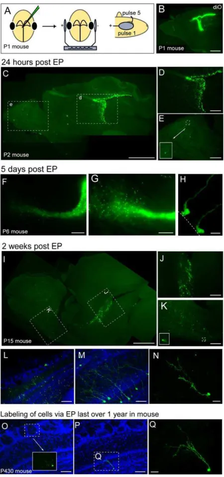
Selective gene expression by postnatal electroporation during olfactory interneuron neurogenesis.
Texto
Imagem
Documentos relacionados
translation as a function of the time after electroporation; capped reporter mRNA (R-RNA) was electroporated into BSRT7 cells expressing NSP3, and the Renilla luciferase activity
Cells grown in both the media demonstrated a similar expression of endothelial cell markers when assessed by immunohistochemistry, although HCEC marker gene expression was higher
Recently, it has been demonstrated that the low levels of viral gene expression induced by a candidate HDACi may be insufficient to cause the death of infected cells by viral
Figure 5.1 - Heatmap of gene expression of all tested genes in all patients, including both cell. cultures (in both passages), and tissue
The results showed that the physical exercise program during the postnatal development increased the mossy fibers density and hippocampal expression of parvalbumin,
Label-and-chase experiments, in which cells are imaged immediately after fluorescent labeling of OM proteins and again during growth without further labeling, show cells that
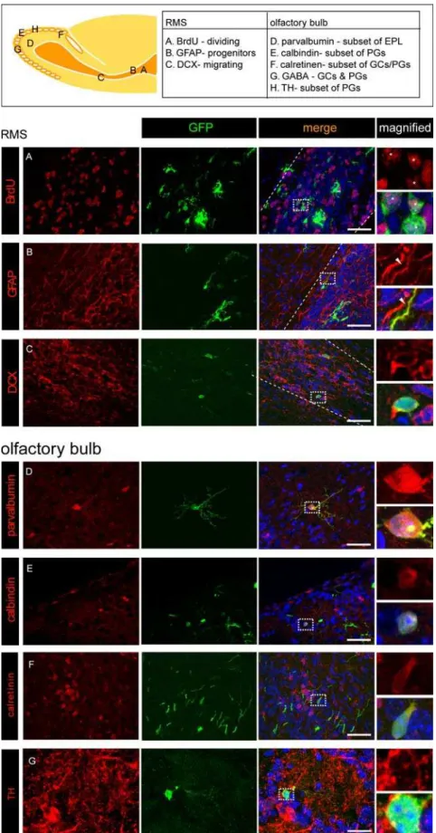
of PK2 gene. Immunofluorescence staining showed EGFP-ir cells in B ) the granule layer (GL), periglomerular layer (PGL) and olfactory ventricle (OV) of the olfactory bulb; C)
cinnamomi zoospores by electroporation was successfully performed and the genome integration of the silencing construct has been verified by genotypic tests (PCR
