FACULDADE DE ODONTOLOGIA
EFEITOS DOS BISFOSFONATOS NITROGENADOS SOBRE O OSSO ALVEOLAR
PONTIFÍCIA UNIVERSIDADE CATÓLICA DO RIO GRANDE DO SUL FACULDADE DE ODONTOLOGIA
NICOLE DE MELLO RAHDE
EFEITOS DOS BISFOSFONATOS NITROGENADOS SOBRE O OSSO ALVEOLAR
NICOLE DE MELLO RAHDE
EFEITOS DOS BISFOSFONATOS NITROGENADOS SOBRE O OSSO ALVEOLAR
Tese apresentada como requisito para obtenção do título de Doutor pelo Programa de Pós-Graduação em Odontologia, Área de Concentração Estomatologia Clínica, Faculdade de Odontologia, Pontifícia Universidade Católica do Rio Grande do Sul
Orientador(a): Profa. Karen Cherubini
Dados Internacionais de Catalogação na Publicação (CIP)
R147e Rahde, Nicole de Mello
Efeitos dos bisfosfonatos nitrogenados sobre o osso alveolar. / Nicole de Mello Rahde – Porto Alegre, 2010.
108 f.
Tese (Doutorado em Estomatologia Clínica) – Faculdade de Odontologia, PUCRS.
Orientação: Profa. Karen Cherubini.
1. Odontologia. 2. Estomatologia Clínica. 3. Bisfosfonatos. 4. Alendronato. 5. Ácido Zoledrônico. 6. Osteonecrose dos Maxilares. I . Cherubini, Karen. I I . Título.
CDD 617.66
Uma janela aberta ao conhecimento científico é caminho árduo, que se percorre com amor à verdade, sabedoria, coragem e liberdade, com o qual se conquista a paz interior e o progresso do mundo.
Agradeço à professora Dra. Karen Cherubini, cuja sabedoria, coragem e
dedicação à ciência são admiráveis. Agradeço pela acolhida carinhosa e comprometimento com o desenvolvimento da Tese. Nesses quatro anos de convívio e amizade, aprendi não só a desenvolver uma pesquisa científica, mas também, que com estudo, empenho e ética, é possível alcançar objetivos aparentemente inatingíveis.
Aos meus pais, Antonio e Dioneia, a quem admiro muito e tenho como
exemplo de caráter e perseverança. Ambos sempre respeitaram e apoiaram todas as minhas decisões desde muito cedo. Com amor e muito esforço puderam proporcionar a realização dos meus sonhos.
Ao meu irmão Rafael, pelo carinho, generosidade e disponibilidade
incansáveis em todos os momentos de minha vida em que precisei de ajuda.
À minha irmã Shalimar, minha grande companheira, a quem admiro por seu
caráter e competência e em quem tento me espelhar. Agradeço o amor e a torcida incondicionais desde sempre.
À minha afilhada Pietra, nossa alegria de viver, pelo carinho, companhia e
amizade; e por compreender quando não era possível dormir na casa da Dinda.
À minha sobrinha Manoela, pelos sorrisos, brincadeiras e intervenções
surpreendentes.
Ao Rodrigo, por seu amor, paciência e tranquilidade, que serviram de alicerce
para a realização desta Tese. Agradeço o carinho, o cuidado, as ideias pertinentes e o interesse real pelo meu trabalho desde que nos conhecemos.
Às professoras do Doutorado em Estomatologia Clínica, Dra. Liliane Soares Yurgel, Dra. Fernanda Salum e Dra. Maria Antonia Zancanaro de Figueiredo, pela
À professora Dra. Maria Martha Campos pela constante disposição e
ensinamentos fundamentais para a concepção do trabalho.
Ao professor Dr. Eraldo Batista Jr, por desvendar os “mistérios” da biologia
molecular e pelo trabalho realizado como Presidente da Comissão Científica e de Ética desta Faculdade.
À Dra. Márcia Maahs, cujo desprendimento, disponibilidade e participação
foram imprescindíveis para a efetivação da presente pesquisa.
Aos colegas do Curso de Doutorado Aline Morosolli, Carla Girardi, Frederico Saueressig, Juliana Romanini e Tatiana Pinto pelo companheirismo,
apoio mútuo e momentos de descontração.
Ao professor Marcos Túlio Mazzini Carvalho, diretor desta Faculdade, por
ensinar-nos, desde cedo, a importância da relação paciente/cirurgião-dentista e pelo incentivo constante na busca do aprimoramento profissional.
Ao professor Dr. José Antonio Poli de Figueiredo, meu grande Mestre, cuja
dedicação pelo que faz foi e sempre será fonte de inspiração e incentivo.
À professora Dra. Nilza Pereira da Costa pela disposição e entusiasmo com
que conduziu o ensino da Radiologia e a Coordenação da Pós-Graduação.
À professora Dra. Julieta Tavares, meu exemplo de profissionalismo, pela
amizade de toda a vida, apoio, incentivo, “puxões de orelha” e parceria incansável.
Aos funcionários do Programa de Pós-Graduação em Odontologia Marcos, Ana Lúcia, Davenir e Carlos pela simpatia e competência no trabalho realizado.
À professora Dra. Edela Puricelli pelo exemplo profissional e pessoal, que
Aos colegas do Serviço de Odontologia da Santa Casa de Misericórdia de Porto Alegre: Isabel, Adriana, Gustavo, Baraldi, Mauro, Marcel, Henrique, Jéssica, Vica, Patrícia, Fernanda, Julieta, Clarissa, Mario, Humberto, Liane, Mari, Alessandro, Deise e Letícia, pela seriedade no trabalho, troca de experiências e espírito
de equipe.
Aos colegas do Curso de Especialização em Endodontia da ABO-RS, meus grandes mestres, professores João Ferlini, Lílian Neuvald Zanatta, Anelise Ligocki, Jussara Mallmann e Régis Burmeister dos Santos, pelo carinho, convivência alegre e
ensinamentos transmitidos desde a graduação até hoje.
Às minhas verdadeiras amigas: Dani Ody, Deisi, Aninha, Simone, Fernanda Saffer, Fernanda Glitz, Ana Zaffari, Cristiane Pires e Juliana Tigre pelo apoio e
torcida constantes.
A Angela Rosa de Souza e a Carlos Augusto de Souza, que torceram por
mim e souberam compreender minha ausência nos momentos cruciais.
E, enfim, dedico esta Tese aos pacientes usuários de bisfosfonatos, que
RESUMO
Os bisfosfonatos são drogas inibidoras da reabsorção óssea e têm sido associados a uma forma peculiar de osteonecrose dos maxilares. O efeito dessas drogas é investigado principalmente em tíbia e fêmur, sendo poucos os estudos conduzidos em maxila e mandíbula. A presente pesquisa teve por objetivo investigar, ao exame microscópico, o efeito dos bisfosfonatos nitrogenados alendronato e ácido zoledrônico sobre o osso alveolar. Trinta e um ratos fêmeos (Rattus norvegicus, Wistar) foram
distribuídos em 3 grupos: (1) 11 animais tratados com alendronato (gavagem oral); (2) 10 animais tratados com ácido zoledrônico (intraperitoneal) e (3) 10 animais que não receberam bisfosfonato. Completado o período de 150 dias do início da terapia, os animais foram submetidos à eutanásia. As maxilas foram processadas e cortes histológicos foram corados por hematoxilina e eosina (HE) e picrossírius. Nos cortes corados por HE, foram realizadas a contagem de osteoclastos e a avaliação da densidade trabecular óssea. Nos cortes corados por picrossírius, a densidade de fibras colágenas dos espaços medulares foi determinada. Também foi realizado processamento imunoistoquímico para avaliação da expressão de osteoprotegerina (OPG). As variáveis foram quantificadas com o auxílio dos programas Adobe Photoshop CS3 e Image Pro Plus 4.5.1. O grupo ácido zoledrônico apresentou densidade trabecular significativamente maior que o grupo-controle (ANOVA, teste de Tukey, P<0,001), e o
grupo alendronato não apresentou diferença significativa quando comparado aos demais grupos (P>0,05). Não houve diferença significativa para contagem de osteoclastos,
densidade de fibras colágenas dos espaços medulares e expressão de OPG entre os grupos (ANOVA, P>0,05). Os resultados permitem concluir que (1) o ácido
zoledrônico promove aumento da densidade trabecular do osso alveolar, enquanto o alendronato não produz esse efeito; (2) alendronato e ácido zoledrônico não estão associados à fibrose dos espaços medulares do osso alveolar. Os efeitos do alendronato e do ácido zoledrônico sobre o número de osteoclastos, bem como sobre a expressão imunoistoquímica de OPG, necessitam ser avaliados por novas pesquisas.
Palavras-chave: Bisfosfonatos, Alendronato, Ácido zoledrônico, Osteonecrose
SUMMARY
Bisphosphonates inhibit bone resorption and have been related to a peculiar form of osteonecrosis of the jaws. Nevertheless, bisphosphonate effects have been investigated mainly in bones like tibia and femur, with just few studies conducted on maxilla and mandible. The present research aimed to investigate bisphosphonates microscopic effects on alveolar bone. Thirty one female rats (Rattus norvegicus, Wistar)
were allocated into 3 groups: (1) 11 animals treated with oral alendronate; (2) 10 animals treated with intraperitoneal zoledronic acid; and (3) 10 animals without bisphosphonate treatment. One hundred and fifty days after the beginning of the treatment, the animals were euthanized. Maxillae were processed and histological sections were stained with hematoxilin and eosin (H&E) to evaluate bone trabecular density and osteoclast count; and with picrosirius to evaluate collagen fiber density in medullary spaces. Immunohistochemical expression of osteoprotegerin (OPG) was also evaluated. The variables were quantified with Adobe photoshop CS3 and Image Pro Plus 4.5.1 softwares. Zoledronic acid group showed higher trabecular density than control group (ANOVA, Tukey’s test, P<0.001), and alendronate group did not show
significant difference when compared to the other groups (P>0.05). There was no
significant difference in osteoclast count, collagen fiber density in medullary spaces and OPG expression among the groups (ANOVA, P>0.05). According to the results, (1)
zoledronic acid promotes trabecular density increase while alendronate does not; (2) alendronate and zoledronic acid use is not associated to alveolar bone marrow fibrosis; (3) alendronate and zoledronic acid effects on osteoclast number and immunohistochemical expression of OPG need to be evaluated by other investigations.
Key-words: Bisphosphonates, Alendronate, Zoledronic acid, Osteonecrosis of
SUMÁRIO
1. INTRODUÇÃO ... 17
2. ARTIGO 1 ... 21
2.1. INTRODUCTION ... 24
2.2. CHEMICAL FEATURES AND INDICATIONS OF BISPHOSPHONATES. 25 2.3. MECHANISM OF ACTION OF BISPHOSPHONATES ... 27
2.3.1. DIRECT EFFECTS ON OSTEOCLASTS ... 27
2.3.2. INDIRECT EFFECTS ... 28
2.3.3. ANTI-ANGIOGENIC EFFECT ... 29
2.3.4. EFFECTS ON EPITHELIAL CELLS AND COLLAGEN FIBERS ... 30
2.4. BISPHOSPHONATE-RELATED OSTEONECROSIS OF THE JAWS ... 30
2.4.1. ETIOPATHOGENESIS ... 31
2.4.2. CLINICAL FEATURES ... 32
2.4.3. IMAGINOLOGICAL FEATURES ... 33
2.4.4. HISTOLOGICAL FEATURES ... 34
2.4.5. RISK FACTORS ... 34
2.4.6. TREATMENT ... 35
2.5. DISCUSSION ... 36
3. ARTIGO 2 ... 43
3.1. INTRODUCTION ... 46
3.2. MATERIAL AND METHODS ... 48
3.3. RESULTS ... 56
3.4. DISCUSSION ... 59
3.5. REFERENCES ... 67
4. DISCUSSÃO GERAL ... 72
REFERÊNCIAS ... 77
ANEXOS ... 91
1. INTRODUÇÃO
Os bisfosfonatos são a mais potente classe de drogas inibidoras da atividade
osteoclástica (RUSSELL et al., 2008) e têm como principal indicação o tratamento de
enfermidades do metabolismo ósseo, tais como a osteoporose pós-menopausa e
induzida por corticoterapia. Também são empregados no manejo de complicações
relacionadas a neoplasias, como hipercalcemia maligna, lesões osteolíticas do mieloma
múltiplo e metástases ósseas associadas ao câncer de mama, próstata, pulmão e a outros
tumores de tecidos moles. Condições menos prevalentes, como a doença de Paget e a
osteogênese imperfeita, podem ser igualmente tratadas por essas drogas (MARX, 2007;
LANDESBERG et al., 2008; RUGGIERO et al., 2009).
Por serem análogos sintéticos do pirofosfato inorgânico, os bisfosfonatos
apresentam alta afinidade por cristais de fosfato de cálcio (hidroxiapatita). Têm como
alvo, portanto, a porção mineral do tecido ósseo, sítio em que permanecem por longo
período de tempo, e seus efeitos podem persistir por, aproximadamente, dez anos
(RUSSELL et al., 2008; MARX; CILLO; ULLOA, 2007). O mecanismo de ação
baseia-se na inibição da reabsorção óssea por meio de efeitos diretos e indiretos sobre os
osteoclastos. Após a administração, acumulam-se na superfície óssea, em locais de
intensa reabsorção, sendo englobados diretamente pelos osteoclastos durante o processo
normal de remodelamento. Uma vez no citoplasma da célula, promovem perda de
função ou apoptose da mesma, por inibição de sistemas enzimáticos ou produção de
metabólitos citotóxicos (SATO et al., 1991; LIN, 1996; ROGERS et al., 2000;
THOMPSON et al., 2006; SARIN; de ROSSI; AKINTOYE, 2008). Também inibem a
diferenciação das células da linhagem monócito-macrófago em osteoclastos (SAHNI et
(RANK), seu ligante (RANKL) e receptor decoy osteoprotegerina (OPG) (VIERECK et
al., 2002; PAN et al., 2004; ZHOU et al., 2005). Outros efeitos como inibição da
angiogênese (SANTINI et al., 2002; ZERVAS et al., 2006), da proliferação e do reparo
tecidual de células epiteliais orais in vitro (LANDESBERG et al., 2008),
comprometimento da cicatrização da mucosa oral in vivo (MAAHS, 2008) e fibrose dos
espaços medulares (HANSEN et al., 2006; BEDOGNI et al., 2008) foram relatados.
A osteonecrose maxilar associada ao uso de bisfosfonatos é um efeito adverso
dessas drogas e constitui causa de significativa morbidade entre os pacientes
acometidos. Para a confirmação diagnóstica da enfermidade, o paciente deve apresentar
as seguintes características: tratamento atual ou prévio com bisfosfonatos, exposição do
tecido ósseo do complexo maxilo-mandibular ao meio bucal persistente por mais de oito
semanas e ausência de história de radioterapia na região de cabeça e pescoço
(RUGGIERO et al., 2009). As lesões, geralmente, ocorrem após procedimentos
cirúrgicos invasivos nos ossos maxilares, tais como exodontias, colocação de implantes,
cirurgias periodontais e periapicais. Casos de exposição óssea espontânea e após trauma
causado por próteses parciais removíveis também foram relatados e são atribuídos a
características anatômicas e fisiológicas, pois são mais frequentes na região posterior da
mandíbula, que apresenta mucosa de espessura fina (MARX et al., 2005;
MIGLIORATI; SIEGEL; ELTING, 2006). Os principais sinais e sintomas incluem
eritema, edema e ulceração da mucosa, supuração, sequestros ósseos, suscetibilidade à
fratura patológica, dor e parestesia. A condição é refratária ao tratamento, e tentativas de
debridamento local levam à piora do quadro (RUGGIERO et al., 2004; BAGAN et al.,
2005; BAMIAS et al., 2005; MARX et al., 2005; MIGLIORATI; SIEGEL; ELTING,
Apesar de a osteonecrose maxilar associada ao uso de bisfosfonatos resultar de
efeitos recíprocos entre metabolismo ósseo, trauma local, aumento da demanda de
reparo ósseo, infecção e hipovascularização (MIGLIORATI et al., 2005), sua
patogênese ainda não é completamente conhecida. Além disso, os fatores responsáveis
pelo acometimento exclusivo dos ossos maxilares são ignorados (MARX, 2007). A
maioria das pesquisas in vivo que avaliam os efeitos dos bisfosfonatos são conduzidas
em ossos como tíbia e fêmur. A investigação das repercussões microscópicas do uso de
bisfosfonatos, especificamente sobre o osso alveolar, se faz necessária para o
esclarecimento dessas questões.
O presente estudo compreende dois trabalhos apresentados sob a forma de
artigos científicos. O primeiro teve como objetivo fundamentar o experimento por meio
de uma revisão da literatura a respeito do mecanismo de ação dos bisfosfonatos e sua
relação com a osteonecrose maxilar. O segundo artigo descreve o experimento, cujo
objetivo foi investigar, ao exame microscópico, os efeitos dos bisfosfonatos
2. ARTIGO 1
O artigo a seguir intitula-se “Mechanism of action of bisphosphonates and its relation to osteonecrosis of the jaws: A review of the literature” e foi formatado de
MECHANISM OF ACTION OF BISPHOSPHONATESAND ITS RELATION TO OSTEONECROSIS OF THE JAWS: A REVIEW OF THE LITERATURE
Nicole de Mello Rahde, DDS, MS. Fernanda Salum, DDS, PhD.
Maria Antonia Zancanaro de Figueiredo, DDS, PhD. Karen Cherubini, DDS, PhD.
Porto Alegre, Brazil
PONTIFICAL CATHOLIC UNIVERSITY OF RIO GRANDE DO SUL
Division of Stomatology and Prevention of Oral and Maxillofacial Cancer, School of Dentistry, Hospital São Lucas, Pontifical Catholic University of Rio Grande do Sul
Corresponding author:
Karen Cherubini
Serviço de Estomatologia – Hospital São Lucas
Pontifícia Universidade Católica do Rio Grande do Sul
Av Ipiranga, 6690 Sala 231 CEP: 90610-000
Porto Alegre – RS – Brazil
E-mail: karen.cherubini@pucrs.br
kebini.ez@terra.com.br
Abstract
In the last years, many reports have been published on the occurrence of
osteonecrosis of the jaws in patients using nitrogen-containing bisphosphonates who
had not had radiotherapy inthe craniofacial region. The disease is characterized by
non-healing exposure of bone to the oral cavity, associated with pain, paresthesia,
suppuration, bone sequestration and susceptibility to pathological fracture. This article
addresses chemical features of bisphosphonates, indications and mechanism of action
and also reviews one of their major side effects: osteonecrosis of the jaws.
INTRODUCTION
Bisphosphonates are chemical compounds with high affinity for calcium
phosphate crystals, exerting their effects on bone tissue. This group of drugs has been
widely used in the treatment of diseases of bone metabolism.1 Their mechanism of
action is based on the inhibition of bone resorption and thus of bone remodeling. There
are direct and indirect effects on osteoclasts, which undergo apoptosis 2 or become
unable to differentiate from hematopoietic stem cells.3 Impairment of angiogenesis4 and
damage to epithelial cells have also been reported.5 After administration,
bisphosphonates accumulate on the bone surface, at sites of intense resorption, being
engulfed by osteoclasts during normal remodeling. They exert intracellular effects, such
as loss of function and apoptosis, mediated by the inhibition of enzymatic systems.1
Jaws are considered the exclusive site for the occurrence of
bisphosphonate-related osteonecrosis.6 The presence of teeth, which makes the jaws susceptible to bone
exposure, along with the high rate of bone turnover, and the need for adequate bone
the jaws.7, 8 However, the factors responsible for the exclusive involvement of the jaws
are still unknown.
The purpose of this work was to review the mechanism of action of
bisphosphonates and its relation to osteonecrosis of the jaws.
CHEMICAL FEATURES AND INDICATIONS OF BISPHOSPHONATES
Bisphosphonates are considered the most potent class of drugs responsible for
the inhibition of osteoclast activity. They have a chemical structure (Fig. 1) similar to
that of inorganic pyrophosphate, an endogenous regulator of bone mineralization. The
chemical structure is based on the presence of two phosphonate groups linked by
phosphoether bonds to a central carbon (a P-C-P structure). The P-C-P structure is
resistant to pyrophosphatases and acid hydrolysis. The phosphonate groups provide
anchoring to divalent cations such as calcium. They form a three-dimensional structure
in a bidentate manner, by coordination of one oxygen from each phosphonate group
with the divalent cation. Two additional covalent bonds to the central carbon atom of
bisphosphonates form two side chains, R1 and R2. The affinity for the hydroxyapatite
can be increased further if one of the side chains (R1) is a hydroxyl or primary amino
group, because this allows the formation of a tridentate conformation that is able to bind
calcium more effectively.1, 9 This property explains the long half-life of these drugs in
bone tissue, being as long as approximately ten years.6 R2 components, on the other
hand, are responsible for antiresorptive potency, which is magnified in the presence of
nitrogen. Thus, nitrogen-containing bisphosphonates (e.g., alendronate, ibandronate,
pamidronate, risedronate and zoledronic acid) are more potent than non
Fig.1. Bisphosphonate chemical structure (HA= hydroxyapatite).
Bisphosphonates are mainly indicated to treat postmenopausal and
corticosteroid-induced osteoporosis, as well as to manage complications related to
malignancies, such as hypercalcemia, osteolytic lesions of multiple myeloma, and
breast, prostate and lung cancer bone metastases. Off-label use of bisphosphonates has
also been reported in Paget’s disease and osteogenesis imperfecta.5, 6, 11 Oral
bisphosphonates are generally indicated for the treatment of osteoporosis, where
alendronate is the most common followed by risedronate and ibandronate.6, 12, 13,14
Parenteral bisphosphonate formulations such as zoledronic acid, used once yearly and
ibandronate, administered every three months can also be employed for this purpose.11
The bisphosphonates indicated for the control of hypercalcemia and osteolytic lesions
associated with malignancies are administered intravenously. The drug most used in
these situations is zoledronic acid followed by pamidronate, both as monthly infusions.6,
14
MECHANISM OF ACTION OF BISPHOSPHONATES
The main mechanism of action of bisphosphonates is based on the inhibition of
bone resorption and thus of bone remodeling through effects on osteoclasts which
undergo apoptosis.1 An increase in bone mineral density has been demonstrated, 6, 13, 15,
16 since some bisphosphonates such as etidronate, alendronate, pamidronate and
olpadronate exert an anti-apoptotic effect on osteoblasts and osteocytes.1, 17-19
P C O OH P OH OH OH O
R1 R2
Direct effects on osteoclasts
After administration, part of the bisphosphonate is available for incorporation
into the bone matrix at sites of intense osteoclastic activity.2 The bisphosphonate
remaining is excreted unmetabolized in the urine. Once incorporated into bone tissue,
the drug is removed slowly and can remain in place for up to ten years.20 During
resorption, in the acidic environment of the Howship lacuna, bisphosphonate bound to
bone is released, the drug reaches high concentrations in solution or in the form of
calcium salts, and enters the osteoclast by endocytosis.1
Specific intracellular effects occur depending on the presence or absence of
nitrogen in the side chain of the molecule. The non nitrogen-containing bisphosphonates
are metabolized into cytotoxic analogues of adenosine triphosphate (ATP) and
accumulate intracellularly interfering with osteoclast function through inhibition of
ATP-dependent enzymes.6 On the other hand, nitrogen-containing bisphosphonates are
considered more potent, and impair the intracellular mevalonate pathway, which is
associated with cholesterol biosynthesis. This effect by the latter bisphosphonates
occurs through the inhibition of farnesylpyrophosphate synthase, which catalyzes the
synthesis of isoprenoid lipids such as farnesylpyrophosphate and geranylgeranyl
farnesylpyrophosphate. These molecules modulate prenylation, a structural change of
small guanosine triphosphate-binding proteins (GTPases), such as Rhas and Rho. The
lack of prenylated protein prevents the anchoring of other molecules of the signaling
cascade, causing different changes in cellular function, such as loss of the ruffled
border, cytoskeleton breakage, impairment of adhesion proteins and proton pump and
Fig. 2. Effects of bisphosphonates on mevalonate pathway, schematic representation. N-BPs inhibit FPP synthase, resulting in lack of prenylated proteins, which are essential to osteoclast activity and survival (N-BPs = nitrogen-containing bisphosphonates; FPP = farnesyl pyrophosphate).
Indirect effects
The mechanism of action of bisphosphonates is not only related to direct actions
on osteoclastic activity. Indirect effects, mediated by osteoblasts, have also been
observed. Sahni et al.24 conducted a study in which cells of osteoblast lineage,
previously treated with non nitrogen- and nitrogen-containing bisphosphonate for five
minutes, were incubated in coculture with untreated osteoclasts attached to a mineral
surface (ivory) for 24 hours. There was a decrease in osteoclastic activity resulting in a
reduction in the number and diameter of resorption pits. This study was the first to
launch the hypothesis that the effects of bisphosphonates on bone resorption also
depend on osteoblasts, since they secrete proteins which either stimulate or impair
osteoclastogenesis.3
RANK/RANKL/OPG pathway
One of the main regulators of the molecular mechanisms involved in the
development and function of osteoclasts is the pathway of receptor activator of nuclear
factor-B ligand (RANKL) and osteoprotegerin (OPG) secreted by osteoblast lineage N-BPs
FPP synthase
Ost eoclast act ivit y and survival farnesyl pyrophosphat e
geranylgeranyl pyrophosphat e
f arnesyl at ed prot eins
geranyl geranyl at ed prot eins PRENIYLATED PROTEINS
ruf f l ed border cyt oskelet on adhesion prot eins prot on pump
lysossomal enzymes
apoptosis
dimet hyl allyl diphosphat e
MEVALONATE
isopent enyl diphosphat e (IPP)
FPP synthase
cells. OPG is a member of the tumor necrosis factor receptor (TNFR) superfamily and
acts as a decoy receptor for RANKL, the osteoclast differentiation-stimulating protein.
The binding of RANKL to receptor activator of nuclear factor-B (RANK) present on
the surface membrane of monocyte-macrophage lineage cells, which are osteoclast
precursors, produces the signal that leads to their differentiation into osteoclasts. This
signal may be interrupted by the secretion of OPG, which binds to RANKL and thereby
blocks the interaction with RANK.6, 25-28
Some cell culture studies demonstrated an increase in OPG expression in
osteoblasts treated with bisphosphonates.3, 27 Similar results were obtained by Zhou et
al.29 in an in vivo study. The authors evaluated OPG immunohistochemical expression
in the tibia of mice bearing osteolytic tumors, treated with zoledronic acid. They found
increased OPG expression in animals treated with the bisphosphonate in comparison to
those that did not receive treatment.
Anti-angiogenic effect
Besides suppressing bone remodeling, bisphosphonates are reported to exert an
anti-tumor effect by interfering with angiogenesis. The effect occurs when more potent
drugs, such as zoledronic acid, are used. These compounds presumably modulate the
secretion of specific growth factors, such as vascular endothelial growth factor (VEGF),
and inhibit the proliferation, migration and adhesion of endothelial cells and the
consequent formation of capillary tubes.30, 31 Nevertheless, different results were found
by Pampu et al.32 in rabbits given a single dose of zoledronic acid and submitted 5 days
later to mandibular distraction osteogenesis for 5 more days. After 32 days of a
consolidation period, a histomorphometric evaluation was performed in the regenerated
number of blood vessels. Moreover, Maahs33 reported that the administration of neither
alendronate nor zoledronic acid was associated with lower VEGF immunohistochemical
expression in rat maxillae.
Effects on epithelial cells and collagen fibers
Other actions of bisphosphonates have been reported. Deleterious effects on
epithelial cells in vitro,5 as well as on oral mucosa healing in vivo33 and increase in
fibrous connective tissue (collagen fibers) in medullary spaces of bone tissue15, 34 were
observed.
Landesberg et al.5 evaluated the effect of pamidronate on oral keratinocytes of
mice in vitro and found inhibition of cell proliferation and tissue repair when these cells
were exposed to therapeutic concentrations of the drug. The effects, however, did not
occur at the expense of apoptosis. It is believed that pamidronate promotes necrosis.
BISPHOSPHONATE-RELATED OSTEONECROSIS OF THE JAWS
There are many cases of bisphosphonate-related osteonecrosis of the jaws
(BRONJ) reported in the literature. The condition is characterized by exposed bone in
the maxillofacial region that persists for more than eight weeks in patients treated with
bisphosphonates who do not have a history of radiotherapy in the head and neck region.
The lesions develop spontaneously or after trauma on bone areas covered with a thin
mucosal layer or after dento-alveolar surgery.6, 7, 35 Although a cause and effect
relationship has not yet been established, several epidemiological and some
experimental studies provide evidence to support a strong correlation between monthly
intravenous bisphosphonate therapy associated with tooth extractions, and the
the estimated cumulative incidence ranges from 0.8% to 12%. Lower risk is observed
with the use of oral bisphosphonates for the treatment of osteoporosis in comparison to
intravenous bisphosphonates for the treatment of osteolytic tumors. However, the
association of oral bisphosphonate use with co-factors could play a key role in the
development of osteonecrosis.11
BRONJ is limited to the jaws,6 since it has not so far been reported in other
bones of the skeleton. The factors identified as possible reasons for the uniqueness of
the maxilla and mandible in hosting the injury are: (1) the presence of teeth, which
exposes bone to the external environment; (2) periodontal disease, abscesses,
endodontic treatment as well as the occurrence of injuries7 requiring bone metabolism
and blood supply to maintain the appropriate balance;37 and (3) the high bone turnover
rate at these sites, which causes a higher drug uptake.8
Etiopathogenesis
BRONJ is the result of reciprocal complex effects between bone metabolism,
local trauma, increased demand for bone repair, infection and hypovascularity.8 Bone
homeostasis depends on the balance between osteoblasts and osteoclasts. When bone
resorption is impaired by inhibition of osteoclastic activity, the bone matrix is degraded
and non-vital bone, which is characterized by the absence of osteocytes, accumulates.15
In the jaws, the constant need for repair due to masticatory forces, coupled with the
presence of infection when the bone is exposed to the oral environment, increases the
demand for resorption to such a level that exceeds the response capacity of the tissue
whose metabolism has been modified by bisphosphonates. The result is
Hansen et al.,34 using H&E, Grocott and periodic acid-Schiff (PAS), found areas
of bone erosion covered with Actinomyces sp. in all samples of osteonecrosis from
patients receiving bisphosphonates. One sample showed fungi consistent with Candida
sp. Osteoclasts were observed within resorption lacunae in five out of eight specimens
evaluated.
The damage on epithelial cells caused by bisphosphonates, as demonstrated in vitro by Landesberg et al.5, could contribute to the persistent exposure of underlying
bone and the development of osteonecrosis.
Clinical features
Clinically, BRONJ occurs as one or multiple areas of alveolar bone exposed to
the oral cavity (Fig. 3), which can display sequestration, purulent discharge, ulceration
and/or swelling of the adjacent mucosa,fistula or pathological fracture. The patient may
have pain or paresthesia; the mandible is more affected than the maxilla, and although
less frequently, both bones can be affected simultaneously. The mandibular posterior
region is the site of greatest prevalence of osteonecrosis due to compressive forces
induced by the occlusion in that area.6, 7, 11, 20, 37-44
Fig.3. BRONJ clinical features. (a) 82-year-old male patient under zoledronic acid therapy for multiple myeloma, who developed osteonecrosis at the tooth extraction site; (b) 58-year-old
female patient treated for breast cancer bone metastasis with zoledronic acid, who developed osteonecrosis at implant placement sites.
Imaginological features
The radiographic examination shows ill-defined radiolucent areas in alveolar
bone which has a mottled appearance suggestive of osteolytic lesion (Fig. 4) with or
without radiopaque areas consistent with bone sequestration. Occasionally, the
radiographic signs may be absent.37, 41, 44 Sclerosis and loss of integrity of the lamina
dura (Fig. 4), as well as thickening of the periodontal ligament space could be
radiographic signs of subclinical damage caused by bisphosphonates.6, 7, 11, 45
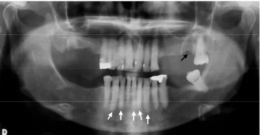
Fig. 4. Panoramic radiograph from a female patient under pamidronate therapy, who developed osteonecrosis after extraction of 2nd upper left molar. The black arrow shows osteolytic area in left maxilla; white arrows show sclerosis of lamina dura in lower teeth.
Computed tomography (CT) scans are used to identify alterations difficult to
discern on radiographs. They provide three-dimensional information and better
delineation of the lesion. Areas of clinically exposed bone in panoramic radiographic
images tend to be smaller than the areas of injured bone shown by CT. CT also shows
focal sclerosis in early disease with the presence of a disorganized trabecular pattern
The findings of magnetic resonance imaging in six patients with a diagnosis of
BRONJ showed, in early disease, loss of the normal hyperintensity of fatty marrow in
the mandible and maxilla. More advanced BRONJ cases demonstrated bone destruction,
soft tissue swelling, inferior alveolar nerve thickening, and pterygoid muscle swelling.47
O’Ryan et al.48 evaluated bone scintigraphy imaging in patients receiving
intravenous nitrogen-containing bisphosphonates. They identified positive tracer uptake
in maxilla and mandible areas which subsequently developed BRONJ. According to this
finding, bone scintigraphy could be useful in the early subclinical detection of BRONJ.
Histological features
On light microscopy, BRONJ appears as multiple areas of non-vital bone, partly
confluent, interspersed with residual vital bone. Inflammatory infiltrate, Actinomyces sp.
colonies and Candida sp. are frequent findings.15, 33, 34, 49 Perinecrotic bone shows
inflammatory reaction in the marrow spaces, with marrow fibrosis, inflammatory cell
infiltration and blood vessels. Osteoclasts, in contact or not with the bone surface, are
also observed.15, 49
Sonis et al.,50 in an animal model of zoledronic acid-related osteonecrosis, found
areas of acellular necrotic bone regularly associated with mucosal ulceration and a
robust inflammatory response. Proliferation of small blood vessels and absence of
Actinomyces sp. as determined by PAS staining were also observed.
Risk factors
Based on the latest findings available in the literature, the main risk factors for
the development of osteonecrosis is the use of intravenous bisphosphonates and surgical
demographic and systemic factors, should also be considered.10, 11 Drug potency and
duration of therapy are directly related to the risk of developing this injury. Zoledronic
acid is more potent than pamidronate, which in turn is more potent than oral
bisphosphonates.11
Local risk factors include dentoalveolar procedures such as tooth extractions,
periodontal surgery involving bone, periapical surgery and implant placement. Specific
anatomical sites, such as the mylohyoid ridge as well as the maxillary and mandibular
tori, have a thin mucosa predisposing bisphosphonate users to the development of
osteonecrosis. The concomitant presence of inflammatory dental disease such as
periodontal and periapical abscesses also increases the risk.11
Age and race are demographic factors associated with the risk of developing
osteonecrosis. The lesions occur most frequently in older Caucasian patients over 60
years of age. Systemic factors such as renal failure,11, 51 diabetes,8, 11 obesity, metastatic
disease, low hemoglobin levels,11 steroids, chemotherapy,11, 37, 42, 52, 53 smoking11, 13, 54
and alcohol ingestion11, 45 can also predispose bisphosphonate users to osteonecrosis.
Treatment
Since the occurrence of the first reports of BRONJ until today, it has not been
possible to determine a treatment strategy that promotes the cure of the disease. There is
difficulty in obtaining adequate surgical margins, because the entire bone is exposed to
bisphosphonate effects. Thus, surgical interventions can often exacerbate the injury. In
mild cases, the lesion is controlled by the use of antimicrobial rinses. When there are
signs of infection in soft tissues and associated symptoms, systemic antibiotics are used,
often for extended periods. Mobile segments of bone sequestration can be removed
region should be avoided. The other dental procedures that do not involve direct
manipulation of bone tissue should be performed in order to prevent the need for tooth
extractions.6, 11
In some situations, temporary discontinuation of bisphosphonate therapy under
the guidance of the attending physician, along with monitoring serum biochemical
markers of bone metabolism, may help therapeutic decisions regarding the management
of the injury, especially in patients who have taken or are taking oral bisphosphonates.6
Hyperbaric oxygen therapy has been evaluated through randomized studies.
Preliminary results indicate some improvement in symptoms and partial healing.
However, it does not promote the complete repair of the lesions when used as single
therapy.55
DISCUSSION
Bisphosphonate mechanism of action is based on impairment of bone resorption
through direct and indirect effects on osteoclasts.1 These effects are inhibition of
osteoclastogenesis and osteoclastic function, as well as inducing apoptosis of mature
cells, causing a reduction in osteoclast number.2 The role of bone marrow stromal cells
or their osteoblast progeny in the maturation of monocyte-macrophage lineage cells into
osteoclasts is essential. These precursor cells secrete both RANKL, which stimulates
osteoclastogenesis, and OPG, which inhibits it.56 Bisphosphonates may stimulate OPG
secretion and contribute, consequently, to impaired bone resorption, as indicated by
some in vitro3, 27 and in vivo studies.29 Anti-angiogenic properties of bisphosphonates
were also reported and are associated with anti-tumor activities.31 However, the
consequences of these effects in the jaws are not completely understood.
rate in this skeletal region may result in higher drug uptake.6 The complex microbial
flora of the oral cavity associated with the low biological response of
bisphosphonate-treated bone leads to several structural changes.37 Osteonecrosis of the jaws is a serious
side effect of bisphosphonates that impairs the quality of life of the patient. The major
obstacle lies in the difficulties of developing an adequate therapeutic approach.11
Furthermore, lack of information among physicians and dentists regarding
bisphosphonate mechanism of action and osteonecrosis onset is disturbing, and
prevention is still the best strategy available. Not only epidemiological studies, but also
experimental investigations, using specific BRONJ animal models, are needed to
REFERENCES
1. Russell RG, Watts NB, Ebetino FH, Rogers MJ. Mechanisms of action of bisphosphonates: similarities and differences and their potential influence on clinical efficacy. Osteoporos Int 2008;19:733-59.
2. Sarin J, DeRossi SS, Akintoye SO. Updates on bisphosphonates and potential pathobiology of bisphosphonate-induced jaw osteonecrosis. Oral Dis 2008;14:277-85.
3. Viereck V, Emons G, Lauck V, Frosch KH, Blaschke S, Grundker C, et al. Bisphosphonates pamidronate and zoledronic acid stimulate osteoprotegerin production by primary human osteoblasts. Biochem Biophys Res Commun 2002;291:680-6.
4. Wood J, Bonjean K, Ruetz S, Bellahcene A, Devy L, Foidart JM, et al. Novel antiangiogenic effects of the bisphosphonate compound zoledronic acid. J Pharmacol Exp Ther 2002;302:1055-61.
5. Landesberg R, Cozin M, Cremers S, Woo V, Kousteni S, Sinha S, et al. Inhibition of oral mucosal cell wound healing by bisphosphonates. J Oral Maxillofac Surg 2008;66:839-47.
6. Marx RE, Cillo JE, Jr., Ulloa JJ. Oral bisphosphonate-induced osteonecrosis: risk factors, prediction of risk using serum CTX testing, prevention, and treatment. J Oral Maxillofac Surg 2007;65:2397-410.
7. Marx RE, Sawatari Y, Fortin M, Broumand V. Bisphosphonate-induced exposed bone (osteonecrosis/osteopetrosis) of the jaws: risk factors, recognition, prevention, and treatment. J Oral Maxillofac Surg 2005;63:1567-75.
8. Migliorati CA, Schubert MM, Peterson DE, Seneda LM. Bisphosphonate-associated osteonecrosis of mandibular and maxillary bone: an emerging oral complication of supportive cancer therapy. Cancer 2005;104:83-93.
9. Rogers MJ, Gordon S, Benford HL, Coxon FP, Luckman SP, Monkkonen J, et al. Cellular and molecular mechanisms of action of bisphosphonates. Cancer 2000;88:2961-78.
10. Boonyapakorn T, Schirmer I, Reichart PA, Sturm I, Massenkeil G. Bisphosphonate-induced osteonecrosis of the jaws: prospective study of 80 patients with multiple myeloma and other malignancies. Oral Oncol 2008;44:857-69.
12. Dalle Carbonare L, Bertoldo F, Valenti MT, Zenari S, Zanatta M, Sella S, et al. Histomorphometric analysis of glucocorticoid-induced osteoporosis. Micron 2005;36:645-52.
13. Yarom N, Yahalom R, Shoshani Y, Hamed W, Regev E, Elad S. Osteonecrosis of the jaw induced by orally administered bisphosphonates: incidence, clinical features, predisposing factors and treatment outcome. Osteoporos Int 2007;18:1363-70.
14. Kyle RA, Yee GC, Somerfield MR, Flynn PJ, Halabi S, Jagannath S, et al. American Society of Clinical Oncology 2007 clinical practice guideline update on the role of bisphosphonates in multiple myeloma. J Clin Oncol 2007;25:2464-72.
15. Bedogni A, Blandamura S, Lokmic Z, Palumbo C, Ragazzo M, Ferrari F, et al. Bisphosphonate-associated jawbone osteonecrosis: a correlation between imaging techniques and histopathology. Oral Surg Oral Med Oral Pathol Oral Radiol Endod 2008;105:358-64.
16. Mahl CR, Fontanella V. Evaluation by digital subtraction radiography of induced changes in the bone density of the female rat mandible. Dentomaxillofac Radiol 2008;37:438-44.
17. Manolagas SC. Birth and death of bone cells: basic regulatory mechanisms and implications for the pathogenesis and treatment of osteoporosis. Endocr Rev 2000;21:115-37.
18. Naidu A, Dechow PC, Spears R, Wright JM, Kessler HP, Opperman LA. The effects of bisphosphonates on osteoblasts in vitro. Oral Surg Oral Med Oral Pathol Oral Radiol Endod 2008;106:5-13.
19. Plotkin LI, Weinstein RS, Parfitt AM, Roberson PK, Manolagas SC, Bellido T. Prevention of osteocyte and osteoblast apoptosis by bisphosphonates and calcitonin. J Clin Invest 1999;104:1363-74.
20. Migliorati CA, Siegel MA, Elting LS. Bisphosphonate-associated osteonecrosis: a long-term complication of bisphosphonate treatment. Lancet Oncol 2006;7:508-14.
21. Lin JH. Bisphosphonates: a review of their pharmacokinetic properties. Bone 1996;18:75-85.
22. Sato M, Grasser W, Endo N, Akins R, Simmons H, Thompson DD, et al. Bisphosphonate action. Alendronate localization in rat bone and effects on osteoclast ultrastructure. J Clin Invest 1991;88:2095-105.
23. Thompson K, Rogers MJ, Coxon FP, Crockett JC. Cytosolic entry of bisphosphonate drugs requires acidification of vesicles after fluid-phase endocytosis. Mol Pharmacol 2006;69:1624-32.
25. Crotti TN, Smith MD, Findlay DM, Zreiqat H, Ahern MJ, Weedon H, et al. Factors regulating osteoclast formation in human tissues adjacent to peri-implant bone loss: expression of receptor activator NFkappaB, RANK ligand and osteoprotegerin. Biomaterials 2004;25:565-73.
26. Manolagas SC, Weinstein RS. New developments in the pathogenesis and treatment of steroid-induced osteoporosis. J Bone Miner Res 1999;14:1061-6.
27. Pan B, Farrugia AN, To LB, Findlay DM, Green J, Lynch K, et al. The nitrogen-containing bisphosphonate, zoledronic acid, influences RANKL expression in human osteoblast-like cells by activating TNF-alpha converting enzyme (TACE). J Bone Miner Res 2004;19:147-54.
28. Wada T, Nakashima T, Hiroshi N, Penninger JM. RANKL-RANK signaling in osteoclastogenesis and bone disease. Trends Mol Med 2006;12:17-25.
29. Zhou Z, Guan H, Duan X, Kleinerman ES. Zoledronic acid inhibits primary bone tumor growth in Ewing sarcoma. Cancer 2005;104:1713-20.
30. Santini D, Vincenzi B, Avvisati G, Dicuonzo G, Battistoni F, Gavasci M, et al. Pamidronate induces modifications of circulating angiogenetic factors in cancer patients. Clin Cancer Res 2002;8:1080-4.
31. Zervas K, Verrou E, Teleioudis Z, Vahtsevanos K, Banti A, Mihou D, et al. Incidence, risk factors and management of osteonecrosis of the jaw in patients with multiple myeloma: a single-centre experience in 303 patients. Br J Haematol 2006;134:620-3.
32. Pampu AA, Dolanmaz D, Tuz HH, Avunduk MC, Kisnisci RS. Histomorphometric evaluation of the effects of zoledronic acid on mandibular distraction osteogenesis in rabbits. J Oral Maxillofac Surg 2008;66:905-10.
33. Maahs MAP. Association between bisphosphonate use and osteonecrosis of the jaws: Study in rats. Dentistry - Post Graduate Program. Porto Alegre: Pontifical Catholic University of Rio Grande do Sul, 2008:89.
34. Hansen T, Kunkel M, Weber A, James Kirkpatrick C. Osteonecrosis of the jaws in patients treated with bisphosphonates - histomorphologic analysis in comparison with infected osteoradionecrosis. J Oral Pathol Med 2006;35:155-60.
35. Hewitt C, Farah CS. Bisphosphonate-related osteonecrosis of the jaws: a comprehensive review. J Oral Pathol Med 2007;36:319-28.
36. Khosla S, Burr D, Cauley J, Dempster DW, Ebeling PR, Felsenberg D, et al. Bisphosphonate-associated osteonecrosis of the jaw: report of a task force of the American Society for Bone and Mineral Research. J Bone Miner Res 2007;22:1479-91.
38. Bagan JV, Murillo J, Jimenez Y, Poveda R, Milian MA, Sanchis JM, et al. Avascular jaw osteonecrosis in association with cancer chemotherapy: series of 10 cases. J Oral Pathol Med 2005;34:120-3.
39. Dannemann C, Zwahlen R, Gratz KW. Clinical experiences with bisphopsphonate induced osteochemonecrosis of the jaws. Swiss Med Wkly 2006;136:504-9.
40. Pires FR, Miranda A, Cardoso ES, Cardoso AS, Fregnani ER, Pereira CM, et al. Oral avascular bone necrosis associated with chemotherapy and biphosphonate therapy. Oral Dis 2005;11:365-9.
41. Sanna G, Preda L, Bruschini R, Cossu Rocca M, Ferretti S, Adamoli L, et al. Bisphosphonates and jaw osteonecrosis in patients with advanced breast cancer. Ann Oncol 2006;17:1512-6.
42. Van den Wyngaert T, Huizing MT, Vermorken JB. Bisphosphonates and osteonecrosis of the jaw: cause and effect or a post hoc fallacy? Ann Oncol 2006;17:1197-204.
43. Walter C, Grotz KA, Kunkel M, Al-Nawas B. Prevalence of bisphosphonate associated osteonecrosis of the jaw within the field of osteonecrosis. Support Care Cancer 2007;15:197-202.
44. Woo SB, Hellstein JW, Kalmar JR. Narrative [corrected] review: bisphosphonates and osteonecrosis of the jaws. Ann Intern Med 2006;144:753-61.
45. Agarwala S, Sule A, Pai BU, Joshi VR. Alendronate in the treatment of avascular necrosis of the hip. Rheumatology (Oxford) 2002;41:346-7.
46. Arce K, Assael LA, Weissman JL, Markiewicz MR. Imaging findings in bisphosphonate-related osteonecrosis of jaws. J Oral Maxillofac Surg 2009;67:75-84.
47. Krishnan A, Arslanoglu A, Yildirm N, Silbergleit R, Aygun N. Imaging findings of bisphosphonate-related osteonecrosis of the jaw with emphasis on early magnetic resonance imaging findings. J Comput Assist Tomogr 2009;33:298-304.
48. O'Ryan FS, Khoury S, Liao W, Han MM, Hui RL, Baer D, et al. Intravenous bisphosphonate-related osteonecrosis of the jaw: bone scintigraphy as an early indicator. J Oral Maxillofac Surg 2009;67:1363-72.
49. Philippe L, Simon AN, Jean-Pierre C, Brigitte B, Tommaso L, Jean-Pierre W, et al. Bisphosphonate-associated osteonecrosis of the jaw: A key role of inflammation? Bone.
51. Nase JB, Suzuki JB. Osteonecrosis of the jaw and oral bisphosphonate treatment. J Am Dent Assoc 2006;137:1115-9; quiz 69-70.
52. Hellstein JW, Marek CL. Bisphosphonate osteochemonecrosis (bis-phossy jaw): is this phossy jaw of the 21st century? J Oral Maxillofac Surg 2005;63:682-9.
53. Purcell PM, Boyd IW. Bisphosphonates and osteonecrosis of the jaw. Med J Aust 2005;182:417-8.
54. Wessel JH, Dodson TB, Zavras AI. Zoledronate, smoking, and obesity are strong risk factors for osteonecrosis of the jaw: a case-control study. J Oral Maxillofac Surg 2008;66:625-31.
55. Freiberger JJ. Utility of hyperbaric oxygen in treatment of bisphosphonate-related osteonecrosis of the jaws. J Oral Maxillofac Surg 2009;67:96-106.
3. ARTIGO 2
MICROSCOPIC AND IMMUNOHISTOCHEMICAL EVALUATION OF EFFECTS OF NITROGEN-CONTAINING BISPHOSPHONATES ON THE RAT ALVEOLAR BONE TISSUE
Nicole de Mello Rahde, DDS, MSca
Márcia Angélica Peter Maahs, DDS, PhDa Alan Arrieira Azambuja, MScb
Fernanda Salum, DDS, PhDa
Maria Antonia Zancanaro de Figueiredo, DDS, PhDa Karen Cherubini, DDS, PhDa
Porto Alegre, Brazil
PONTIFICAL CATHOLIC UNIVERSITY OF RIO GRANDE DO SUL
a
Division of Stomatology and Prevention of Oral and Maxillofacial Cancer, School of Dentistry, Hospital São Lucas, Pontifical Catholic University of Rio Grande do Sul
b
Division of Oncology, School of Medicine, Hospital São Lucas, Pontifical Catholic University of Rio Grande do Sul
Corresponding author:
Karen Cherubini
Serviço de Estomatologia – Hospital São Lucas; Av Ipiranga, 6690 Sala 231
CEP: 90610-000; Porto Alegre – RS – Brazil
E-mail: kebini.ez@terra.com.br
karen.cherubini@pucrs.br
ABSTRACT
Objective. This study aimed to investigate effects of nitrogen-containing
bisphosphonates on alveolar bone.
Study design. Thirty-one female Wistar rats were allocated into 3 groups: oral
alendronate treatment (n=11); intraperitoneal zoledronic acid treatment (n=10); and
control (n=10). After 150 days of treatment, the animals were euthanized, maxillae were
processed, and histological sections of alveolar bone were stained with H&E (osteoclast
count; trabecular density) and picrosirius (collagen fiber density in medullary spaces).
Immunohistochemical expression of osteoprotegerin (OPG) was also evaluated.
Results. Osteoclast count, collagen fiber density and OPG expression were not
significantly different between groups. Trabecular density was statistically higher in the
zoledronic acid group than control (ANOVA; Tukey; p=0.038), but was not
significantly different between alendronate and the other groups.
Conclusions. Zoledronic acid administration increased trabecular density of alveolar
bone, while alendronate did not. Neither bisphosphonate tested caused marrow fibrosis;
their effects on osteoclast number and OPG expression need further investigations.
INTRODUCTION
Bisphosphonates have been widely used in the treatment of bone metabolism
disorders, such as osteoporosis and bone metastases.1 These therapeutic agents inhibit
bone resorption through direct and indirect effects on osteoclasts. After administration,
circulating bisphosphonates bind to exposed hydroxyapatite crystals at resorption sites21
bisphosphonates, such as chlodronate, etidronate and tiludronate are metabolized into
cytotoxic analogues of ATP, which cause inhibition of osteoclastic activity and
apoptosis.9 Nitrogen-containing bisphosphonates, such as alendronate, risedronate,
ibandronate, pamidronate and zoledronic acid, are considered more potent and inhibit
the mevalonate pathway. These drugs prevent the formation of prenylated proteins,
which regulate a variety of cellular processes involved in osteoclast function.
Consequently, loss of osteoclast activity and apoptosis occur.2, 9, 23
Indirect effects have also been reported.1, 24 In vitro studies showed that
pamidronate3 and zoledronic acid3, 27 promoted osteoprotegerin (OPG) secretion by
osteoblasts. Immunohistochemical expression of OPG was increased in bone tissue of
mice bearing Ewing sarcoma, treated with zoledronic acid.29 OPG acts as a soluble
antagonist for receptor activator of nuclear factor-B ligand (RANKL), the osteoclast
differentiation stimulating protein. When RANKL binds to its receptor RANK on bone
marrow mononuclear precursors, they differentiate into osteoclasts. However, when
OPG interacts with RANKL, binding to RANK is prevented, and osteoclast
differentiation does not occur.28
Direct and indirect actions of bisphosphonates reduce the number of
osteoclasts available on the bone surface. As a consequence, bone mineral density and
trabecular bone density increase.6, 12, 16 Other effects, such as impairment of both
angiogenesis31 and epithelial cell proliferation,5 as well as increase in the fibrous
component (collagen fibers) of bone medullary space15, 34 have also been reported.
The occurrence of osteonecrosis of the jaw in patients who have received
nitrogen-containing bisphosphonates without history of radiotherapy in the head and
neck region has brought concern. The disease is characterized by exposure of bone to
susceptibility to pathological fracture. The condition is refractory to treatment, since
debridement attempts may lead to injury exacerbation.7, 20, 37, 38, 57
It is important to consider that the majority of studies evaluating the effects of
bisphosphonates on bone have been conducted on the tibia and femur.58-63 The lack of
knowledge about the effects of these drugs on alveolar bone, especially in regard to
bone resorption and formation, as well as the restriction of osteonecrosis to the jaws
reinforces the need for further investigations.
This study aimed to investigate effects of alendronate and zoledronic acid on
the rat alveolar bone tissue, regarding osteoclast number, trabecular bone density,
collagen fiber density in medullary spaces and immunohistochemical expression of
OPG.
MATERIALS AND METHODS
Animals
The present study was approved by the Ethics Committee of the Pontifical
Catholic University of Rio Grande do Sul, and the procedures were carried out in
accordance with institutional guidelines for animal care and use. The sample was
composed of 31 female Wistar rats (Rattus novergicus albinus) aged 140 days and
weighing 241 g on average; they were obtained from the animal facility of the Federal
University of Pelotas (UFPEL, RS, Brazil). Animals were individually numbered on the
tail and housed in plastic cages placed in ventilated racks (Alesco, Monte Mor, SP,
Brazil) at a temperature of 22°C with a 12-h light/dark cycle. Animals were fed a
No experimental procedures were carried out in the place where the animals were kept
to avoid any type of behavioral stress.
Study design
The animals were randomly allocated into three groups, according to the
bisphosphonate used: group 1 (n=11): alendronate (0.05 mg/kg, oral gavage, once a
week); group 2 (n=10): zoledronic acid (0.6 mg/kg, intraperitoneally, every 28 days);
and group 3 (n=10): control (no bisphosphonate used). After completing a period of 150
days of drug administration, the animals were euthanized by inhalation of isoflurane in
an appropriate anesthesia chamber64 (Cristalia, Porto Alegre, RS, Brazil), and the
maxillae were removed and fixed in 10% buffered formalin for 24 h. Maxillae were
transversally split in the region of the first and second molars, obtaining two fragments,
called A and B.
Histological processing
Fragments A and B (n=62) were decalcified in formic acid solution, composed
of 780 ml of 10% tribasic sodium citrate P.A. (Cromoline, Diadema, SP, Brazil) and
220 ml of 85% formic acid P.A. (Synth, Diadema, SP, Brazil), for 24 h. They were then
paraffin-embedded, cut into 4-m sections and stained with hematoxylin and eosin
(H&E) and picrosirius, as well as submitted to immunohistochemical analysis for OPG
detection.
Immunohistochemistry
Antigen retrieval was performed with Tris/EDTA buffer, pH 9 (20 mM Tris/0.65
mM EDTA) in a 99ºC water-bath for 30 min. Endogenous peroxidase was blocked with
incubated in anti-OPG goat polyclonal antibody (SC8468 – Santa Cruz Biotechnology,
Santa Cruz, CA, USA), diluted at 1:100. The Dako LSAB kit was used as the detection
system. Reaction products were visualized by immersing the sections in 0.03%
diaminobenzidine solution containing 0.002% hydrogen peroxide. Hematoxylin was
used for counterstaining. Negative control sections were treated identically, except that
the primary antibody was substituted with phosphate-buffered saline.
Histological evaluation
The sections were digitized using a light microscope (Olympus BX-50;
Olympus America Inc., Miami, FL, USA) coupled to a video camera (CCD-IRIS Sony
DXc 107 A/107 AP; Sony, Park Ridge, NJ, USA), with Image Pro Plus 4.5.1 software
(Media Cybernetics Inc., Bethesda, MD, USA). The images were analyzed by a
calibrated and blinded examiner using Image Pro Plus 4.5.1 and Adobe Photoshop CS3
(Adobe Systems Inc., San Jose, CA, USA) softwares. Intra-examiner agreement was
determined with a paired t test (P<0.05), Pearson's correlation coefficient and intraclass
correlation coefficient, according to the method tested. No significant difference and a
strong correlation, as demonstrated by the Pearson correlation coefficient, were
observed as follows: trabecular density - P= 0.87, R=0.90; collagen fiber density - P=0.84, R=0.90; and OPG expression - P=0.47; R=0.92. Intraclass correlation
coefficient for osteoclast count was R=0.99.
Study endpoints
Osteoclast count
In the H&E stained sections, the number of osteoclasts was determined on the
Large cells containing three or more nuclei located on the bone surface and displaying
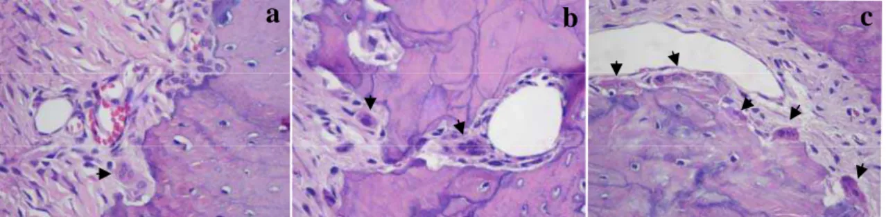
eosinophilic cytoplasm, were considered osteoclasts (Fig. 1).
Fig.1. Osteoclast count. (a) H&E (40x); black line points out the surface, from buccal (B) to palatal crest (P), where osteoclasts were counted. (b) H&E (400x); osteoclast (arrow) attached to alveolar bone (AB) surface; (PL) periodontal ligament.
Trabecular bone density
Trabecular bone density was evaluated in the H&E sections in four alveolar
bone fields (apical, interradicular, buccal and palatal). The histological images were
digitized with a x10 objective and analyzed according to Mahl and Fontanella,16 using
Adobe Photoshop CS3 software (Adobe Systems Inc., San Jose, CA, USA). The extract
filter discarded the structures not corresponding to alveolar bone, and the histogram
function calculated total bone area in pixels. The extract filter was again used to select
only the bone trabeculae, discarding the image of medullary spaces. The histogram
function calculated trabecular area (Fig. 2). Trabecular bone density was considered the
percentage of trabecular area in each field, calculated using the following formula:
trabecular area x 100 / total bone area.
P
PL
AB
B
Fig.2. Trabecular density, H&E (x10 objective). (a), (b) Elimination of areas not corresponding to alveolar bone; (c) elimination of image of medullary spaces; (d) trabecular area quantification.
Collagen fiber density in the medullary spaces of alveolar bone
Collagen fiber density in the medullary spaces was evaluated in the
picrosirius-stained sections in three bone fields (apical, buccal and palatal), using a x20 objective.
Adobe Photoshop CS3 software was used to select only the medullary spaces in each
field, with the extract filter. The semi-automated segmentation method65, 66 was used
with Image Pro Plus 4.5.1 software (Media Cybernetics Inc., Bethesda, MD, USA) for
collagen fiber area quantification. As collagen fibers stained with picrosirius show a red
color, the red areas were selected with the function measure – count/size. After that, a
mask applied to the image converted it into black and white, and the white areas,
corresponding to collagen fibers, were quantified in 2. The same procedure was
performed to quantify total area (Fig. 3). Collagen fiber density was considered the
b
d
a
percentage of collagen area in each field, calculated using the following formula:
collagen area x 100 / total area of medullary spaces.
Fig.3. Collagen fiber density, picrosirius (x20 objective). (a) Medullary spaces previously selected; (b) selection of collagen-positive areas; (c) quantification of collagen-positive areas.
a
b
OPG expression
OPG expression was assessed in four alveolar bone fields (apical, interradicular,
buccal and palatal), using a x20 objective, with the semi-automated method.30 Alveolar
bone areas expressing OPG showed an intense brown color. The brown areas were
selected with the function measure – count/size. A mask applied to the image converted
it into black and white. The white areas, corresponding to OPG-positive structures, were
quantified in 2. The same procedure was performed to quantify total area (Fig. 3).
Immunohistochemical expression of OPG in alveolar bone was calculated using the
Fig.4. OPG immunohistochemical expression (x20 objective). (a) Measure-count/size function; (b) selection of OPG-positive areas; (c) quantification of OPG-positive areas.
b
a
Statistical Analysis
The data were analyzed by descriptive statistics, and the comparison of the variables
tested between the three groups was performed using ANOVA and Tukey’s test, at a
5% level of significance.
RESULTS Osteoclast count
Table I displays the results of osteoclast counts in the H&E stained sections of alveolar
bone surface. Although the number of osteoclasts was higher in the control group than
in alendronate and zoledronic acid groups, there was no statistically significant
difference between the three groups analyzed (ANOVA; P=0.208). Figure 5 illustrates
osteoclast counting on alveolar bone.
Table I: Osteoclast count on alveolar bone surface
Group Osteoclast number
n Mean SD Minimum Maximum
Alendronate 11 9.09 6.25 0.00 18.00
Zoledronic Acid 10 11.20 5.25 0.00 17.00
Control 10 14.0 9.87 1.00 19.00
SD=Standard deviation; n=Sample size ANOVA (=0.05); P=0.208
Fig.5. Histological sections showing osteoclast counting (arrows) on alveolar bone surface from (a) alendronate, (b) zoledronic acid and (c) control groups (H&E, 400x).
c
b
Trabecular bone density
Regardless of the bone fields evaluated, trabecular density was significantly higher in
the zoledronic acid group, when compared to control (ANOVA and Tukey’s test;
P=0.038). There was no significant difference between the zoledronic acid and
alendronate groups, nor between the alendronate and control groups. Regardless of the
group evaluated, the apical field showed significantly lower trabecular density when
compared to the interradicular, buccal and palatal ones (ANOVA; P<0.001) (Table II;
Fig. 6).
Table II: Trabecular density of alveolar bone
Bone field
Group
Total Alendronate Zoledronic Acid Control
Mean (%) SD Mean (%) SD Mean (%) SD Mean (%) SD
Apical 91.20 5.13 91.94 3.01 87.67 5.86 90.39B 4.97
Interradicular 94.70 5.80 98.07 1.48 95.07 3.22 95.74A 4.25
Buccal 98.07 1.18 98.03 1.14 93.57 13.01 96.66A 7.33
Palatal 98.85 0.55 98.08 1.40 98.36 0.64 98.46A 0.95
Total 95.57ab 4.91 96.45a 3.30 93.67b 8.03 95.27 5.73
SD=Standard deviation
ANOVA; Tukey’s test (=0.05) - Means followed by different upper case letters in the column are significantly different (P<0.001). Means followed by different lower case letters in the row are
significantly different (P=0.038).
Fig.6. Histological sections showing alveolar bone trabecular density in (a) alendronate (b) zoledronic acid, and (c) control groups (H&E, 100x).
Collagen fiber density in medullary spaces of alveolar bone
There was no significant difference in collagen fiber density in medullary spaces
between the alendronate, zoledronic acid and control groups, nor between the apical,
buccal and palatal bone fields (ANOVA; P>0.05) (Table III; Fig. 7).
Table III: Collagen fiber density in medullary spaces of alveolar bone
Bone field
Group
Total Alendronate Zoledronic Acid Control
Mean(%) SD Mean(%) SD Mean(%) SD Mean(%) SD
Apical 42.05 10.91 39.88 13.08 37.45 9.11 39.87 10.85 Buccal 50.4 14.17 38.59 11.61 42.75 13.56 44.02 13.45 Palatal 46.72 17.75 45.84 23.84 37.38 8.90 42.96 16.49 Total 45.67 13.99 40.98 15.36 39.06 10.48 41.98 13.38 SD=Standard deviation
ANOVA (=0.05); P>0.05
Fig.7. Histological sections showing collagen fiber in medullary spaces stained by picrosirius (red) in (a) alendronate, (b) zoledronic acid and (c) control groups (picrosirius, 200x).
OPG expression
There was no significant difference in immunohistochemical expression of OPG on
alveolar bone between the alendronate, zoledronic acid and control groups (ANOVA;
P>0.05). Regardless of the group evaluated, the interradicular field demonstrated
significantly higher OPG expression than the other fields (ANOVA and Tukey’s test;
P=0.003) (Table IV; Fig. 8).