225
Estudo da regeneração nervosa em nervos tibiais de ratos
Wistar utilizando o Fluoro-Gold
®
como marcador neuronal.
A study on nerve regeneration in tibial nerves of Wistars rats,
using Fluoro-Gold
®as a neuronal tracer
MARCO AURÉLIO SERTÓRIO GRECCO
1, VILNEI MATTIOLI LEITE
2, WALTER MANNA ALBERTONI
3,
JOÃO BAPTISTA GOMES DOS SANTOS
4, CELSO KIYOSHI HIRAKAWA
5, FLÁVIO FALOPPA
6, SANDRA GOMES VALENTE
7.
A
RTIGOO
RIGINALTrabalho recebido em 21/05/2003. Aprovado em 27/07/2003
Trabalho realizado no Laboratório de Microcirurgia da Disciplina de Cirurgia da Mão e Membro Superior do Departamento de Ortopedia e Traumatologia e nos Laboratórios da Disciplina de Neurologia Experimental do Departamento de Neurologia e Neurocirurgia da Unifesp-EPM.
1. Médico; Mestre em Ortopedia e Traumatologia
2. Professor Livre-Docente; Chefe da Disciplina de Cirurgia da Mão e Membro Superior
3. Professor Titular; Chefe do Departamento de Ortopedia e Traumatologia 4. Médico assistente da Disciplina de Cirurgia da Mão e Membro Superior; Mestre em Ortopedia e Traumatologia
5. Professor Livre-Docente, coordenador do Curso de Pós-graduação
6. Doutor em Medicina, Chefe de Clínica na Disciplina de Cirurgia da Mão e Membro Superior do Departamento de Ortopedia e Traumatologia da UNIFESP/EPM 7. Bióloga; Mestre em Neurociências.
Endereço para correspondência: Rua Borges Lagoa, 786 – Cep 04038-002 – São Paulo, SP. E-mail: [email protected]
RESUMO
Os autores estudaram em ratos a regeneração nervosa no enxerto nervoso tradicional, comparando-os com grupo contro-le, através da contagem no nível da medula espinhal, entre L3 e S1, de motoneurônios marcados por meio de exposição do ner-vo tibial ao Fluoro-Gold® (FG). No grupo controle, ambos os
nervos tibiais foram expostos ao FG®, e em 48 horas, após a
perfusão, os motoneurônios foram contados. No grupo experi-mental, foi ressecado um segmento de 8mm do nervo tibial cri-ando uma falha segmentar. O segmento de nervo de um foi usado para reparar o lado contralateral, utilizando sutura epi-neural. Após quatro meses, o nervo tibial direito foi exposto ao FG após o enxerto nervoso e, o esquerdo, antes do enxerto. Os motoneurônios foram contados após a perfusão. Ambas as medulas foram cortadas em segmentos de 40µm. Para a
aná-lise estatística foram utilizados testes de Wilcoxon e Student. O grupo controle apresentou um significativo aumento de moto-neurônios quando comparado ao experimental. No grupo ex-perimental, o número de motoneurônios foi significantemente maior, quando a exposição do nervo tibial ao FG®. era feita
dis-talmente ao enxerto nervoso. O enxerto nervoso funciona como um bloqueio parcial para a migração dos axônios em regenera-ção.
Descritores Descritores Descritores Descritores
Descritores: Transplante autólogo; Regeneração nervosa; Ner-vos periféricos; Ratos wistar; Corantes fluorescentes.
INTRODUÇÃO
As lesões do sistema nervoso periférico têm sido alvo de inúmeros estudos para compreensão e entendimento do
pro-SUMMARY
The authors studied, in rats, of nerve regeneration in nerve grafts comparing with control group using Fluoro-Gold® (FG)
la-beled motor neurons count in spinal cord. In control group both tibial nerves were exposed to FG® and motor neurons counted in spinal cord in 48 hours, after perfusion, in a medular segment from L3 to S1. In experimental group, ressecting 8 mm, a gap was created in both tibial nerves and nerve segment of one ner-ve was used to repair contralateral side in a traditional nerner-ve graft suture. After four months, right tibial nerve was exposed to FG® distal to graft and left tibial nerve exposed to FG proximal to graft. In 48 hours, after perfusion, motoneurons were counted in a medular segment from L3 to S1. In both groups medular seg-ment was cut in 40 µm slices and all labeled cells counted. Wil-coxon and Student tests were used for statistical analysis. Con-trol group presented a significative increased number of moto-neurons when compared to experimental group. In experimental group number of motoneurons was significantly decreased when tibial nerve was exposed to FG® distal to nerve graft. Nerve graft promoted a partial block to axon migration after four months of surgery.
Key words: Key words: Key words: Key words:
Key words:Transplantation autologous; Nerve regeneration; Pe-ripheral nerves; Wistar rats; Dyes fluorescents.
Work performed at the Laboratory of Microsurgery, Discipline of Hand and Upper Limb Surgery, Department of Orthopedics and Traumatology, and at the Laboratories of the Discipline of Experimental Neurology, Department of Neurology and Neurosurgery, Unifesp - EPM. Abstract of Master’s Thesis within the Post-Graduation Program in Orthopedics and Traumatology, Unifesp - EPM
1. Doctor; Master’s degree in Orthopedics and Traumatology 2. Full Professor; Head, Discipline of Hand and Upper Limb Surgery 3. Chairman Professor; Head, Department of Orthpedics and Traumatology 4. Assistant Doctor, Discipline of Hand and Upper Limb Surgery; Master’s degree in Orthopedics and Traumatology
5. Full Professor; Coordinator, Post Graduation Course
6. PhD of Medicine; Discipline of Hand and Upper Limb Surgery, Department of Orthopedics and Traumatology, UNIFESP/EPM
7. Biologist; Master’s degree in Neuroscience.
Address: Rua Borges Lagoa, 786 - São Paulo, SP 04038-002 - Brasil. E-mail: [email protected]
INTRODUCTION
226
cesso de reparação nervosa, pois podem ocasionar transtor-nos importantes à função dos membros superiores e inferiores. O método de sutura epineural foi descrito por Hueter (1873) e o primeiro enxerto nervoso experimental por Philipeau, Vulpi-an (1886)(10).
Graças à implantação de técnicas e materiais microcirúrgi-cos, ocorreu um aperfeiçoamento na reconstrução das estrutu-ras lesadas, com melhora da recuperação funcional do nervo(14)
O enxerto nervoso foi apresentado como uma das formas de tratamento de lesões nervosas com falha segmentar e sutu-ras epineurais sob tensão(10,11,12) e a sua utilização tem
melhora-do significativamente com o emprego de técnicas microcirúrgi-cas(5). Entretanto, outros autores(13), não
acreditam nos resultados do emprego de tais técnicas.
Como vimos, o enxerto nervoso é em-pregado desde o final do século XIX. Por esse motivo, resolvemos estudar a regene-ração nervosa através da contagem de mo-toneurônios marcados pelo FG® por meio
de: comparação entre o número de moto-neurônios marcados em animais do grupo controle e o número de motoneurônios em animais do grupo experimental, onde foi fei-to o enxerfei-to nervoso; comparação entre o número de motoneurônios marcados atra-vés da exposição ao FG®, realizada
proxi-mal e distalmente ao enxerto nervoso nos animais do grupo experimental, e avaliação para verificar se há bloqueio da regenera-ção nervosa através do enxerto nervoso
MATERIAL E MÉTODOS
Usamos 28 ratos da raça Wistar, ma-chos, como grupo controle. Foram usados 14 animais adultos com peso médio de 298g e com idade média de cinco meses e 14 ratos com peso médio de 155g, ecom idade média de um mês, como grupo ex-perimental.
Cada animal recebeu anestesia intrape-ritoneal com solução anestésica compos-ta por hidrato de cloral (4,25g), sulfato de magnésio (2,25g), propilenoglicol (4,28ml), álcool etílico absoluto (11,5ml) e água des-tilada (45,7ml). (Figura 1).
Fez-se uma incisão póstero-lateral em ambas as coxas de cada pata. Utilizou-se microscópio e instrumental microcirúrgico para dissecar o nervo ciático e suas ramifi-cações sural, peroneiro e tibial. (Figura1). No grupo controle, operamos os dois mem-bros inferiores, expondo o nervo tibial de cada perna, seccionando-os e expondo-os imediatamente ao marcador neuronal FG®
a 3%, por um período de uma hora e 30 minutos. No grupo experimental, operar-mos os dois membros inferiores, expondo
Figura 1 – Nervo ciático com seus ramos sural, peroneiro e tibial.
Figure 1 - Sciatic nerve showing its sural, peroneal and tibial ramifications.
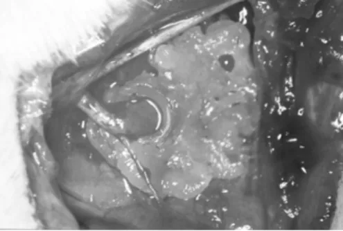
Figura 2 –Sutura do enxerto de nervo com ampliação de 25x ao microscópio cirúrgico.
Figure 2 -Nerve graft suture (surgical microscope, 25x).
Figura 3 – Nervo tibial exposto ao FG®. Observa-se isolamento de vaselina com calha de polietileno ao centro e com a extremidade
do nervo mergulhado em FG® .
Figure 3 - FG® -exposed tibial nerve. Vaseline was isolated using a polyethylene trough in the center, with the nerve end immersed in FG®.
The epineural suture method was described by Hueter in 1873, and the first experimental nerve grafting was performed by Vulpi-an Philipeau in 1886 (10).
Thanks to the implementation of microsurgical techniques and materials, the reconstruction of injured structures could be im-proved, and nerve functions could be recovered(14).
Nerve graft was introduced as one of ways of managing ner-ve injuries with segment defect and epineural sutures under stress
(10,11,12) and its use has significantly improved using microsurgical
techniques (5). However, other authors (13) do not believe in the
results of such techniques.
As mentioned before, nerve grafting has been employed sin-ce the end of 19th sin-century. This is the rea-son why we decided to investigate nerve regeneration by count FG®-labeled
moto-neurons and comparing the number of la-beled motoneurons in animals of the con-trol group and the experimental group that subjected to nerve grafting; also, a compa-rison was made between the number of la-beled motoneurons through exposure to FG® performed proximally and distally to the
nerve graft in the animals of the experimen-tal group, and also an evaluation to see whether the nerve regeneration was blocked by the nerve graft.
MATERIAL AND METHODS
Twenty-eigth male Wistar rats were used as a control group. The experimental group consisted of 14 adult animals weighing on average 298 g, with mean age of five mon-ths, plus 14 rats weighing on average 155 g, with mean age of one monthl.
Each animal was intraperitoneally ane-thesized with an anaesthetic solution formed by 4.25 g choral hydrate, 2.25 g magnesium sulfate, 4.28 ml propylenglycol, 11.5 ml ethyl alcohol, anhydrous, and 45.7 ml distilled water (Figure 1).
Posterolateral incisions were performed, one in each thigh of the rats. To dissect the sciatic nerve and its sural, peroneal and ti-bial ramifications, a microscope and surgi-cal instrumentation were used (Figure 1). We operated both lower limbs of each rat in the control group, exposing the tibial nerve of each hind leg, sectioning and immedia-tely exposing them to a 3% solution of neu-ronal tracer FG® for one hour and thirty
227
o nervo tibial de cada lado e ressecando um segmento de 8mm do nervo, simulando, ao mesmo tempo, a perda de substância e a obtenção do enxerto nervoso autólogo. O segmento de nervo de um foi usado para reparar o lado contralateral, utilizando-se su-tura epineural (Figura 2). Após quatro me-ses, os animais foram submetidos à nova cirurgia para exposição dos nervos tibiais ao marcador neuronal FG® (Figura 3).O
vo tibial direito foi corado após o enxerto ner-voso e o esquerdo antes do enxerto. Após 48 horas os animais de ambos os grupos foram novamente anestesiados e perfundi-dos com 50ml de solução salina isotônica intra-cardíaca para limpeza do sistema vas-cular, 200ml de “solução de perfusão” (4% paraformaldeído/“buffer” fosfato) rapida-mente em 8 a 10 minutos, 300ml de “solu-ção de perfusão”, lentamente, em 20 minu-tos, 200ml de “buffer” sacarose a 10%, ra-pidamente, em oito a 10 minutos, 300ml de “buffer” sacarose a 10%, lentamente, em 20 minutos.
Ao término desse procedimento, com o animal em decúbito ventral, realizou-se a la-minectomia dorsal e lombar. Após a exposi-ção da medula espinhal, ressecou-se o seg-mento correspondente de L3 a S1. Marcou-se, então, um sulco na região dorsal deste segmento para indicar o lado direito.
A peça foi colocada em micrótomo e fa-tiada em cortes de 40 mm, montadas em lâminas, deixadas secando na geladeira por 24 horas. As lâminas prontas foram exami-nadas em microscópio ZEISS-AXIOLAB com luz fluorescente para evidenciar o FG®.
Exa-minou-se todos os cortes em ampliações de 100 e 400 vezes e contou-se os neurônios motores marcados(Figura 4).
Realizou-se a análise estatística aplican-do-se os testes não-paramétrico de Wilco-xon sinalizado, e o teste paramétrico t-Stu-dent. O nível de significância considerado para os testes foi de 5% (p< 0,05).
RESULTADOS
O número de motoneurônios, marcados com FG®, nos animais do grupo controle é
maior que no grupo experimental submeti-do ao enxerto nervoso, tanto proximal como distal. Não há diferença no número de mo-toneurônios marcados, entre os lados direi-to e esquerdo, nos animais do grupo con-trole (Tabela 2). O número de motoneurôni-os, nos animais do grupo experimental, é maior quando a marcação com FG® é feita
proximal ao enxerto nervoso (Tabela 1).
Figura 4 – Secção de medula com ampliação de 25X ao microscópio. Observam-se células
marcadas pelo FG® .
Figure 4 - Medullar section under the microscope (25x). FG-labeled cells can be seen.
Tabela 1 – Número de células absolutas contadas no lado direito e esquerdo; valores
totais e média; em animais do grupo experimental
Table 1 - Absolute number of cells counted on the right and left sides of animals in the experimental group; total values and average
value
Tabela 2 – Número de células absolutas contadas no lado direito e esquerdo; valores totais e média; em animais do grupo controle
Table 2 - Absolute number of cells counted on the right and left sides of animals in the
control group; total and average values
p< 0,001 há diferença estatística entre eles, sendo que o lado esquerdo é maior. Fonte: DOT-UNIFESP/EPM
p< 0,490, portanto não existe diferença estatística entre os lados. Fonte: DOT-UNIFESP/EPM
suture (Figure 2). Four months later, the animals underwent another surgery in or-der to have their tibial nerves exposed to neuronal tracer FG® (Figure 3). The right
tibial nerve was stained after the nerve grafting, while the left tibial nerve was stai-ned before the nerve grafting. 48 hours later the animals of both groups were anesthetized again and received a quick (8 to 10-minute) perfusion of a mixture of 50 ml of intracardiac isotonic saline solu-tion (to clean their vascular systems) and 200 ml of “perfusion solution” (4% para-formaldehyde/phosphate buffer); a slow (20-minute) perfusion of 300 ml of “perfu-sion solution”; a quick (8 to 10-minute) perfusion of 200 ml of 10% saccharin bu-ffer; and a slow (20-minute) perfusion of 300 ml of 10% saccharin buffer solution. After this procedure, dorsal and lum-bar laminectomies were performed with the animal in the ventral decubitus positi-on. After exposing the spinal cord, the segment corresponding to L3-S1 was re-sected. A groove was inscribed in the dorsal region of this segment, to indicate the right side.
The specimen was placed in a micro-tome and sliced into 40-mm sections that were mounted and left for 24 hours in the refrigerator to dry. A Zeiss Axiolab fluo-rescence biological microscope was used to examine the sections in order to microscopically evidence the FG. All sec-tions were magnified 100x and 400x, and the count of labeled motoneurons was performed(Figure 4).
The statistical analysis included the signalized non-parametric Wilcoxon test and the Student’s t parametric test. The significance level considered for the tes-ts was of 5% (p< 0.05).
RESULTS
The number of FG® -labeled
motoneu-rons in the animals of the control group is higher as compared with the experimen-tal group that underwent nerve graftings, both proximal and distal. The animals in the control group showed no difference in the number of labeled motoneurons between the left and right sides (Table 2). The number of motoneurons in the expe-rimental group animals is higher when FG® labeling is performed proximal to the
228
DISCUSSÃO
Comumente, em reparo nervoso de lesões de nervos perifé-ricos, a neurorrafia término-terminal é a ideal e a que apresenta melhores resultados, com o que praticamente todos os auto-res, citados em nosso trabalho, concordam. Entretanto, no que tange à lesão nervosa com perda de substância e à sutura sob tensão, vários tipos de enxertos, biológicos e não biológicos, podem ser empregados. Dentre esses, o enxerto nervoso autó-geno é o mais utilizado desde o final do século XIX, considera-do assim o “Gold Standard”(8) .
A despeito da sua ampla utilização, os resultados funcio-nais têm sido inferiores aos da sutura término-terminal(10,11,12),,,,,
mesmo com o desenvolvimento e emprego de técnicas micro-cirúrgicas(14).
Foram usados ratos da raça Wistar, por serem conhecidos biologicamente e estudados há muito tempo pela comunidade científica(4).
Fatores resultantes da lesão do nervo ciático, tais como, úl-ceras de pele(7), autofagia das partes insensíveis(6) e a
dificulda-de do rato em manter-se em pé para a alimentação e por se tratar de nervo puramente motor, nos levaram à utilização do nervo tibial. Utilizou-se enxerto nervoso de 8mm de comprimen-to, respeitando-se o limite de 10mm da regeneração em tubos para ratos(6,9).
O método utilizado para estudar a regeneração nervosa foi através da marcação neuronal retrógrada com FG®, na
concen-tração de 3%, Este marcador é fluorescente e permite sua ob-servação direta ao microscópio sob uso de filtros(6).
Ao analisarmos os resultados (Tabela 3), constatamos dife-renças nas contagens celulares em todos os animais, porém, evidenciamos proporções semelhantes, coincidindo com os achados da literatura(1,6,9),com alguns autores concordando que
a individualidade de cada animal influencia a contagem(1,6)
Utilizaram-se dois métodos estatísticos para definir se havia diferença significativa entre as técnicas empregadas. Os testes empregados foram t-Student e Wilcoxon sinalizado.
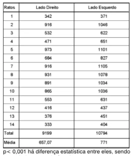
Na contagem neuronal dos animais do grupo de experiên-cia, averiguamos que o lado esquerdo apresentou média de 771 motoneurônios (máximo de 1078 e mínimo de 371) e o di-reito 657,07 (máximo de 931 e mínimo de 342), ou seja, o lado direito apresentou, em média, 14,78% menos motoneurônios que o esquerdo (Tabela 3).
De acordo com estudo realizado por Sweet et al.(15), o
nú-mero de motoneurônios do nervo tibial foi de 982 ± 36, utilizan-do coloração retrógrada com HRP, o que está de acorutilizan-do com o observado no grupo controle (Tabela 1).
Encontramos, em média, 20%, menos motoneurônios mar-cados do nervo tibial do lado esquerdo dos animais do grupo experimental (com a marcação neuronal realizada proximal ao enxerto nervoso), quando comparamos com o número do gru-po controle.
Isso significa que houve uma diminuição no número de cé-lulas, em decorrência de uma lesão nervosa isolada, na qual não incidiu interferência técnica do cirurgião, mostrando uma possível morte celular. Portanto, este nervo não será igual ao lado não lesado (nervo tibial normal), havendo uma dificuldade para o retorno à função normal do nervo após uma neurorrafia. Altissimi et al.(2) analisaram a recuperação da sensibilidade em
DISCUSSION
Usually, as regards nerve repair of peripheral nerves, a termi-no-terminal neurorrhaphy is the ideal process, presenting the best results, a fact agreed by most authors quoted in this study. Ho-wever, as regards nerve injuries with loss of substance and sutu-re under stsutu-ress, various types of grafts may be used, either biolo-gical or non-biolobiolo-gical. Among these, since the end of nineteen-th century nineteen-the most frequently used is nineteen-the nerve allograft, which is then considered the gold standard (8).
Although these grafts are widely employed, their functional results have shown to be inferior to those achieved with termino-terminal suture (10,11,12), even after the development and use of of
microsurgical techniques (14).
We used Wistar rats because they are biologically known and studied for a long time by the scientific community (4).
Factors resulting from the sciatic nerve injury such as skin ulcers (7), autophagia of the non-sensible parts (6) and the
difficul-ty in standing up on their hind legs in order to eat, besides the fact that this is a purely motor nerve, led us to employ the tibial nerve. An 8-mm long nerve graft was used, in accordance to the limit of 10 mm for tubular regeneration in rats (6,9).
The method employed to study nerve regeneration consisted of retrograde labeling of neurons with a 3% FG® solution Since
this is a fluorescent tracer, it may be directly observed at the mi-croscope through filters (6).
Although the analysis of results (Table 3) showed certain di-fferences in the cell counts in all animals, similar proportions were evidenced that agreed with results found in literature (1,6,9),where
some authors agree that the individuality of the animals will influ-ence the count(1,6).
Two statistical methods were used to define whether a signi-ficant difference would be found between the techniques em-ployed. Such tests were: Student’s t test and the signalized Wil-coxon test.
The neuronal count performed in the experimental group ani-mals showed an average of 771 motoneurons (maximum 1,078, minimum 371) on the left side, and an average of 657.07 (maxi-mum 931, mini(maxi-mum 342) on the right side; this means that, on the average, the right side presented 14.78% less motoneurons as compared with the left side (Table 3).
According to Sweet et al (1986), who used the retrograde HRP (horseradish peroxydase) staining method, the tibial nerve presented 982 ± 36 motoneurons, in agreement with the num-ber observed in the control group (Table 1).
On the average, we found 20% less tibial nerve labeled mo-toneurons on the left side of the animals of the experimental group (in which neuronal labeling was performed proximal to the nerve graft) as compared with the control group.
This means that the number of cells had decreased due to an isolated nerve injury, with no technical interference on the part of the surgeon, showing a possible cell death. Therefore, this nerve will not be equal to that on the non-injured side (which is a normal tibial nerve), so after a neurorraphy the nerve will hardly recover its normal function. Altissimi et al(2) analyzed the
reco-very of sensibility in digital nerves, when only 13% of patients returned to normal.
229
nervos digitais e apenas 13% dos pacientes chegaram ao nor-mal.
No que se refere à contagem de motoneurônios do nervo tibial normal (perna esquerda do controle), comparada com a contagem realizada após o enxerto (perna esquerda do animal de experiência), observamos uma perda aproximada de 35%. Esta perda seria decorrente de 14,78% do próprio enxerto, so-mado à possível morte de motoneurônios (20%) ocasionada pela própria lesão.
Isso denota uma perspectiva ruim de regeneração nervosa após a utilização do enxerto nervoso. Mediante esses resulta-dos por nós obtiresulta-dos, parece-nos que o “Gold Standard” ainda não é ideal.
Fatores como tensão no sítio de sutura, tecido cicatricial e dois sítios de suturas podem ter influenciado o resultado da re-paração nervosa(10,11,12).
Talvez, o mais importante deste trabalho diz respeito aos 20% de morte neuronal observada após quatro meses da le-são, quando comparamos a contagem de neurônios marcados proximais ao enxerto com os neurônios marcados no grupo controle. Essa redução de número de neurônios associada às alterações topográficas de reinervação devem ser os responsá-veis pelos maus resultados das neurorrafias, o que nos leva a propor a utilização de substâncias neurotróficas e neurotrópi-cas, com o objetivo de minimizar essa perda celular e reorientar os axônios.
CONCLUSÕES
Observou-se maior número de motoneurônios marcados em nervos tibiais, não submetidos a enxerto nervoso, expostos ao FG®, quando comparados àqueles submetidos ao enxerto
ner-voso. Notou-se menor número de motoneurônios na medula espinhal, quando a exposição do nervo tibial ao FG® era feita
distalmente ao enxerto nervoso. Essa diminuição de motoneu-rônios, quando a exposição do nervo tibial ao FG® era feita
dis-talmente ao enxerto nervoso, mostra que o enxerto nervoso fun-ciona como um bloqueio parcial para a migração dos axônios em regeneração.
REFERÊNCIAS BIBLIOGRÁFICAS
1. Aldskogius H, Thomander L. Selective reinnervation of somatotopically appro-priate muscles after facial nerve transection and regeneration in the neonatal rat. Brain Res 375:126-134, 1986.
2. Altissimi M, Mancini GB, Azzarà A. Results of primary repair of digital nerves. J Hand Surg Br 16:546-547, 1991.
3. Fields RD, Le Beau JM, Longo FM, Ellisman MH. Nerve regeneration through artificial tubular implants. Prog Neurobiol 33:87-134, 1989.
4. Greene EC. Anatomy of the rat. New York: Hafner, 1963. p. 370.
5. Kalomiri DE, Soucacos PN, Beris AE. Nerve grafting in peripheral nerve micro-surgery of upper extremity. Micromicro-surgery 15:506-511, 1994.
6. Leite VM. Estudo do comportamento das lesões altas do nervo ciático através da utilização de Fluoro-Gold e Horseradish Peroxidase como marcadores neuro-nais. [Livre Docência]. São Paulo: Escola Paulista de Medicina; 1994. 7. Lolley RD, Bose WJ, Bastian F, Bassam B, Meyer FN, Anderson LD. Vein silas-tic, and polyglycolic acid fine mesh: a comparative study in peripheral nerve re-pair. Ann Plast Sur35:266-271, 1995.
8. Mackinnon SE, Dellon AL. Comparison of nerve regeneration across a sural nerve graft and a vascularized pseudosheath. J Hand Surg Am 13:935-942, 1988. 9. Madorsky SJ, Swett JE, Crumley RL. Motor versus sensory neuron regenerati-on through collagen tubules. Plast Recregenerati-onstr Surg 102:430-436, 1998. 10. Millesi H. The interfascicular nerve grafting of the median and ulnar nerves. J Bone Joint Surg Am 54:727-748, 1972.
11. Millesi H. Further experience with interfascicular grafting of the median, ulnar and radial nerves. J Bone Joint Surg Am 58:209-218, 1976.
12. Millesi, H. Forty-two years of peripheral nerve surgery. Microsurgery 14:228-233, 1993.
13. Omer GE. Injuries to nerves of the upper extremity. J Bone Joint Surg Am 56:1615-1624, 1974.
14. Smith JW. Microsurgery of nerves peripheral. Plast ReconstrSurg 33:317-329, 1964.
15. Sweet JE, Wikholm RP, Blanks RHI, Sweet A, Conley L. Motoneurons of rat sciatic nerve. Exp Neurol 93:227-252,1986.
as compared with the post-grafting count (left arm of the experi-mental animal). Such loss might be due to 14.78% of the graft, added to the possible death of motoneurons (20%) caused by the injury itself.
This indicates a poor perspective of nerve regeneration after the use of a nerve graft. Based on our findings, it seems to us that the gold standard is not yet the optimum standard.
Factors such as stress at the suture site, cicatricial tissue, and two suture sites may have influenced the result of the nerve repair (10,11,12).
Maybe the most important aspect of this study is the percen-tage of neuronal death (20%) observed four months after the in-jury, if we make a comparison between the count of labeled neu-rons proximal to the graft and the labeled neuneu-rons in the control group. This reduction in the number of neurons, plus the topo-graphical alterations in reinnervation must account for the poor neurorraphic results obtained, which led us to propose the use of neutrophic and neurotropic substances, with the purpose of minimizing such cell loss and redirect the axons.
CONCLUSIONS
The number of tibial nerve labeled motoneurons that did not receive nerve grafts and were exposed to Fluoro-Gold® was
hi-gher as compared to those that did receive nerve grafts. The spinal cord showed less motoneurons when the tibial nerve was exposed to Fluoro-Gold® distal to the nerve graft. This reduction
in the number of motoneurons, when the exposure of the tibial nerve to Fluoro-Gold® was performed distally to the nerve graft