The Molecular Epidemiology and Evolution of
Murray Valley Encephalitis Virus: Recent
Emergence of Distinct Sub-lineages of the
Dominant Genotype 1
David T. Williams1
*, Sinéad M. Diviney2¤a
*, Aziz-ur-Rahman Niazi2¤b, Peter A. Durr1, Beng
Hooi Chua3, Belinda Herring4¤c, Alyssa Pyke5, Stephen L. Doggett6, Cheryl A. Johansen7,
John S. Mackenzie2
1CSIRO, Australian Animal Health Laboratory, Geelong, Victoria, Australia,2Faculty of Health Sciences, Curtin University, Perth, Western Australia, Australia,3Office of Research and Development, Curtin University, Perth, Western Australia, Australia,4Infectious Diseases and Immunology, University of Sydney, New South Wales, Australia,5Public Health Virology, Queensland Health Forensic and Scientific Services, Coopers Plains, Queensland, Australia,6Department of Medical Entomology, Westmead Hospital, University of Sydney and Institute for Clinical Pathology and Medical Research, New South Wales, Australia,
7Arbovirus Surveillance and Research Laboratory, School of Pathology and Laboratory Medicine, University of Western Australia, Perth, Western Australia, Australia
¤a Current address: CSIRO, Australian Animal Health Laboratory, Geelong, Victoria, Australia ¤b Current address: Department of Infectious Diseases, Faculty of Medicine, Herat University, Herat, Afghanistan
¤c Current address: School of Biomedical Sciences, Faculty of Health, Queensland University of Technology, Brisbane, Australia
*d.williams@csiro.au(DW);sinead.diviney@googlemail.com(SMD)
Abstract
Background
Recent increased activity of the mosquito-borne Murray Valley encephalitis virus (MVEV) in Australia has renewed concerns regarding its potential to spread and cause disease.
Methodology/Principal Findings
To better understand the genetic relationships between earlier and more recent circulating strains, patterns of virus movement, as well as the molecular basis of MVEV evolution, com-plete pre-membrane (prM) and Envelope (Env) genes were sequenced from sixty-six MVEV strains from different regions of the Australasian region, isolated over a sixty year period (1951–2011). Phylogenetic analyses indicated that, of the four recognized
geno-types, only G1 and G2 are contemporary. G1 viruses were dominant over the sampling period and found across the known geographic range of MVEV. Two distinct sub-lineages of G1 were observed (1A and 1B). Although G1B strains have been isolated from across mainland Australia, Australian G1A strains have not been detected outside northwest Aus-tralia. Similarly, G2 is comprised of only Western Australian isolates from mosquitoes, sug-gesting G1B and G2 viruses have geographic or ecological restrictions. No evidence of
OPEN ACCESS
Citation:Williams DT, Diviney SM, Niazi A-u-R, Durr PA, Chua BH, Herring B, et al. (2015) The Molecular Epidemiology and Evolution of Murray Valley Encephalitis Virus: Recent Emergence of Distinct Sub-lineages of the Dominant Genotype 1. PLoS Negl Trop Dis 9(11): e0004240. doi:10.1371/journal.pntd.0004240
Editor:David W.C. Beasley, University of Texas Medical Branch, UNITED STATES
Received:May 22, 2015
Accepted:October 26, 2015
Published:November 24, 2015
Copyright:© 2015 Williams et al. This is an open access article distributed under the terms of the
Creative Commons Attribution License, which permits unrestricted use, distribution, and reproduction in any medium, provided the original author and source are credited.
Data Availability Statement:All sequence data are available from the NCBI GenBank database (http:// www.ncbi.nlm.nih.gov/genbank). Accession numbers are listed inS1 Tableof the manuscript. All other relevant data are within the paper and its Supporting Information files.
recombination was found and a single amino acid substitution in the Env protein (S332G) was found to be under positive selection, while several others were found to be under direc-tional evolution. Evolutionary analyses indicated that extant genotypes of MVEV began to diverge from a common ancestor approximately 200 years ago. G2 was the first genotype to diverge, followed by G3 and G4, and finally G1, from which subtypes G1A and G1B diverged between 1964 and 1994.
Conclusions/Significance
The results of this study provides new insights into the genetic diversity and evolution of MVEV. The demonstration of co-circulation of all contemporary genetic lineages of MVEV in northwestern Australia, supports the contention that this region is the enzootic focus for this virus.
Author Summary
Murray Valley encephalitis virus is the most significant cause of mosquito-borne encepha-litis in humans in Australia, and can also cause neurological disease in horses. This study reports an expanded phylogenetic study of this virus and the first molecular evolutionary analysis. Of the four recognized genotypes of Murray Valley encephalitis virus, only two were found to be actively circulating (genotypes 1 and 2), and genotype 1 was dominant. Distinct genetic sub-lineages within genotype 1 were found to have recently emerged. Molecular clock analysis indicated that genotype 2 viruses are the oldest genetic lineage while genotype 1 viruses are the most recent to diverge. The co-circulation of distinct genetic lineages of this virus in northwestern Australia, comprising the oldest and youn-gest lineages, supports previous findings that MVEV circulates endemically in this region.
Introduction
Murray Valley encephalitis virus (MVEV; genusFlavivirus, familyFlaviviridae) is the most
important cause of arboviral encephalitis in Australia. It is a member of the Japanese encephali-tis (JE) serocomplex, along with other medically important flaviviruses such as JE virus (JEV) and West Nile virus (WNV), which also circulate in the Australasian region [1]. MVEV exists in zoonotic cycles betweenCulexspecies mosquitoes and water birds (reviewed in [2]). It is
enzootic in northern parts of Australia, including the Kimberley region of Western Australia and northern areas of the Northern Territory, with frequent spillovers into the Pilbara and Gascoyne regions of Western Australia, and occasional spread to southern areas of the North-ern Territory and northNorth-ern Queensland [3–5]. MVEV very occasionally spreads into south-eastern Australia following periods of heavy and prolonged rainfall, where it has caused major epidemics in the last century, centred in the Murray Valley region [2,3,6]. Since the last nation-wide epidemic of MVE in 1974, there have been approximately 127 human cases, with the majority occurring in Western Australia or the Northern Territory. Outside Australia, MVEV is believed to be endemic in Papua New Guinea (PNG), where human cases, serological evi-dence of infection, and virus isolations have been reported [7–9]. In addition to human disease, MVEV can cause fatal encephalitis in horses [10,11].
Australia Premier’s Research Fellowship (https:// www.dpc.wa.gov.au/science/FundingPrograms/ Pages/WAFellowshipsProgram.aspx); CAJ received funding from the Western Australian Department of Health (ww2.health.wa.gov.au); SLD was funded by the NSW Ministry of Health (www.health.nsw.gov.au). The funders had no role in study design, data collection and analysis, decision to publish, or preparation of the manuscript.
In recent years, MVEV has become increasingly active in south-eastern Australia, raising concerns that the virus may become established in enzootic transmission cycles in populous regions. Seroconversions to MVEV were found in sentinel chicken flocks in New South Wales in 2001 and 2003 [12], and in 2008 the first human case since the 1974 epidemic occurred in this state along with sentinel chicken seroconversions [13]. Similarly, detections of virus activ-ity in sentinel chicken flocks in Victoria in 2008 were the first since 1974 [14]. An equine case of MVE also occurred in southeast Queensland in 2008 [10]. In 2011, following record levels of rainfall, widespread virus activity was once again observed across Australia, with MVEV detected in all mainland states, resulting in seventeen cases of human disease and numerous equine cases [11,15].
Four genotypes (G1-G4) of MVEV have been identified, based on limited genetic analyses using RNase oligonucleotide mapping [16] or gene sequencing of the E gene [17,18], 5’ termi-nal non-coding region [19], and NS5-3’non-coding region [20]. These studies showed that G1 is the predominant type on mainland Australia. The most recent isolates of MVEV from PNG also belong to G1 [18]. Genotype 2 consists of mosquito isolates from the northeast Kimberley region of northern Western Australia. G2 viruses have not been found outside this area, sug-gesting that this lineage occupies a unique and/or rarely sampled ecological niche [18,21], or may represent an incursion from the Indonesian archipelago [3,19]. Genotypes 3 and 4 com-prise single strains of MVEV from PNG isolated from a human case in 1956 [8] and from mos-quitoes in 1966 [22], respectively.
The single-stranded positive sense RNA genome of MVEV encodes three structural proteins (capsid, pre-membrane [prM] and envelope [Env]) and seven non-structural proteins (NS1, NS2A, NS2B, NS3, NS4A, NS4B and NS5) in the ORF. The prM and Env proteins have impor-tant biological roles in virus particle assembly and virus entry during infection of cells [23]. The Env protein is also the major target of neutralising antibodies during infection [24], and uncleaved prM, present in immature virus particles and released from infected cells, can elicit host immunity [25]. Both prM and Env genes exhibit relatively high levels of sequence varia-tion, allowing robust phylogenetic resolution [26–29]. In the present study, we performed an expanded phylogenetic analysis of MVEV, including sixty-four new MVEV structural gene (prM-Env) sequences, in order to better understand the genetic relationships of earlier isolates with more recent circulating strains. In addition, to gain insights into the temporal and biologi-cal basis of MVEV evolution, molecular clock and selection analyses were undertaken.
Materials and Methods
Viruses
Virus strains were sourced from the culture collections of the authors. In addition, MVEV strains PNG6910, PNG6523 and CY1189 were kindly provided by Prof Roy Hall (University of Queensland). When required, one to two additional passages of virus stocks were performed in porcine stable equine kidney cells (Arbovirus Surveillance and Research Laboratory, University of Western Australia). MVEV strain 611W/WA/08 was identified in post-mortem clinical specimens taken from the lymph node, brain stem, cervical cord and cerebrum of a fatal human MVE case from Kununurra, Western Australia, in April 2008. Virus isolation from these specimens in cell culture was unsuccessful. Clinical specimens were kindly provided by Dr David Smith (PathWest Laboratory Medicine WA).
RT-PCR and sequencing
RNA was then performed using Superscript III (Invitrogen) and the oligonucleotide primer VD8, specific for the 3’untranslated region (3’UTR) of the virus genome [30]. Amplification of the complete prM and Env genes of MVEV was performed from viral cDNA using PCR Super-Mix (Invitrogen) and overlapping oligonucleotide primers (S2 Table). Amplicons were directly sequenced at the Australian Genome Research Facility (Brisbane) using an AB3730xl capillary sequencer.
Phylogenetic analyses
Viral sequences were aligned using ClustalW as implemented in MEGA6 [31]. Phylogenetic trees were constructed from either combined prM and Env genes (2004 nt) or individual prM (501 nt) or Env genes (1503 nt). Maximum Likelihood (ML) trees were estimated using PhyML v3.0 [32] using substitution models and rates among sites selected with JModelTest v2.1.5 [33]. Neighbour-Joining (NJ) trees were constructed using a maximum composite likeli-hood model with a gamma distribution using MEGA6. Reliability of the inferred trees was tested by the bootstrap method using 1000 replicates. All trees were rooted with analogous genes from JEV (GenBank accession no. AF217620), and visualised using FigTree v1.4.0. Pair-wise distances were determined at the nucleotide and amino acid level for prM and/or Env gene sequences using the p-distance model in MEGA6.
In order to assess the temporal signal of the ML prM-Env phylogeny, which does not assume the operation of a molecular clock, a root-to-tip regression analysis was first performed using the Path-O-Gen (v1.3) program (http://tree.bio.ed.ac.uk/software/pathogen/). The Bayesian Markov Chain Monte Carlo (MCMC) method available in BEAST v1.8 [34] was then employed to estimate the evolutionary rates and divergence times from the most recent com-mon ancestors (MRCA) using prM-Env sequences. BEAST analyses used a relaxed molecular clock (uncorrelated lognormal), the GTR nucleotide substitution model with no site heteroge-neity and partitions at each codon position (1+2+3), and a Bayesian skyline coalescent tree prior [35]. MCMC chains were run for 50 million generations and convergence was assessed using Tracer software (v1.6;http://beast.bio.ed.ac.uk). Effective sample size values were greater than 200 for all parameters. A maximum clade credibility (MCC) tree across all sample trees generated by BEAST was computed using TreeAnnotator available in the BEAST package, with the first 10% of trees removed as burn-in. Posterior probability (PP) indicates the degree of support for each node on the tree, while statistical uncertainty in parameter estimates are reflected as the 95% highest probability density (HPD) values.
Recombination analyses
Evidence of recombination between MVEV prM-Env genes was tested using the RDP3 pro-gram, which employs the RDP, Chimeara, BootScan, 3Seq, GENECONV, MaxChi, SiScan, and LARD methods to detect and characterise recombination events [36]. Sequences below a mini-mum genetic distance threshold of 0.006 (p-distance) were removed from the analyses since recombination between similar sequences is not detectable. Default settings were used for each method.
Selection pressures within MVEV
nonsynonymous (amino-acid change)/synonymous (silent) ratio (dN/dS) for codons using NJ phylogeny and the general reversible nucleotide substitution (REV) model (Env and prM-Env) or TrN93 model (prM). In addition, the mixed effects model of evolution (MEME) [39] and fast unbiased Bayesian approximation (FUBAR) [40] methods were used to detect episodic and pervasive diversifying selection, respectively. Evidence of directional selection was also assessed using the directional evolution in protein sequences (DEPS) method [41] in HyPhy. The Jones, Taylor, Thornton (JTT) amino acid substitution model and NJ phylogeny were employed in the DEPS method.
Homology modelling
Since the MVEV Env protein crystal structure has not yet been solved, we constructed a homology model to map amino acid residues and sequence motifs of interest. As there is a high degree of amino acid sequence identity between MVEV and JEV (~80%), the crystal structure of the JEV Env protein ectodomain was used as the template [PDB accession code 3P54; [42]]. This was performed using the Swissmodel structure homology server [43]. To visualise and identify selected residues and motifs on the resulting homology model, we used the UCSF Chi-mera package, v1.9 [44].
Nucleotide sequence accession numbers
All sequences generated in this study were deposited in GenBank (Accession no. JN119755 to JN119814 for prM-Env gene sequences;S1 Table).
Ethics statement
The animal work shown inS8 Tablewas conducted with the approval of the Curtin University Animal Ethics Committee (approval number: AEC_2011_64). All procedures were conducted according to the Australian code for the care and use of animals for scientific purposes [45]. Mice were euthanised by intraperitoneal injection of a lethal dose of pentobarbitol sodium (150 mg/kg body weight) followed by cervical dislocation.
Results
PrM and Env gene sequencing of selected MVEV strains
Complete prM and Env genes encoded by sixty-six strains of MVEV were sequenced. Virus strains originated from the known geographic range of MVEV, encompassing north-western, eastern and south-eastern Australia, as well as PNG, and were isolated over a sixty year period from 1951 (S1 Table;Fig 1). Analogous gene sequences of the previously reported prototype strain MVE-1-51 [46] and the equine isolate V11-10 [47] were also included in this study. Pair-wise comparison of partial or complete prM or Env gene sequences previously submitted to GenBank with sequences generated in this study revealed minor sequence inconsistencies for some strains; namely, NG156 (EF015076), 145663 (FJ037717), 145694 (FJ037721) and 145705 (FJ037722). In addition, the partial Env sequences for PNG6523 (EF015065) and PNG6910 (EF015066) matched with PNG6910 and PNG6523 sequences, respectively, from this study. In all cases, discrepancies were confirmed by re-sequencing from the earliest passage of virus cul-ture available.
Phylogenetic analysis of MVEV prM and Env genes
G2, which was not well resolved using the prM gene (S1 Fig). The different phylogenetic meth-ods employed estimated similar trees. However, the NJ and ML methmeth-ods differed with respect to the branching positions of the G2, G3/G4 lineages in the Env and prM-Env trees (S1 Fig). Bootstrap support at the corresponding nodes was also poor (<70%), such that the relative
branching positions of G2 and G3/G4 could not be reliably inferred. The phylogenetic tree esti-mated using the ML method and combined prM-Env sequences is shown inFig 2, as represen-tative of this analysis. G2 occupies the most divergent lineage and is made up of isolates from mosquitoes trapped in the Kimberley region of Western Australia between 1973 and 2009.
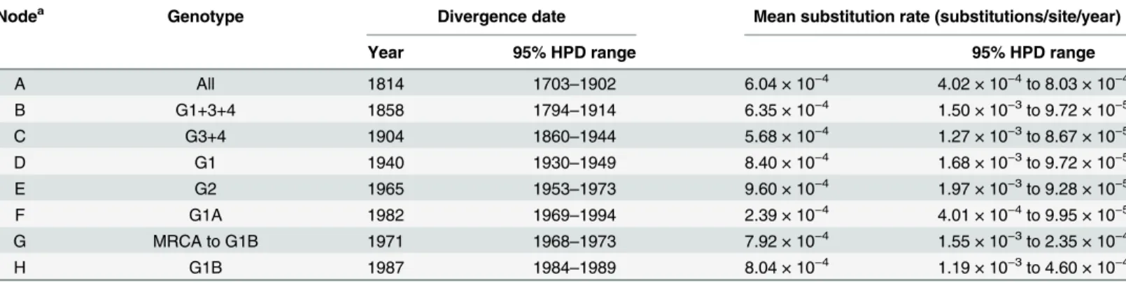
Fig 1. Map of Australia and Papua New Guinea showing the geographic origins of the MVEV strains used in this study.Australian states and territories are indicated, as well as selected regions or provinces.
Genotypes 3 and 4 form a well-supported clade, internal to G1 and G2 in the ML tree; no new members of G3 or G4 were found. G1 is the largest group, comprising 54 strains (82% of the sample set), indicating its dominance over the sampling period.
Pairwise distances within and between genotypes calculated using the prM and Env genes reflect the inferred phylogenies (S3 Table). Genotype 1 strains have high levels of genetic iden-tity to each other within the prM (96.2% nt) and Env genes (93.7% nt). Strains within G2 have slightly higher levels of nt identity (prM:97.8%; Env:96.2%), but are the most diver-gent when compared to other genotypes (prM:86.8%; Env:84.8%). As for previous studies [16,18], G3 and G4 are the most closely-related of the genotypes (prM: 94% nt, 99.4% aa; Env: 93% nt, 99.2% aa), and have comparable levels of identity to G1 and G2. Genotypes could be defined by nt p-distance (%) cut-off values of 6.0% (prM), 7.0% (Env), and 6.7% (prM-Env).
Recent circulating strains of MVEV belong to distinct sub-lineages of G1
Phylogenetic analyses revealed two distinct sub-lineages within G1, designated G1A and G1B, based on their relative age of divergence (seeEvolutionary history, below). These sub-lineages comprise the most recent strains of MVEV. G1A includes Western Australian strains from mosquitoes collected in 2008 and 2009 and one human case of MVE from 2008, as well as the most recent strains from PNG, isolated in 1998. G1A shares common ancestry with early iso-lates from Victoria (MVE-1-51 and TC123130). As for G2 viruses, all recent Australian strains in the G1A sub-lineage were isolated from the Kimberley region. In contrast, G1B is the most geographically diverse genotype and comprises strains isolated from Western Australian, Queensland, New South Wales and Victoria between 1989 and 2011. Both G1A and G1B sub-lineages are genetically homogenous (S3 Table): G1A has98.0% nt (99.4% aa) identity between strains for the prM gene and97.3% nt (99.6% aa) identity for Env; G1B strains share98.8% nt (98.2% aa) identity for prM and97.6% nt (99.0% aa) for Env.
Several intermediate sub-lineages within MVEV G1 were also observed, comprising Queensland isolates from 1996 and early isolates (1969–84) from Western Australia (Figs2
andS1). Of these, only two could be reliably resolved: the first comprised four strains from the Kimberley region of Western Australia, isolated between 1994 and 2003 (K16383, K16825, K16963 and K50609), the other was made-up of three strains from the Kimberley and Pilbara regions, isolated between 1977 and 1984 (AN505, OR1109 and PH491).
Sequence analysis of the prM and Env proteins: Genotype-defining
amino acid differences and conservation in biologically important motifs
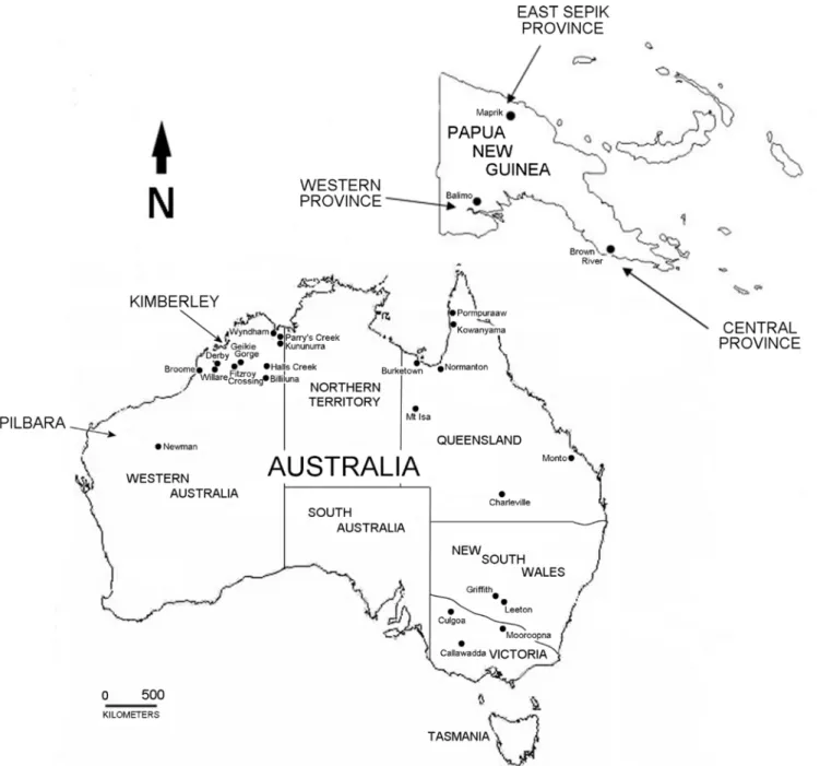
For each genotype, one or more amino acid residues were found to be unique to, and defining of, all virus strains belonging to that genotype (S4 Table). Using the Env protein crystal struc-ture of the closely-related JEV [42], the locations of these amino acids were modeled (Fig 3). The flavivirus Env protein is comprised of three structural domains: a centralβ-barrel (DI), an elongated dimerisation region (DII) and a C-terminal immunoglobulin-like domain (DIII) [48]. The soluble ectodomain of the Env protein is tethered to the viral membrane by a trans-membrane region.
For G1, V165 (DI), A229 (DII) and V240 (DII) of the Env protein are unique to this geno-type. Sub-lineages 1A and 1B can be further differentiated by single conservative amino acids estimated using a general time-reversible model of nucleotide substitution with a gamma distribution and invariant sites. Numbers at the nodes represent bootstrap support as a percentage of 1000 replicates; only values50% are shown. The scale bar indicates 2 nucleotide substitutions per site. The tree was rooted with the prM-Env sequence of JEV, however this has been removed to improve visual resolution of the tree.
substitutions in DI of Env (L180) or the prM (V24), respectively. Fifteen amino acids located in each of the structural domains and the transmembrane region of the Env protein of G2 viruses are defining of this genotype (S4 Table). Genotype 3 is defined by four amino acids encoded in the prM (H77) and Env protein in domains I and II and the stem (A187, V270 and I442), while G4 encodes only a single unique amino acid (I64) in DII. Two regions within the Env protein also encode sequences that are differentiating of the genotypes (S2 Fig). The region encompass-ing positions 228 to 232 in DII corresponds to a hypervariable domain previously identified for flaviviruses [49]. Within this region, G1 encodes the sequence motif PAST/SE, G2 encodes PSN/STD, and G3 and G4 encode PSSTE (S2A Fig).
Other regions and motifs of the prM and Env proteins known to be biologically important or to encode virulence determinants of MVEV or other flaviviruses were conserved within this species. In the prM protein, these included the flavivirus conserved region at position 61 to 69 [50] and tyrosine 78 [51], both involved in virus assembly. The canonical furin cleavage site [52], and the helix domain (prM 114–130), which interacts with the Env protein in immature particles [53], were also conserved. In the Env protein, the glycosylation site (NYS) at positions 154–157 [54,55], the receptor binding site in the F-G loop of DIII [positions 387–392; [48,56]], and the fusion peptide in DII [57] were all conserved among the MVEV strains sequenced. His-tidine residues proposed to be involved in the structural transition leading to membrane fusion [58] were also conserved.
Positive selection at position S332G of the Env protein
Selection analyses were performed to assess whether the genotype-defining amino acids identi-fied above were selected during the evolution of MVEV. The dN/dS ratio of nucleotide substi-tutions was estimated to be 0.052 in the DataMonkey analysis, indicating predominantly negative (purifying) selection (S5 Table), consistent with findings for other flaviviruses [59–
62]. A single (non-conservative) amino acid substitution in the Env protein (S332G) was found to be under positive (diversifying) selection in five of the six methods employed. This position is located in the B-C loop of DIII and is encoded by fourteen strains of G1 viruses isolated from
Fig 3. Location of genotype-defining amino acid residues and motifs in the ectodomain of the MVEV Env protein, based on the crystal structure of JEV [42].The three domains of the Env protein are shown in red (DI), yellow (DII) and blue (DIII). The fusion loop is coloured green, and the N-linked glycosylation site at position 154, the hypervariable domain in DII and the hinge connecting theαB helix of DII and the Ioβ-strand of DI are indicated.
pools ofCx.annulirostrisandAe.normanensisin Western Australia and Queensland between
1977 and 2009 (S6 Table). The S332G substitution is found in strains belonging to the recently emerged G1A and G1B, as well as intermediate lineages within G1, and does not therefore cor-relate with phylogenetic sub-lineage. One site in the prM protein and seventeen sites in the Env protein were found to be under directional evolution (S7 Table), including S332G; thirteen of these were identified as genotype-defining.
No evidence for recombination in the MVEV prM-Env genes
Evidence for homologous recombination, with breakpoints in the Env gene, has been reported for JEV and other flaviviruses, such as dengue virus (DENV) and Saint Louis encephalitis virus, based on database-derived sequences [63,64] or from cloned cDNA from clinical and mosquito isolates [65,66]. Experimental evidence of recombination between JEV genomes has also been reported [67,68]. In the present study, no evidence of recombination was detected in MVEV prM-Env genes using eight different computational methods implemented in the RDP3 program [36]. This is consistent with the finding of no major incongruence in phyloge-netic groupings when trees generated from prM or Env genes were compared (S1 Fig).
Evolutionary history of MVEV
The root-to-tip regression analysis of the MVEV prM-Env sequences demonstrated a high level of clock-like behavior in the tree (R2= 0.65) and therefore justified a more in-depth
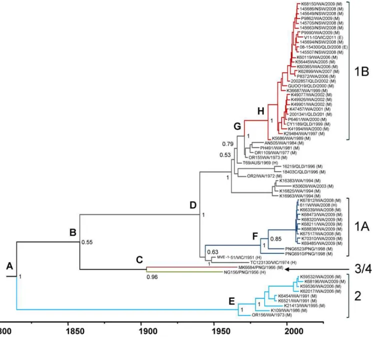
analy-sis of lineage evolution. To do this, a Bayesian MCMC analyanaly-sis was performed to estimate time to MRCA and evolutionary rates for MVEV genotypes. The MCC tree for the prM-Env sequences is shown inFig 4. The MRCA for all genotypes of MVEV was estimated to circulate between 1703 and 1902 (mean: 1814;Table 1). G2 emerged directly from this ancestral virus, with further evolution of recent isolates occurring between 1953 and 1973 (mean: 1965). The ancestral lineage of genotypes 1, 3 and 4 evolved between 1794 and 1914 (mean: 1858), and G3 and G4 later diverged into distinct lineages between 1860 and 1944 (mean: 1904). The MRCA of G1 evolved between 1930 and 1949 (mean: 1940), just prior to the first isolation of MVEV, belonging to this genotype, in the 1950–51 epidemic in southeast Australia [69]. The sub-line-ages of G1A and G1B were the most recent to emerge: G1A between 1969 and 1994 (mean: 1982) and G1B between 1984 and 1989 (mean: 1987).
Nucleotide substitution rates were also estimated from the Bayesian MCMC analysis. The mean rate of nucleotide substitutions within the prM-Env genes for all MVEV strains was 6.04 × 10⁻⁴substitutions per site per year (95% HPD: 4.02 × 10⁻⁴to 8.03 × 10⁻⁴). A low level of heterogeneity in evolutionary rates, as estimated by the MCMC analysis, was observed between individual genotypes and ancestral lineages (Table 1). Although rates varied from 2.39 × 10⁻⁴ (G1A) to 9.60 × 10⁻⁴(G2) substitutions per site per year, uncertainty intervals (95% HPD) for all lineages were overlapping. These rates are comparable to those estimated for other flavivi-ruses belonging to the JEV serogroup [60,70–72].
Regression analysis of the prM-Env gene phylogeny supported the above results from the MCMC analysis. The estimated time to the MRCA was the year 1800 (R2= 0.65) for all strains
and the substitution rate was 6.68 × 10⁻⁴nucleotide substitutions per site per year.
Discussion
sub-lineages (designated G1A and G1B) that have recently emerged. G2 was shown to be the oldest lineage, while no additional members of G3 and G4 were found. This study clearly dem-onstrated that all contemporary lineages of MVEV have been isolated from Kimberley region of Western Australia, which supports the previous contention that the enzootic focus for this virus resides in northwestern Australia, at least since the early 1970s following construction of the Ord River irrigation area [3,73].
Of the two G1 sub-lineages of MVEV identified in this study, G1B is the most widely trans-mitted and, for Australian strains, the only contemporary genotype found outside northwest-ern Australia; viruses belonging to this group have been isolated from the eastnorthwest-ern Australian
Fig 4. Bayesian MCC phylogenetic tree for MVEV prM-Env sequences.Divergence times and branch lengths are drawn according to the time scale bar (in years), such that the tips of the tree correspond to sampling date of each virus strain. Branches and ancestral lineage of each genotype and sub-lineage are coloured blue (G1A), red (G1B), grey (remaining G1 strains), light blue (G2), green (G3) and dark red (G4). Posterior probability values are shown at the nodes. Letters at the upper left of each node correspond to divergence times and substitution rates shown inTable 1.
states. G1A comprised both Australian and PNG strains, the latter forming separate lineages within this group (Fig 2). Phylogenetic analyses showed that these viruses diverged from a common ancestral strain, further supporting evidence for virus movement between northwest-ern Australia and southnorthwest-ern PNG [18]. Despite this, and in contrast to G1B, the Australian G1A viruses have only been isolated from the Kimberley region of WA. A similar geographic restric-tion was observed for G2 viruses, and it has been suggested that this lineage occupies an eco-logical niche that underlies its range [18,21]. Australian G1A viruses may also be subjected to ecological or biological restrictions. Similarly, G3 or G4 viruses appear to be either confined to the New Guinea Island or have become extinct. No new members of these types were identified in this study. However, in this regard, it is notable that surveillance activities for MVEV and other flaviviruses in PNG are rarely undertaken, and due to the paucity of isolates from this country, the circulation of additional genotypes cannot be discounted.
The observation of dominant and minority circulating genotypes for MVEV has also been found for other members of the JEV group. Notably, GIII of JEV was dominant throughout northern and western Asia for several decades until it was displaced in the early 1990s by GI, which is now the major circulating lineage [74–78]. In contrast, JEV GIV viruses have only been isolated from mosquitoes and appear to be geographically confined to Indonesia [78], analogous to the apparent restricted circulation of MVEV G2 in the Kimberley region of West-ern Australia. Thus, these minority lineages, which have no known disease association in either humans or animals, may represent low pathogenic types. However, given their close relation-ships with pathogenic viruses, and the propensity of flaviviruses to rapidly evolve and adapt to new environments and hosts, they may represent potential pathogens.
One or more unique amino acids encoded in the prM and Env proteins were found to define each MVEV genotype (S4 Table). Given the importance of these proteins in the virus replica-tion cycle and host immune response, these differences may underlie the observed variareplica-tion in geographic distribution of genotypes and sub-lineages. Of potential significance for G1 viruses is Env 229, which is located on the‘h’loop of DII within the flavivirus hypervariable domain [49], a region containing both type- and group reactive flavivirus epitopes [79,80]. Thus, A229 may have been selected as an escape from neutralization mutation that has conferred a fitness advantage to G1 MVEV. Since A229 is conserved within both G1A and G1B sub-lineages, the factors determining the observed differences in their transmission patterns in Australian may lie in other regions of the genome. Full genome sequencing of G1A strains may reveal biologi-cally significant differences to the genome of G1B MVEV [47]. It may be speculated that G1B
Table 1. Divergence times and evolutionary rates for genotypes and sub-lineages of MVEV estimated by Bayesian MCMC analysis.
Nodea Genotype Divergence date Mean substitution rate (substitutions/site/year)
Year 95% HPD range 95% HPD range
A All 1814 1703–1902 6.04 × 10⁻⁴ 4.02 × 10⁻⁴to 8.03 × 10⁻⁴
B G1+3+4 1858 1794–1914 6.35 × 10⁻⁴ 1.50 × 10⁻³to 9.72 × 10⁻⁵
C G3+4 1904 1860–1944 5.68 × 10⁻⁴ 1.27 × 10⁻³to 8.67 × 10⁻⁵
D G1 1940 1930–1949 8.40 × 10⁻⁴ 1.68 × 10⁻³to 9.72 × 10⁻⁵
E G2 1965 1953–1973 9.60 × 10⁻⁴ 1.97 × 10⁻³to 9.28 × 10⁻⁵
F G1A 1982 1969–1994 2.39 × 10⁻⁴ 4.01 × 10⁻⁴to 9.95 × 10⁻⁵
G MRCA to G1B 1971 1968–1973 7.92 × 10⁻⁴ 1.55 × 10⁻³to 2.35 × 10⁻⁴
H G1B 1987 1984–1989 8.04 × 10⁻⁴ 1.19 × 10⁻³to 4.60 × 10⁻⁴
aNodes are as indicated in the MCMC tree shown inFig 3.
viruses have a replicative advantage enabling more rapid and widespread transmission. Viruses of this genotype may be more efficiently transmitted by vector mosquitoes. This is believed to be a contributing factor to the emergence of the dominant WNV WN02 genotype in North America between 2001 and 2004, which was found to be transmitted earlier and more effi-ciently than the pre-existing NY99 genotype virus [81].
For G2 viruses, several of the observed unique amino acids have potential biological signifi-cance. Residues at positions 72, 126, 275, 276, 330 and 369 are predicted to be exposed on the virus surface (Fig 3) [42,48] and have been associated with biological function and antigenicity. Env 126 has been shown experimentally to be a determinant of neurovirulence in mice for DENV2 [82]; and residues 330 and 369 are located in loops of DIII involved in receptor attach-ment [48,83]. Moreover, amino acids at positions 275 and 276 are located in one of four putative hinges connecting DI and DII that are thought to play a critical structural role during pH-dependent membrane fusion [42,48,84]. Mutations at the adjacent position 277 have also been shown to reduce MVEV haemagglutination and attenuate neuroinvasiveness in mice [85,86]. It has been previously proposed that the unique amino acids encoded by G2 viruses may affect their replicative fitness and virulence, thereby restricting their transmission via vec-tor mosquitoes and/or water birds [18]. Our findings support this hypothesis, as doin vivo
studies using the mouse model of neurotropism: we and others have shown that G2 viruses, including early (OR156) and recent (K59532) strains, have a low virulence phenotype following intraperitoneal inoculation ([87];S8 Table).
The unique amino acid residues encoded by G2 virus strains at positions 126, 275, 276 and 330 make-up neutralizing epitopes of MVEV (126, 276), WNV (330) and JEV (126, 275) [80,88–91]. We predict these residues may also distinguish G2 viruses antigenically from viruses belonging to other genotypes. Similarly, position 332 in the Env protein, found to be under positive selection for some G1 strains (S6 Table), is found on the distal lateral surface of DIII, which protrudes from the virion surface and is a known hot-spot for potent flavivirus neutralising epitopes [90,92–95]. Mutations at position 332 of WNV and JEV (MVEV num-bering) can reduce or abolish antibody binding [90–92]. MVEV strains encoding the S332G substitution may have escaped from antibody neutralisation and are antigenically different at this site compared to other MVEV strains.
became the dominant and most widespread lineage of MVEV in Australia. We propose that the changes to the ecosystem in the northeast Kimberley facilitated the emergence of this line-age, via increased populations of waterbirds andCulexspecies mosquitoes, and the creation of
year-round conditions suitable for maintaining virus transmission cycles.
Currently, only four full length genomes of MVEV have been published [86,98]. Additional full length genomes are expected to allow more accurate and comprehensive evolutionary anal-yses of MVEV. Further sequencing of earlier isolates may also provide a greater level of confi-dence to the MCMC analysis, particularly for older divergence points. Continued surveillance will also be necessary to track the ongoing evolution of this virus. It is expected that sub-line-ages G1A and G1B will continue to diverge genetically and potentially antigenically, with possi-ble consequences to the sensitivity of existing laboratory diagnostics. Of interest will be the relative pattern of spread of each of these lineages. Are G1A and G2 viruses found outside northwestern Australia? And if not, what are the biological and ecological drivers of this phe-nomenon? The isolation and characterisation of MVEV strains from regions north of Austra-lia, such as PNG and Indonesia, will also shed light on the diversity of MVEV circulation and patterns of spread to mainland Australia.
Supporting Information
S1 Table. Details of MVEV strains, detected or isolated in Australia and PNG between 1951 and 2011, used for genetic analyses in this study.
(DOCX)
S2 Table. Oligonucleotides employed for amplification of pre-membrane and envelope genes of Murray Valley encephalitis virus.
(DOCX)
S3 Table. Pairwise distances between genotypes and subgenotypes of Murray Valley encephalitis virus for the pre-membrane (prM) and envelope genes.
(DOCX)
S4 Table. Unique amino acids in the pre-membrane and envelope protein sequences of MVEV that define genotype or sub-lineage.
(DOCX)
S5 Table. Evidence for positive and negative selection in the prM and Env genes (668 amino acids) of Murray Valley encephalitis using six different methods implemented in the DataMonkey server of the HyPhy software package.
(DOCX)
S6 Table. Murray Valley encephalitis virus strains encoding the S332G amino acid substitu-tion in the envelope protein.
(DOCX)
S7 Table. Amino acid sites within the prM and Env proteins under directional selection.
Amino acids corresponding to genotype-defining residues are indicated by ( ). (DOCX)
S8 Table. Virulence of Murray Valley encephalitis virus strains, representing genotypes 1 and 2, in 18-day old Swiss mice following intraperitoneal inoculation.
(DOCX)
model with a gamma distribution. ML trees (B, D and F) were estimated using a general time-reversible model of nucleotide substitution with a gamma distribution and invariant sites. Numbers at the nodes represent bootstrap support as a percentage of 1000 replicates; only val-ues50% are shown. The scale bar indicates nucleotide substitutions per site. Each tree was rooted with the analogous sequence of JEV, however this has been removed to improve visual resolution of the tree.
(PDF)
S2 Fig. The hypervariable region (A) and DI-DII hinge motif (B) of the Envelope protein encoded by representative strains of Murray Valley encephalitis virus.Identical amino acids are colour-shaded. The symbols below the alignments indicate identical amino acids (
), strongly conserved (:), and weakly conserved (.) amino acids.
(PDF)
Acknowledgments
The authors thank Mr. John Haniotis (Department of Medical Entomology, Westmead Hospi-tal, University of Sydney and ICPMR, New South Wales, Australia) for laboratory support. The Chimera package was developed by the Resource for Biocomputing, Visualization, and Informatics at the University of California, San Francisco.
Author Contributions
Conceived and designed the experiments: DTW SMD AuRN PAD BHC BH SLD CAJ JSM. Performed the experiments: DTW SJD AuRN PAD BHC BH AP SLD CAJ. Analyzed the data: DTW SJD AuRN PAD BHC BH CAJ. Contributed reagents/materials/analysis tools: AP SLD CAJ. Wrote the paper: DTW SMD AuRN PAD BHC BH AJ SLD CAJ JSM.
References
1. Mackenzie JS, Williams DT (2009) The zoonotic flaviviruses of southern, south-eastern and eastern Asia, and Australasia: the potential for emergent viruses. Zoonoses Public Health 56: 338–356. doi: 10.1111/j.1863-2378.2008.01208.xPMID:19486319
2. Marshall ID (1989) Murray Valley and Kunjin encephalitis. In: Monath TP, editor. The arboviruses: epi-demiology and ecology. Florida: CRC Press, Boca Raton. pp. 151–189.
3. Mackenzie J, Lindsay M, Coelen R, Broom A, Hall R, et al. (1994) Arboviruses causing human disease in the Australasian zoogeographic region. Archives of Virology 136: 447–467. PMID:8031248 4. Burrow J, Whelan P, Kilburn C, Fisher D, Currie B, et al. (1998) Australian encephalitis in the Northern
Territory: clinical and epidemiological features, 1987–1996. Australian and New Zealand journal of
medicine. pp. 590–596. PMID:9847946
5. Brown A, Bolisetty S, Whelan P, Smith D, Wheaton G (2002) Reappearance of human cases due to Murray Valley encephalitis virus and Kunjin virus in central Australia after an absence of 26 years. Com-municable diseases intelligence 26: 39–44.
6. Forbes JA (1978) Murray Valley encephalitis 1974, also the epidemic variance since 1914 and predis-posing rainfall patterns. Sydney, Australia: Australasian Medical publishing.
7. Anderson SG, Price Av N-K, Slater K (1960) Murray Valley encephalitis in Papua and New Guinea. II. Serological survey, 1956–1957. Med J Aust 47(2): 410–413.
8. French EL, Anderson SG, Price AV, Rhodes FA (1957) Murray Valley encephalitis in New Guinea. I. Isolation of Murray Valley encephalitis virus from the brain of a fatal case of encephalitis occurring in a Papuan native. Am J Trop Med Hyg 6: 827–834. PMID:13470202
10. Gordon AN, Marbach CR, Oakey J, Edmunds G, Condon K, et al. (2012) Confirmed case of encephali-tis caused by Murray Valley encephaliencephali-tis virus infection in a horse. J Vet Diagn Invest 24: 431–436. doi: 10.1177/1040638711433325PMID:22379060
11. Roche SE, Wicks R, Garner MG, East IJ, Paskin R, et al. (2013) Descriptive overview of the 2011 epi-demic of arboviral disease in horses in Australia. Aust Vet J 91: 5–13. doi:10.1111/avj.12018PMID: 23356366
12. Doggett S (2004) Population health aspects of mosquito-borne disease in New South Wales. N S W Public Health Bull 15: 193–199. PMID:15711613
13. Doggett SL, Clancy J., Haniotis J., Webb C. E., Hueston L., Marchetti M., Blyth F., Howard S., Dwyer D. E., and Russell R. C. (2009) Arbovirus and vector surveillance in New South Wales, 2004/05-2007/8. Arbovirus Research in Australia 10: 28–37.
14. Fitzsimmons GJ, Wright P, Johansen CA, Whelan PI, National A, et al. (2009) Arboviral diseases and malaria in Australia, 2007/08: annual report of the National Arbovirus and Malaria Advisory Committee. Commun Dis Intell Q Rep 33: 155–169. PMID:19877534
15. Selvey LA, Dailey L, Lindsay M, Armstrong P, Tobin S, et al. (2014) The changing epidemiology of Mur-ray Valley encephalitis in Australia: the 2011 outbreak and a review of the literature. PLoS Negl Trop Dis 8: e2656. doi:10.1371/journal.pntd.0002656PMID:24466360
16. Coelen RJ, Mackenzie JS (1988) Genetic variation of Murray Valley encephalitis virus. Journal of Gen-eral Virology 69: 1903–1912. PMID:2841405
17. Lobigs M, Marshall ID, Weir RC, Dalgarno L (1988) Murray Valley encephalitis virus field strains from Australia and Papua New Guinea: studies on the sequence of the major envelope protein gene and vir-ulence for mice. Virology 165: 245–255. PMID:2838962
18. Johansen C, Susai V, Hall R, Mackenzie J, Clark D, et al. (2007) Genetic and phenotypic differences between isolates of Murray Valley encephalitis virus in Western Australia, 1972–2003. Virus Genes
35: 147–154. PMID:17393295
19. Coelen RJ, Mackenzie JS (1990) The 5'-terminal non-coding region of Murray Valley encephalitis virus RNA is highly conserved. Journal of General Virology 71: 241–245. PMID:2154536
20. Poidinger M, Hall R, Mackenzie J (1996) Molecular characterization of the Japanese encephalitis sero-complex of the flavivirus genus. Virology 218: 417–421. PMID:8610471
21. Niazi A (2014) The role of genetic diversity in the replication, pathogenecity and virulence of Murray Val-ley encephalitis virus. Perth, Western Australia: Curtin University.
22. Lobigs M, Marshall ID, Weir RC, Dalgarno L (1986) Genetic differentiation of Murray Valley encephalitis virus in Australia and Papua New Guinea. Australian Journal of Experimental Biology and Medical Sci-ence 64: 571–585. PMID:2884985
23. Lindenbach BD, Murray C.L., Thiel H.-J., Rice C.M. (2013) Flaviviridae. In: Knipe DM, Howley P.M., edi-tor. Fileds Virology Sxith Edition. 6th ed. Philadelphia, USA: Lippincott Williams & Wilkins. pp. 712–
746.
24. Heinz FX, Stiasny K (2012) Flaviviruses and their antigenic structure. J Clin Virol 55: 289–295. doi:10. 1016/j.jcv.2012.08.024PMID:22999801
25. Colpitts TM, Rodenhuis-Zybert I, Moesker B, Wang P, Fikrig E, et al. (2011) prM-antibody renders immature West Nile virus infectiousin vivo. J Gen Virol 92: 2281–2285. doi:10.1099/vir.0.031427-0
PMID:21697345
26. Chen WR, Tesh RB, Rico-Hesse R (1990) Genetic variation of Japanese encephalitis virus in nature. J Gen Virol 71 (Pt 12): 2915–2922. PMID:2273391
27. Davis CT, Ebel GD, Lanciotti RS, Brault AC, Guzman H, et al. (2005) Phylogenetic analysis of North American West Nile virus isolates, 2001–2004: evidence for the emergence of a dominant genotype.
Virology 342: 252–265. PMID:16137736
28. Kramer LD, Chandler LJ (2001) Phylogenetic analysis of the envelope gene of St. Louis encephalitis virus. Arch Virol 146: 2341–2355. PMID:11811684
29. Williams DT, Wang LF, Daniels PW, Mackenzie JS (2000) Molecular characterization of the first Austra-lian isolate of Japanese encephalitis virus, the FU strain. J Gen Virol 81: 2471–2480. PMID:10993935 30. Pierre V, Drouet M, Deubel V (1994) Identification of mosquito-borne flavivirus sequences using
univer-sal primers and reverse transcription/polymerase chain reaction. Research in Virology 145: 93–104.
PMID:7520190
31. Tamura K, Stecher G, Peterson D, Filipski A, Kumar S (2013) MEGA6: Molecular Evolutionary Genet-ics Analysis version 6.0. Mol Biol Evol 30: 2725–2729. doi:10.1093/molbev/mst197PMID:24132122 32. Guindon S, Gascuel O (2003) A simple, fast, and accurate algorithm to estimate large phylogenies by
33. Darriba D, Taboada GL, Doallo R, Posada D (2012) jModelTest 2: more models, new heuristics and parallel computing. Nat Methods 9: 772.
34. Drummond AJ, Rambaut A (2007) BEAST: Bayesian evolutionary analysis by sampling trees. BMC Evol Biol 7: 214. PMID:17996036
35. Drummond AJ, Rambaut A, Shapiro B, Pybus OG (2005) Bayesian coalescent inference of past popu-lation dynamics from molecular sequences. Mol Biol Evol 22: 1185–1192. PMID:15703244
36. Heath L, van der Walt E, Varsani A, Martin DP (2006) Recombination patterns in aphthoviruses mirror those found in other picornaviruses. J Virol 80: 11827–11832. PMID:16971423
37. Pond SL, Frost SD (2005) Datamonkey: rapid detection of selective pressure on individual sites of codon alignments. Bioinformatics 21: 2531–2533. PMID:15713735
38. Kosakovsky Pond SL, Frost SD (2005) Not so different after all: a comparison of methods for detecting amino acid sites under selection. Mol Biol Evol 22: 1208–1222. PMID:15703242
39. Murrell B, Wertheim JO, Moola S, Weighill T, Scheffler K, et al. (2012) Detecting individual sites subject to episodic diversifying selection. PLoS Genet 8: e1002764. doi:10.1371/journal.pgen.1002764PMID:
22807683
40. Murrell B, Moola S, Mabona A, Weighill T, Sheward D, et al. (2013) FUBAR: a fast, unconstrained bayesian approximation for inferring selection. Mol Biol Evol 30: 1196–1205. doi:10.1093/molbev/ mst030PMID:23420840
41. Kosakovsky Pond SL, Poon AF, Leigh Brown AJ, Frost SD (2008) A maximum likelihood method for detecting directional evolution in protein sequences and its application to influenza A virus. Mol Biol Evol 25: 1809–1824. doi:10.1093/molbev/msn123PMID:18511426
42. Luca VC, AbiMansour J, Nelson CA, Fremont DH (2012) Crystal structure of the Japanese encephalitis virus envelope protein. J Virol 86: 2337–2346. doi:10.1128/JVI.06072-11PMID:22156523
43. Arnold K, Bordoli L, Kopp J, Schwede T (2006) The SWISS-MODEL workspace: a web-based environ-ment for protein structure homology modelling. Bioinformatics 22: 195–201. PMID:16301204 44. Pettersen EF, Goddard TD, Huang CC, Couch GS, Greenblatt DM, et al. (2004) UCSF Chimera—a
visualization system for exploratory research and analysis. J Comput Chem 25: 1605–1612. PMID: 15264254
45. National Health and Medical Research Council (2013) Australian code for the care and use of animals for scientific purposes, 8th edition. Canberra: National Health and Medical Research Council.
46. Hurrelbrink RJ, Nestorowicz A, McMinn PC (1999) Characterization of infectious Murray Valley enceph-alitis virus derived from a stably cloned genome-length cDNA. J Gen Virol 80 (Pt 12): 3115–3125.
PMID:10567642
47. Mann RA, Fegan M, O'Riley K, Motha J, Warner S (2013) Molecular characterization and phylogenetic analysis of Murray Valley encephalitis virus and West Nile virus (Kunjin subtype) from an arbovirus dis-ease outbreak in horses in Victoria, Australia, in 2011. J Vet Diagn Invest 25: 35–44. doi:10.1177/ 1040638712467985PMID:23345269
48. Rey FA, Heinz FX, Mandl C, Kunz C, Harrison SC (1995) The envelope glycoprotein from tick-borne encephalitis virus at 2 A resolution. Nature 375: 291–298. PMID:7753193
49. Shiu SY, Jiang WR, Porterfield JS, Gould EA (1992) Envelope protein sequences of dengue virus iso-lates TH-36 and TH-Sman, and identification of a type-specific genetic marker for dengue and tick-borne flaviviruses. J Gen Virol 73 (Pt 1): 207–212. PMID:1339466
50. Yoshii K, Igarashi M, Ichii O, Yokozawa K, Ito K, et al. (2012) A conserved region in the prM protein is a critical determinant in the assembly of flavivirus particles. J Gen Virol 93: 27–38. doi:10.1099/vir.0. 035964-0PMID:21957123
51. Tan TT, Bhuvanakantham R, Li J, Howe J, Ng ML (2009) Tyrosine 78 of premembrane protein is essen-tial for assembly of West Nile virus. J Gen Virol 90: 1081–1092. doi:10.1099/vir.0.007872-0PMID: 19264649
52. Elshuber S, Allison SL, Heinz FX, Mandl CW (2003) Cleavage of protein prM is necessary for infection of BHK-21 cells by tick-borne encephalitis virus. J Gen Virol 84: 183–191. PMID:12533715
53. Peng JG, Wu SC (2014) Glutamic acid at residue 125 of the prM helix domain interacts with positively charged amino acids in E protein domain II for Japanese encephalitis virus-like-particle production. J Virol 88: 8386–8396. doi:10.1128/JVI.00937-14PMID:24829339
55. Prow NA, May FJ, Westlake DJ, Hurrelbrink RJ, Biron RM, et al. (2011) Determinants of attenuation in the envelope protein of the flavivirus Alfuy. J Gen Virol 92: 2286–2296. doi:10.1099/vir.0.034793-0
PMID:21733886
56. Lee E, Lobigs M (2000) Substitutions at the putative receptor-binding site of an encephalitic flavivirus alter virulence and host cell tropism and reveal a role for glycosaminoglycans in entry. J Virol 74: 8867–8875. PMID:10982329
57. Allison SL, Schalich J, Stiasny K, Mandl CW, Heinz FX (2001) Mutational evidence for an internal fusion peptide in flavivirus envelope protein E. J Virol 75: 4268–4275. PMID:11287576
58. Fritz R, Stiasny K, Heinz FX (2008) Identification of specific histidines as pH sensors in flavivirus mem-brane fusion. J Cell Biol 183: 353–361. doi:10.1083/jcb.200806081PMID:18936253
59. Jerzak G, Bernard KA, Kramer LD, Ebel GD (2005) Genetic variation in West Nile virus from naturally infected mosquitoes and birds suggests quasispecies structure and strong purifying selection. J Gen Virol 86: 2175–2183. PMID:16033965
60. Baillie GJ, Kolokotronis SO, Waltari E, Maffei JG, Kramer LD, et al. (2008) Phylogenetic and evolution-ary analyses of St. Louis encephalitis virus genomes. Mol Phylogenet Evol 47: 717–728. doi:10.1016/ j.ympev.2008.02.015PMID:18374605
61. Schuh AJ, Guzman H, Tesh RB, Barrett AD (2013) Genetic diversity of Japanese encephalitis virus iso-lates obtained from the indonesian archipelago between 1974 and 1987. Vector Borne Zoonotic Dis 13: 479–488. doi:10.1089/vbz.2011.0870PMID:23590316
62. Woelk CH, Holmes EC (2002) Reduced positive selection in vector-borne RNA viruses. Mol Biol Evol 19: 2333–2336. PMID:12446826
63. Worobey M, Rambaut A, Holmes EC (1999) Widespread intra-serotype recombination in natural popu-lations of dengue virus. Proc Natl Acad Sci U S A 96: 7352–7357. PMID:10377418
64. Twiddy SS, Holmes EC (2003) The extent of homologous recombination in members of the genus Fla-vivirus. J Gen Virol 84: 429–440. PMID:12560576
65. Craig S, Thu HM, Lowry K, Wang XF, Holmes EC, et al. (2003) Diverse dengue type 2 virus populations contain recombinant and both parental viruses in a single mosquito host. J Virol 77: 4463–4467. PMID: 12634407
66. Aaskov J, Buzacott K, Field E, Lowry K, Berlioz-Arthaud A, et al. (2007) Multiple recombinant dengue type 1 viruses in an isolate from a dengue patient. J Gen Virol 88: 3334–3340. PMID:18024903 67. Chuang CK, Chen WJ (2009) Experimental evidence that RNA recombination occurs in the Japanese
encephalitis virus. Virology 394: 286–297. doi:10.1016/j.virol.2009.08.030PMID:19766282
68. Taucher C, Berger A, Mandl CW (2010) A trans-complementing recombination trap demonstrates a low propensity of flaviviruses for intermolecular recombination. J Virol 84: 599–611. doi:10.1128/JVI. 01063-09PMID:19864381
69. French EL (1952) Murray Valley encephalitis: isolation and characterisation of the aetiological agent. Medical Journal of Australia 1: 100–103. PMID:14909902
70. Jenkins GM, Rambaut A, Pybus OG, Holmes EC (2002) Rates of molecular evolution in RNA viruses: a quantitative phylogenetic analysis. J Mol Evol 54: 156–165. PMID:11821909
71. May FJ, Davis CT, Tesh RB, Barrett AD (2011) Phylogeography of West Nile virus: from the cradle of evolution in Africa to Eurasia, Australia, and the Americas. J Virol 85: 2964–2974. doi:10.1128/JVI. 01963-10PMID:21159871
72. Mohammed MA, Galbraith SE, Radford AD, Dove W, Takasaki T, et al. (2011) Molecular phylogenetic and evolutionary analyses of Muar strain of Japanese encephalitis virus reveal it is the missing fifth genotype. Infect Genet Evol 11: 855–862. doi:10.1016/j.meegid.2011.01.020PMID:21352956 73. Mackenzie JS, Broom AK (1999) Ord River irrigation area: the effect of dam construction and irrigation
on the incidence of Murray Valley encephalitis virus. In: Kay BH, editor. Water resources: Health, Envi-ronment and Development London: E & FN Spon. pp. 108–122.
74. Pyke AT, Williams DT, Nisbet DJ, van den Hurk AF, Taylor CT, et al. (2001) The appearance of a sec-ond genotype of Japanese encephalitis virus in the Australasian region. Am J Trop Med Hyg 65: 747–
753. PMID:11791969
75. Ma SP, Yoshida Y, Makino Y, Tadano M, Ono T, et al. (2003) Short report: a major genotype of Japa-nese encephalitis virus currently circulating in Japan. Am J Trop Med Hyg 69: 151–154. PMID: 13677370
77. Pan XL, Liu H, Wang HY, Fu SH, Liu HZ, et al. (2011) Emergence of genotype I of Japanese encephali-tis virus as the dominant genotype in Asia. J Virol 85: 9847–9853. doi:10.1128/JVI.00825-11PMID: 21697481
78. Schuh AJ, Ward MJ, Brown AJ, Barrett AD (2013) Phylogeography of Japanese encephalitis virus: genotype is associated with climate. PLoS Negl Trop Dis 7: e2411. doi:10.1371/journal.pntd.0002411
PMID:24009790
79. Oliphant T, Nybakken GE, Engle M, Xu Q, Nelson CA, et al. (2006) Antibody recognition and neutraliza-tion determinants on domains I and II of West Nile Virus envelope protein. J Virol 80: 12149–12159.
PMID:17035317
80. Crill WD, Chang GJ (2004) Localization and characterization of flavivirus envelope glycoprotein cross-reactive epitopes. J Virol 78: 13975–13986. PMID:15564505
81. Moudy RM, Meola MA, Morin LL, Ebel GD, Kramer LD (2007) A newly emergent genotype of West Nile virus is transmitted earlier and more efficiently byCulexmosquitoes. Am J Trop Med Hyg 77: 365–370.
PMID:17690414
82. Gualano RC, Pryor MJ, Cauchi MR, Wright PJ, Davidson AD (1998) Identification of a major determi-nant of mouse neurovirulence of dengue virus type 2 using stably cloned genomic-length cDNA. J Gen Virol 79 (Pt 3): 437–446. PMID:9519821
83. Beasley DW, Li L, Suderman MT, Barrett AD (2002) Mouse neuroinvasive phenotype of West Nile virus strains varies depending upon virus genotype. Virology 296: 17–23. PMID:12036314
84. Modis Y, Ogata S, Clements D, Harrison SC (2003) A ligand-binding pocket in the dengue virus enve-lope glycoprotein. Proc Natl Acad Sci U S A 100: 6986–6991. PMID:12759475
85. McMinn PC, Weir RC, Dalgarno L (1996) A mouse-attenuated envelope protein variant of Murray Valley encephalitis virus with altered fusion activity. J Gen Virol 77 (Pt 9): 2085–2088. PMID:8811007 86. Hurrelbrink RJ, McMinn PC (2001) Attenuation of Murray Valley encephalitis virus by site-directed
mutagenesis of the hinge and putative receptor-binding regions of the envelope protein. J Virol 75: 7692–7702. PMID:11462041
87. Lawson MA (1988) Genotypic and phenotypic variation of Murray Valley encephalitis virus. Perth, Western Australia: University of Western Australia.
88. McMinn PC, Lee E, Hartley S, Roehrig JT, Dalgarno L, et al. (1995) Murray valley encephalitis virus envelope protein antigenic variants with altered hemagglutination properties and reduced neuroinva-siveness in mice. Virology 211: 10–20. PMID:7645203
89. Morita K, Tadano M, Nakaji S, Kosai K, Mathenge EG, et al. (2001) Locus of a virus neutralization epi-tope on the Japanese encephalitis virus envelope protein determined by use of long PCR-based region-specific random mutagenesis. Virology 287: 417–426. PMID:11531418
90. Beasley DW, Barrett AD (2002) Identification of neutralizing epitopes within structural domain III of the West Nile virus envelope protein. J Virol 76: 13097–13100. PMID:12438639
91. Oliphant T, Engle M, Nybakken GE, Doane C, Johnson S, et al. (2005) Development of a humanized monoclonal antibody with therapeutic potential against West Nile virus. Nat Med 11: 522–530. PMID: 15852016
92. Lin CW, Wu SC (2003) A functional epitope determinant on domain III of the Japanese encephalitis virus envelope protein interacted with neutralizing-antibody combining sites. J Virol 77: 2600–2606.
PMID:12551998
93. Nybakken GE, Oliphant T, Johnson S, Burke S, Diamond MS, et al. (2005) Structural basis of West Nile virus neutralization by a therapeutic antibody. Nature 437: 764–769. PMID:16193056
94. Brien JD, Austin SK, Sukupolvi-Petty S, O'Brien KM, Johnson S, et al. (2010) Genotype-specific neu-tralization and protection by antibodies against dengue virus type 3. J Virol 84: 10630–10643. doi:10. 1128/JVI.01190-10PMID:20702644
95. Shrestha B, Brien JD, Sukupolvi-Petty S, Austin SK, Edeling MA, et al. (2010) The development of ther-apeutic antibodies that neutralize homologous and heterologous genotypes of dengue virus type 1. PLoS Pathog 6: e1000823. doi:10.1371/journal.ppat.1000823PMID:20369024
96. Mackenzie JS, Johansen CA, Ritchie SA, van den Hurk AF, Hall RA (2002) Japanese encephalitis as an emerging virus: the emergence and spread of Japanese encephalitis virus in Australasia. Curr Top Microbiol Immunol 267: 49–73. PMID:12083000
97. Mackenzie JS, Broom AK (1995) Australian X disease, Murray Valley encephalitis and the French con-nection. Veterinary Microbiology 46: 79–90. PMID:8545982

![Fig 3. Location of genotype-defining amino acid residues and motifs in the ectodomain of the MVEV Env protein, based on the crystal structure of JEV [42]](https://thumb-eu.123doks.com/thumbv2/123dok_br/16360197.190191/9.918.56.816.110.408/location-genotype-defining-residues-ectodomain-protein-crystal-structure.webp)