Article
J. Braz. Chem. Soc., Vol. 25, No. 4, 704-708, 2014. Printed in Brazil - ©2014 Sociedade Brasileira de Química 0103 - 5053 $6.00+0.00
A
http://dx.doi.org/10.5935/0103-5053.20140023*e-mail: [email protected]
A New Flavonoid Derivative from Leaves of
Oxandra sessiliflora
R. E. Fries
Elcilene A. de Sousa,a Armenio A. C. A. da Silva,a Alberto J. Cavalheiro,b João Henrique G. Lagoc and Mariana H. Chaves*,a
aDepartamento de Química, Universidade Federal do Piauí, 64049-550 Teresina-PI, Brazil
bInstituto de Química, Universidade Estadual Paulista, CP 355, 14800-900 Araraquara-SP, Brazil
cInstituto de Ciências Ambientais, Químicas e Farmacêuticas, Universidade Federal de São Paulo, 09972-270 Diadema-SP, Brazil
A fração em acetato de etila (EtOAc) obtida a partir da partição do extrato de etanol (EtOH) das folhas de O. sessiliflora R. E. Fries (Annonaceae) foi submetida a diversos procedimentos cromatográficos, incluindo cromatografia líquida de alta eficiência (HPLC), o que resultou no isolamento dos flavonóides: quercetina-3-O-α-L-ramnopiranosil-(1→4)-β-D-glucopiranosídeo (1), inédito na literatura, canferol-3-O-α-L-ramnopiranosil-(1→4)-β-D-glucopiranosídeo (2), rutina (3) e canferol-3-O-rutinosídeo (4). As estruturas foram definidas através da análise dos espectros de ressonância magnética nuclear (NMR) de 1H e de 13C (1D e 2D) e espectrometria de massas.
The ethyl acetate (EtOAc) phase obtained from the partition of the ethanol (EtOH) extract from leaves of O. sessiliflora R. E. Fries (Annonaceae) was subjected to several chromatographic steps, including high efficiency liquid chromatography (HPLC), to afford the flavonoids: quercetin-3-O-α-L-rhamnopyranosyl-(1→4)-β-D-glucopyranoside (1), unprecedented in the literature, kaempferol-3-O-α-L-rhamnopyranosyl-(1→4)-β-D-glucopyranoside (2), rutin (3), and kaempferol-3-O-rutinoside (4). The structures were elucidated by analysis of their 1H and 13C nuclear magnetic resonance (NMR) (1D and 2D) spectra and mass spectrometry.
Keywords: Oxandra sessiliflora, flavonoids, quercetin-3-O-α-L-rhamnopyranosyl-(1→
4)-β-D-glucopyranoside
Introduction
The genus Oxandra (Annonaceae) consists of about 22 species, 14 of these being found in Brazil and distributed in North, Northeast, Midwest and Southeast regions.1,2 This genus has native origin and phytogeographic domains in the Amazon, Caatinga, Cerrado, and Atlantic Forest.3 There are few articles reporting the chemical composition and pharmacological activity of plants of the genus Oxandra. Alkaloids, triterpenes, monoterpenes, and steroids with anti-inflammatory and antioxidant activities were isolated from O. xylopioides,4-12 while trypanocidal and antileishmanial monoterpenes have been reported from O. espintana.13 Additionally, alkaloids, sesquiterpenes and triterpenes have been isolated from
O. asbeckii.14
Sousa et al. 705 Vol. 25, No. 4, 2014
Results and Discussion
The EtOH extract of the leaves of O. sessiliflora was partitioned between MeOH:H2O 2:1 and hexane, CH2Cl2 and EtOAc successively. The EtOAc fraction was subjected to column chromatography on reverse phase (C18) and Sephadex LH-20, followed by purification of the obtained fractions by high performance liquid chromatography (HPLC) to afford compounds 1-4(Figure 1).
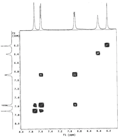
The 1H NMR spectrum of compound 1 showed five signals in the aromatic hydrogen region, consistent with the replacement pattern of the flavonol quercetin: two broad singlets at dH 6.21/6.40, assigned to H-6/H-8, two doublets at dH 6.88 (d, 1H, J 8.0 Hz, H-5’) and 7.58 (d, 1H, J 8.0 Hz, H-6’) as well as one broad singlet at dH 7.71, assigned to H-2’. This spectrum displayed also signals at dH 3.20-3.72 (H-2”-H-6”), which in association to the presence of one doublet at dH 5.24 (d, 1H, J 7.5 Hz, H-1’’), assigned to the anomeric hydrogen trans-diaxial position with H-2, characterize the β-D-glucoside unit. In this spectrum was also observed a broad singlet at dH 5.22 (H-1’’’) assigned to an anomeric di-equatorial hydrogen, which, associated to the doublet at dH 1.25 (d, 3H, J 6.0 Hz, H-6’’’) suggests the presence of α-L-rhamnose.18
The negative HRESIMS of 1 revealed a pseudo-molecular ion at m/z 609.1411 [M-H]–, consistent with the molecular formula C27H30O16. 13C NMR spectra, including DEPT 90° and 135°, displayed 27 carbon signals being one methyl, one methylene, 15 methyne and 10 non-hydrogenated carbons. Oxymethine carbon signals ranging from dC 84 to 69, mainly those at dC 62.5
(C-6”), 17.9 (C-6”’), 102.7 (C-1”’) and 104.3 (C-1”), confirmed the presence of glucose and rhamnose in the molecule of 1.18,19
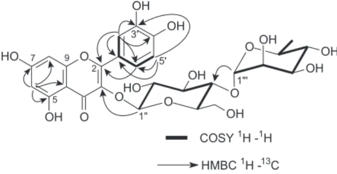
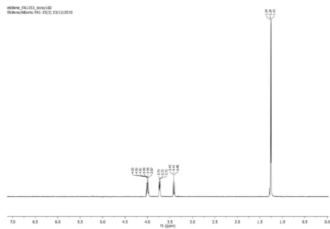
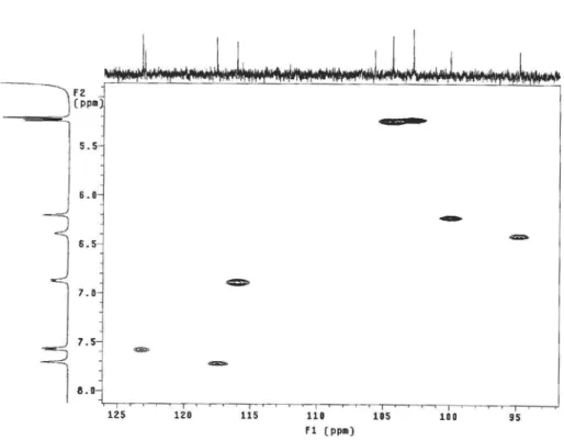
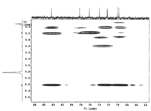
Hydrogen signals of each sugar unit were assigned by analysis of the 1D TOCSY spectrum. Irradiation of the anomeric hydrogen from rhamnose (dH 5.22, H-1”’) allowed the attribution of signals at dH 3.99 (H-2’’/H-5’’’), 3.72 ( H-3’’’), 3.41 (H-4’’’) and 1.25 (H-6’’’) to rhamnose unit and those at dH 3.59 (H-2’’/ H-4’’/ H-6’’a), 3.25 (H-3’’), 3.41 (H-5’’), and 3.72 (H-6’’ b) to glucose unit (Table 1). HMQC, HMBC and DQF-COSY spectra displayed important correlations between hydrogens and carbons of 1 (Figure 2), mainly that of H-1’’ (dH 5.24) with C-3 (dC 135.6), which showed that glucose is linked
to the aglycone at C-3, and that of H-1’’’ (dH 5.22) with C-4’’ (dC 84.4), which indicated that rhamnose is linked at C-4 of glucose. Therefore, analysis of the obtained data was consistent with the new structure quercetin-3-O-β -D-glucopyranosyl-(1→4)-α-L-rhamnopyranoside (1).
The 1H NMR spectrum of 2 showed similarities to that recorded to flavonoid 1, with two broad singlets at dH 6.19/6.38, assigned to H-6/H-8 of ring A. This spectrum displayed also signals at range from dH 3.20 to 5.21 (oxymethine hydrogens) and one doublet at dH 1.24 (J = 6.0 Hz), suggesting the presence of rhamnose in the molecule. The signals superimposed at dH 5.21 (2H) have been assigned to the anomeric protons H-1” and H-1’’’. The main observed difference in the 1H NMR spectrum of 2 is associated to the substitution pattern of kaempferol (1,4-disubstituted B ring), due to the presence of two doublets at dH 6.88 and 8.03 (d, J = 8.0 Hz) integrated to two hydrogens each and thus assigned to H-3’/H-5’ and H-2’/H-6’, respectively. 13C NMR spectra of 2, including DEPT 90° and 135°, showed one carbonyl carbon signal at dC 179.4 (C-4) and aromatic carbon signals at range
dC 166-95, to confirm the kaempferol aglycone moiety. Oxygenated carbons at range dC 84-70, mainly methylene carbons and methyl at dC 62.5 (C-6’’) and 17.9 (C-6’’’), respectively, as well as anomeric carbons at dC 104.2 (C-1”)
Figure 1. Structures of isolated flavonoids from Oxandra sessiliflora
R. E. Fries.
A New Flavonoid Derivative from Leaves of Oxandra sessiliflora R. E. Fries J. Braz. Chem. Soc.
706
and 102.7 (C-1’”), confirming the presence of glucose and rhamnose. These information, associated with literature data for flavonoids with the same aglycone,19 allowed the identification of 2 as kaempferol-3-O-β -D-glucopyranosyl-(1→4)-α-L-rhamnopyranoside, previously isolated from
Acacia pennata Willd (Mimosaceae).20 However, this is the first occurrence in Annonaceae family and the first description of its assigned 13C NMR data.
The structures of flavonoids 3 and 4 were identified by analysis of 1H and 13C NMR as well as HRESIMS and comparison with data described in the literature.18,19
Conclusion
This study contributed to the expansion of the chemical constituents of the Oxandra genus since the compound kaempferol-3-O-β-D-glucopyranosyl-(1→4 )-α
-L-rhamnopyranoside (2) is being described for the first time in Annonaceae while quercetin-3-O-β -D-glucopyranosyl-(1→4)-α-L-rhamnopyranoside (1) is a new compound.
Experimental
General procedures
1H and 13C NMR spectra were obtained on Varian spectrometer-model INOVA, operating at 500 MHz for 1H and 125 MHz for 13C using CD
3OD as a solvent and tetramethylsilane (TMS) as internal reference. HRESIMS spectrum (negative mode) was recorded on a Bruker Daltonics UltrOTOFq-ESI-TOF spectrometer. Silica gel (70-230 mesh, Merck) and Sephadex LH-20 (Amersham Biosciences) were used for column chromatography (CC), whereas silica gel 60 GF254 was employed for analytical
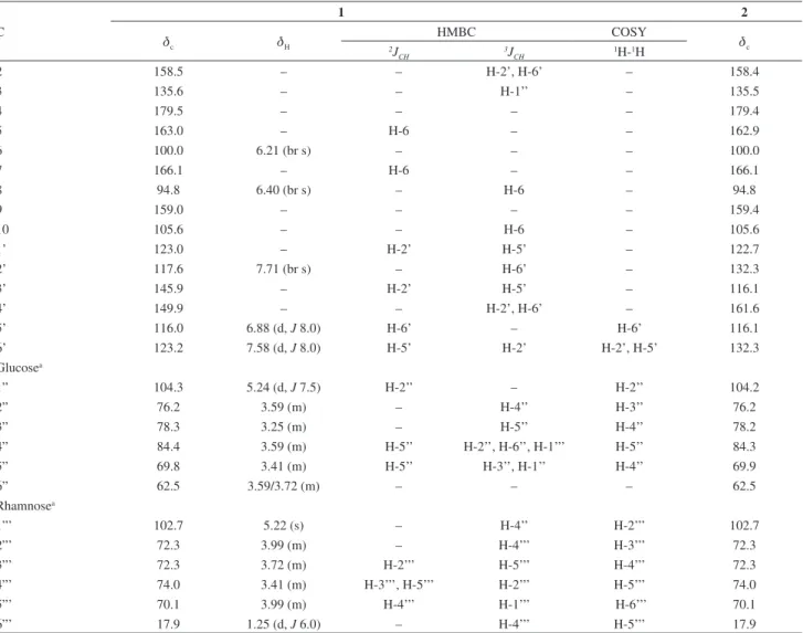
Table 1. NMR data of compounds 1 and 2 (500 MHz and 125 MHz, d in ppm, J in Hz, CD3OD)
C
1 2
dc dH
HMBC COSY
dc 2J
CH 3JCH 1H-1H
2 158.5 – – H-2’, H-6’ – 158.4
3 135.6 – – H-1’’ – 135.5
4 179.5 – – – – 179.4
5 163.0 – H-6 – – 162.9
6 100.0 6.21 (br s) – – – 100.0
7 166.1 – H-6 – – 166.1
8 94.8 6.40 (br s) – H-6 – 94.8
9 159.0 – – – – 159.4
10 105.6 – – H-6 – 105.6
1’ 123.0 – H-2’ H-5’ – 122.7
2’ 117.6 7.71 (br s) – H-6’ – 132.3
3’ 145.9 – H-2’ H-5’ – 116.1
4’ 149.9 – – H-2’, H-6’ – 161.6
5’ 116.0 6.88 (d, J 8.0) H-6’ – H-6’ 116.1
6’ 123.2 7.58 (d, J 8.0) H-5’ H-2’ H-2’, H-5’ 132.3
Glucosea
1” 104.3 5.24 (d, J 7.5) H-2’’ – H-2’’ 104.2
2” 76.2 3.59 (m) – H-4’’ H-3’’ 76.2
3” 78.3 3.25 (m) – H-5’’ H-4’’ 78.2
4” 84.4 3.59 (m) H-5’’ H-2’’, H-6’’, H-1’’’ H-5’’ 84.3
5” 69.8 3.41 (m) H-5’’ H-3’’, H-1’’ H-4’’ 69.9
6” 62.5 3.59/3.72 (m) – – – 62.5
Rhamnosea
1”’ 102.7 5.22 (s) – H-4’’ H-2’’’ 102.7
2”’ 72.3 3.99 (m) – H-4’’’ H-3’’’ 72.3
3”’ 72.3 3.72 (m) H-2’’’ H-5’’’ H-4’’’ 72.3
4”’ 74.0 3.41 (m) H-3’’’, H-5’’’ H-2’’’ H-5’’’ 74.0
5”’ 70.1 3.99 (m) H-4’’’ H-1’’’ H-6’’’ 70.1
6”’ 17.9 1.25 (d, J 6.0) – H-4’’’ H-5’’’ 17.9
Sousa et al. 707 Vol. 25, No. 4, 2014
thin layer chromatography (TLC) (0.50 mm). HPLC analyses were performed on Varian Pro Star with ternary system pumps Model 240, UV-Vis Diode Array Detector (DAD) model 330 and injector model 410 (analytical), and Varian Star Model Prep SD-1 with UV-Vis detector model 320, manual injector Rheodyne model 7725i with sample loop of 2.5 mL (preparative). Phenomenex Gemini C-18 columns (250 × 4.6 mm, 5 µm and 250 × 21 mm, 10 µm) were used to these analyses. Solvents and reagents used were of analytical purity grade and HPLC.
Plant material
The leaves of O. sessiliflora were collected in the Environmental Park of Teresina-PI, in June 2009. The species was identified by Professor Roseli Farias Melo Barros and a voucher specimen with number TEPB 27870 was deposited in the Herbarium Graziela Barroso do Amaral (UFPI).
Extraction and isolation
The leaves of O. sessiliflora were dried at room temperature and then grinded. The obtained material (779 g) was subjected to exhaustive maceration with EtOH at room temperature. After concentration on reduced pressure, 109 g of EtOH extract were obtained (14%). Part of the EtOH extract (86 g) was suspended in MeOH-H2O (2:1) and extracted with hexane, CH2Cl2 and EtOAc successively to afford 21 g (24%), 30 g (35%) and 14 g (17%) of organic phases, respectively.
Part of the EtOAc phase (3.5 g) was suspended in 10 mL of H2O-MeOH 1:1 and the soluble portion was applied in a Stracta column (C18, 10 g), which was eluted with MeOH:H2O 1:1, MeOH and chloroform (CHCl3) successively. The fraction eluted with MeOH-H2O 1:1 (FA1; 1380 mg) was chromatographed on Sephadex LH-20 eluted with MeOH to afford 5 groups (A-E). Group D (345 mg) was analyzed by reverse phase HPLC-UV DAD eluted with exploratory gradient H2O + 0.2% AcOH-MeOH (5% → 100%; 200-600 nm, 1 mL min–1; 50 min) and then subject to a isocratic elution mode. The improved separation of the constituents was achieved with the mobile phase (MeOH-ACN 1:1) / (H2O + 0.2% AcOH) (3:7), resulting in the isolation of flavonoids 1 (20 mg), 2 (21 mg) 3 (11 mg) and 4 (8 mg).
Quercetin-3-O-α-L-rhamnopyranosyl-(1→4)-β -D-glucopyranoside (1)
Yellow amorphous solid; HRESIMS: 609.1411 [M-H]– (calculated to C27H29O16: 609.1455); NMR data: see Table 1.
Kaempferol-3-O-α-L-rhamnopyranosyl-(1→4)-β -D-glucopyranoside (2)
Yellow amorphous solid; 1H NMR (CD
3OD, 500 MHz)
d 6.19 (br s, H-6), 6.38 (br s, H-8), 8.03 (d, J 8.0 Hz, H-2’/H-6’’), 6.88 (d, J 8.0 Hz, H-3’/H-5’), 5.21 (d, J 7.5 Hz, H-1’’), 5.21 (br s, H-1’’’), 1.24 (d, J 6.0 Hz, H-6’’’), 3.20-4.00 (H-2’’ to H-6’’, H-2’’’ to H-5’’’); 13C NMR: see Table 1.
Q u e r c e t i n 3 - O -β- D - g l y c o py r a n o s i l - ( 6→1 ) -α L -rhamnopyranoside (rutin, 3)
Yellow amorphous solid; HRESIMS: 609.1616 [M-H]– (calculated to C
27H29O16: 609.1455) and 301.0851 [M-glucose unit]–; 1H NMR (CD
3OD, 500 MHz) d 6.21 (d,
J 2.0 Hz, H-6), 6.40 (d, J 2.0 Hz, H-8), 7.66 (d, J 2.0 Hz, H-2’), 6.86 (d, J 8.5 Hz, H-5’), 7.60 (dd, J 8.5 and 2.0 Hz, H-6’), 5.11 (d, J 7.5 Hz, H-1’’), 4.52 (d, J 1.5 Hz, H1’’’), 1.18 (d, J 6.0 Hz, H-6’’’), 3.20-3.90 (H-2’’ to H-6’’, H-2’’’ to H-5’’’); 13C NMR (CD
3OD, 125 MHz) d 158.5 (C-2), 135.9 (C-3), 179.5 (C-4), 163.0 (C-5), 100.0 (C-6), 166.1 (C-7), 94.9 (C-8), 159.0 (C-9), 105.6 (C-10), 123.0 (C-1’), 117.9 2’), 145.8 3’), 150.0 4’), 116.1 5’), 123.6 (C-6’), 104.7 (C-1’’), 75.7 (C-2’’), 77.2 (C-3’’), 71.4 (C-4’’), 78.2 (C-5’’), 68.6 (C-6’’), 102.4 (C-1’’’), 72.1 (C-2’’’), 72.3 (C-3’’’), 73.1 (C-4’’’), 69.7 (C-5’’’), 18.0 (C-6’’’).
Kaempferol-3-O-rutinoside (4)
Yellow amorphous solid; HRESIMS: 593.1639, [M-H]– (calculated to C27H29O15: 593.1506), 284.0652 [M-glucose unit]–; 1H NMR (CD
3OD, 500 MHz) d 6.21 (br s, H-6), 6.40 (br s, H-8), 8.06 (d, J 9.0 Hz, H-2’/H-6’), 6.90 (d, J 9.0 Hz, H-3’/H5’), 5.11 (d, J 7.5 Hz, H-1’’), 4.52 (br s, H-1’’’), 1.12 (d, J 6.0 Hz, H-6’’’), 3.27-3.80 (H-2’’ to H-6’’, H-2’’’ to H-5’’’); 13C NMR (CD
3OD, 125 MHz) d 158.7 (C-2), 135.5 (C-3), 179.4 (C-4), 163.1 (C-5), 100.0 (C-6), 166.2 (C-7), 95.0 (C-8), 159.4 (C-9), 105.6 (C-10), 122.8 (C-1’), 132.4 (C-2’/C-6’), 116.2 (C-3’/C-5’), 161.5 (C-4’), 104.6 (C-1’’), 76.8 (C-2’’), 78.2 (C-3’’), 71.5 (C-4’’), 77.2 (C-5’’), 68.6 (C-6’’), 102.4 (C-1’’’), 72.1 (C-2’’’), 72.3 (C-3’’’), 74.0 (C-4’’’), 69.7 (C-5’’’), 17.9 (C-6’’’).
Supplementary Information
Supplementary information (NMR and LRESIMS for compounds 1-4) is available free of charge at http://jbcs. sbq.org.br as PDF file. (Figures S1 to S26).
Acknowledgments
A New Flavonoid Derivative from Leaves of Oxandra sessiliflora R. E. Fries J. Braz. Chem. Soc.
708
References
1. Lobão, A. Q.; Araujo, D. S. D.; Kurtz, B. C.; Rodriguésia2005,
56, 85.
2. Leboeuf, M.; Cavé, A.; Bhaumik, P. K.; Mukherjee, B.; Murkherjee, R.; Phytochemistry1982, 21, 2783.
3. Forzza, R. C.; Leitman, P. M.; Costa, A. F.; Carvalho Jr, A. A.; Peixoto, A. L.; Walter, B. M. T.; Bicudo, C.; Zappi, D.; Costa, D. P.; Lleras, E.; Martinelli, G.; Lima, H. C.; Prado, J.; Stehmann, J. R.; Baumgratz, J. F. A.; Pirani, J. R.; Sylvestre, L. S.; Maia, L. C.; Lohmann, L. G.; Paganucci, L.; Silveira, M.; Nadruz, M.; Mamede, M. C. H.; Bastos, M. N. C.; Morim, M. P.; Barbosa, M. R.; Menezes, M.; Hopkins, M.; Secco, R.; Cavalcanti, T.; Souza, V. C. In Catálogo de Plantas e Fungos do Brasil; vol. 1, Jardim Botânico do Rio de Janeiro: Rio de
Janeiro, 2010.
4. Rojano, B. A.; Gaviria, C. S. A.; Gil, M. A.; Saez, J. A.; Schinella, G.; Tournier, H.; Vitae2008, 15, 173.
5. Rojano, B. A.; Gaviria, M.; Carlos, A.; Saez, V.; Jairo, A.; Yepes, F.; Munoz, F.; Ossa, F.; Vitae2007, 14, 95.
6. Rojano, B.; Pérez, E.; Figadère, B.; Martin, M. T.; Recio, M. C.; Giner, R.; Rios, J. L.; Schinella, G.; Saez, J.; J. Nat. Prod. 2007,
70, 835.
7. Guinaudeau, H.; Leboeuf, M.; Cavé, A.; J. Nat. Prod.1988, 51, 389.
8. Arango, G.; Cortes, D.; Cavé, A.; Phytochemistry1987, 26, 1227.
9. Arango, G. J.; Cortes, D.; Cassels, B. K.; Cavé, A.; Merienne, C.;
Phytochemistry1987, 26, 2093.
10. Zhang, J.; El-Shabrawy, A. O.; El-Shanawany, M. A.; Schiff-Jr, P. L.; Slatkin, D.; J. Nat. Prod. 1987, 50, 800.
11. El-Shanawany, M. A.; Bull. Pharmaceut. Sci.1985, 8, 127. 12. El-Shanawany, M. A.; Slatkin, D. J.; Schiff, P. L.;
El-Shabrawy, A.; Bull. Pharmaceut. Sci.1985, 8, 172. 13. Hocquemiller, R.; Cortes, D.; Arango, G. J.; Myint, S. H.;
Cave, A.; J. Nat. Prod. 1991, 54, 445.
14. Tinto, W. F.; Blair, L. C.; Reynolds, W. F.; Mclean, S.; J. Nat. Prod. 1992, 55, 701.
15. Mesquita, M. R.; Castro, A. A. J. F.; Publ. Avulsas Ciênc. Amb. 2007, 15, 1.
16. Abreu, M. C.; Castro, A. A. J. F.; Publ. Avulsas Ciênc. Amb. 2004, 9, 1.
17. Silva, A. A. C. A.; Souza, E. A.; Matsuo, A. L.; Lago, J. H. G.; Chaves, M. H.; J. Med. Plants Res.2013, 7, 504.
18. Guvenalp, Z.; Kiliç, N.; Kazaz, C.; Kaya, Y.; Demirezer, O.;
Turk. J. Chem.2006, 30, 515.
19. Agrawal, P. K.; Carbon-13 NMR of Flavonoids, Agrawal, P. K., ed.; Elsevier: Amsterdam, 1989.
20. Dongmo, A. B.; Milyamoto, T.; Yoshikawa, K.; Arihara, S.; Lacaille-Dubois, M.; Planta Med. 2007, 73, 1202.
Submitted on: September 24, 2013
Published online: January 31, 2014
Supplementary Information
S
I
J. Braz. Chem. Soc., Vol. 25, No. 4, S1-S13, 2014. Printed in Brazil - ©2014 Sociedade Brasileira de Química 0103 - 5053 $6.00+0.00
*e-mail: [email protected]
A New Flavonoid Derivative from Leaves of
Oxandra sessiliflora
R. E. Fries
Elcilene A. de Sousa,a Armenio A. C. A. da Silva,a Alberto J. Cavalheiro,b
João Henrique G. Lagoc and Mariana H. Chaves*,a
aDepartamento de Química, Universidade Federal do Piauí, 64049-550 Teresina-PI, Brazil
bInstituto de Química, Universidade Estadual Paulista, CP 355, 14800-900 Araraquara-SP, Brazil
cInstituto de Ciências Ambientais, Químicas e Farmacêuticas, Universidade Federal de São Paulo,
09972-270 Diadema-SP, Brazil
A New Flavonoid Derivative from Leaves of Oxandra sessiliflora R. E. Fries J. Braz. Chem. Soc.
S2
Figure S2. 1H NMR spectrum (CD
3OD, 500 MHz) of compound 1 isolated from leaves of Oxandra sessiliflora.
Figure S3.13C NMR spectrum (CD
Sousa et al. S3 Vol. 25, No. 4, 2014
Figure S4. DEPT 135o NMR experiment (CD
3OD, 125 MHz) of compound 1 isolated from leaves of Oxandra sessiliflora.
Figure S5. DEPT 90o NMR experiment (CD
3OD, 125 MHz) of compound 1 isolated from leaves of Oxandra sessiliflora.
A New Flavonoid Derivative from Leaves of Oxandra sessiliflora R. E. Fries J. Braz. Chem. Soc.
S4
Figure S7. TOCSY 1D NMR experiment (CD3OD, 125 MHz) of compound 1 isolated from leaves of Oxandra sessiliflora. Irradiation of the signal δ 5.22.
Sousa et al. S5 Vol. 25, No. 4, 2014
Figure S9. Expansion HMQC NMR experiment (CD3OD, 500 × 125 MHz) of compound 1 isolated from leaves of Oxandra sessiliflora.
A New Flavonoid Derivative from Leaves of Oxandra sessiliflora R. E. Fries J. Braz. Chem. Soc.
S6
Figure S11. gCOSY NMR experiment (CD3OD, 500 MHz) of compound 1 isolated from leaves of Oxandra sessiliflora.
Sousa et al. S7 Vol. 25, No. 4, 2014
Figure S14. Expansion HMBC NMR experiment (CD3OD, 500 × 125 MHz) of compound 1 isolated from leaves of Oxandra sessiliflora.
A New Flavonoid Derivative from Leaves of Oxandra sessiliflora R. E. Fries J. Braz. Chem. Soc.
S8
Figure S15. Expansion HMBC NMR experiment (CD3OD, 500 × 125 MHz) of compound 1 isolated from leaves of Oxandra sessiliflora.
Sousa et al. S9 Vol. 25, No. 4, 2014
Figure S17. 1H NMR spectrum (CD
3OD, 500 MHz) of compound 2 isolated from leaves of Oxandra sessiliflora.
Figure S18. 13C NMR spectrum (CD
A New Flavonoid Derivative from Leaves of Oxandra sessiliflora R. E. Fries J. Braz. Chem. Soc.
S10
Figure S19. DEPT 135o NMR experiment (CD
3OD, 125 MHz) of compound 2 isolated from leaves of Oxandra sessiliflora.
Figure S20. DEPT 90o NMR experiment (CD
Sousa et al. S11 Vol. 25, No. 4, 2014
Figure S22. 1H NMR spectrum (CD
3OD, 500 MHz) of compound 3 isolated from leaves of Oxandra sessiliflora.
A New Flavonoid Derivative from Leaves of Oxandra sessiliflora R. E. Fries J. Braz. Chem. Soc.
S12
Figure S23. 13C NMR spectrum (CD
3OD, 125 MHz) of compound 3 isolated from leaves of Oxandra sessiliflora.
Sousa et al. S13 Vol. 25, No. 4, 2014
Figure S25. 1H NMR spectrum (CD
3OD, 500 MHz) of compound 4 isolated from leaves of Oxandra sessiliflora.
Figure S26. 13C NMR spectrum (CD