Ciprofloxacin Derivatives Affect Parasite Cell
Division and Increase the Survival of Mice
Infected with
Toxoplasma gondii
Erica S. Martins-Duarte1,2, Faustine Dubar3,4, Philippe Lawton5, Cristiane França da Silva6, Maria de Nazaré C. Soeiro6, Wanderley de Souza1,2,7, Christophe Biot3, Rossiane
C. Vommaro1,2*
1Universidade Federal do Rio de Janeiro—Instituto de Biofísica Carlos Chagas Filho, Rio de Janeiro, Brazil, 2Instituto Nacional de Ciência e Tecnologia em Biologia Estrutural e Bioimagens, Rio de Janeiro, Brazil, 3Université de Lille1—Unité de Glycobiologie Structurale et Fonctionnelle, UGSF, F-59650 Villeneuve d'Ascq, France,4CNRS, UMR 8576, F-59650 Villeneuve d'Ascq, France,5Université de Lyon, Université Claude-Bernard Lyon1, ISPB-Faculté de Pharmacie, Lyon, France,6Laboratório de Biologia Celular do Instituto Oswaldo Cruz/Fiocruz, Rio de Janeiro, Brazil,7Instituto Nacional de Metrologia, Qualidade e Tecnologia—Inmetro, Rio de Janeiro, Brazil
*vommaro@biof.ufrj.br
Abstract
Toxoplasmosis, caused by the protozoanToxoplasma gondii, is a worldwide disease whose clinical manifestations include encephalitis and congenital malformations in new-borns. Previously, we described the synthesis of new ethyl-ester derivatives of the antibiotic ciprofloxacin with ~40-fold increased activity againstT.gondii in vitro, compared with the original compound. Cipro derivatives are expected to target the parasite’s DNA gyrase com-plex in the apicoplast. The activity of these compoundsin vivo, as well as their mode of ac-tion, remained thus far uncharacterized. Here, we examined the activity of the Cipro derivativesin vivo, in a model of acute murine toxoplasmosis. In addition, we investigated the cellular effectsT.gondiitachyzoitesin vitro, by immunofluorescence and transmission electron microscopy (TEM). When compared with Cipro treatment, 7-day treatments with Cipro derivatives increased mouse survival significantly, with 13–25% of mice surviving for up to 60 days post-infection (vs. complete lethality 10 days post-infection, with Cipro treat-ment). Light microscopy examination early (6 and 24h) post-infection revealed that 6-h treatments with Cipro derivatives inhibited the initial event of parasite cell division inside host cells, in an irreversible manner. By TEM and immunofluorescence, the main cellular effects observed after treatment with Cipro derivatives and Cipro were cell scission inhibition -with the appearance of‘tethered’parasites–malformation of the inner membrane complex, and apicoplast enlargement and missegregation. Interestingly, tethered daughter cells re-sulting from Cipro derivatives, and also Cipro, treatment did not show MORN1 cap or centrocone localization. The biological activity of Cipro derivatives againstC.parvum, an apicomplexan species that lacks the apicoplast, is, approximately, 50 fold lower than that in
T.gondiitachyzoites, supporting that these compounds targets the apicoplast. Our results show that Cipro derivatives improved the survival of mice acutely infected withT.gondiiand
OPEN ACCESS
Citation:Martins-Duarte ES, Dubar F, Lawton P, França da Silva C, Soeiro MdNC, de Souza W, et al. (2015) Ciprofloxacin Derivatives Affect Parasite Cell Division and Increase the Survival of Mice Infected withToxoplasma gondii. PLoS ONE 10(5): e0125705. doi:10.1371/journal.pone.0125705
Academic Editor:Silvia N Moreno, Univ. Georgia, UNITED STATES
Received:November 17, 2014
Accepted:March 17, 2015
Published:May 7, 2015
Copyright:© 2015 Martins-Duarte et al. This is an open access article distributed under the terms of the Creative Commons Attribution License, which permits unrestricted use, distribution, and reproduction in any medium, provided the original author and source are credited.
Data Availability Statement:All relevant data are within the paper.
inhibited parasite replication early in the first cycle of infectionin vitro, highlighting their ther-apeutic potential for the treatment of toxoplasmosis.
Introduction
Toxoplasma gondii, the causative agent of toxoplasmosis, is a globally spread parasite infecting approximately one third of the human population. Toxoplasmosis has a broad spectrum of clinical presentations. Infection of the central nervous system by this parasite is associated to encephalitis and eye disease. Indeed, the vertical transmission during pregnancy can lead to abortion and congenital malformations in the newborn [1].Whereas infections by this parasite are usually asymptomatic in most individuals,T.gondiiis an important opportunistic pathogen with high mortality and morbidity in immunocompromised patients [2]. Indeed, ocular toxo-plasmosis rates can be extremely high, even in immunocompetent individuals, especially in South American countries [2–4].
Despite the worldwide prevalence of this potentially serious medical condition, toxoplasmo-sis treatment is limited to a small number of drugs that are associated with numerous side ef-fects. The combination of pyrimethamine with sulfadiazine is the first choice of treatment for toxoplasmosis; however, patients often do not tolerate sulfadiazine, and long-term treatment (4–6 weeks) with this drug is commonly associated with gastrointestinal disorders that lead to treatment discontinuation. In patients with intolerance to sulfadiazine, pyrimethamine is com-bined with clindamycin or atovaquone, which also cause gastrointestinal disorders [5,6]. Over-all, it is clear that the development of alternative or replacement treatments for toxoplasmosis is vital for improving disease treatment and control.
The discovery of a‘relic’chloroplast (apicoplast) in apicomplexan parasites–a group that includesT.gondiiand alsoPlasmodium spp., the causative agents of malaria—brought new possibilities for the identification of novel targets for therapy against these parasites [7,8]. Al-though photosynthetic function was lost during apicoplast evolution, this organelle still har-bors many metabolic pathways of prokaryotic origin and whose functions are essential for parasite survival [9,10]. Due to their prokaryotic origins, the apicoplast pathways represent in-teresting targets for the development of specific anti-parasitic compounds with limited toxicity to host cell pathways of eukaryotic origin. The chemotherapeutic potential of the metabolic ac-tivities operating in the apicoplast is well recognized [8,11–13], the antibiotics clindamycin and azithromycin, inhibitors of apicoplast protein synthesis, are already in clinical use as a sec-ond-line therapy for toxoplasmosis treatment [5].
Among apicoplast pathways, DNA replication is an important potential chemotherapeutic target. Fluoroquinolones are known DNA replication inhibitors that target prokaryotic type II topoisomerases (DNA gyrase and topoisomerase IV) [14] These antibiotics have a broad spec-trum of activity against various pathogens, including bacteria, mycoplasma, and protozoan parasites [15,16]. They are generally well tolerated by most patients, and are commonly used in the clinic [17].
DNA gyrases introduce negative supercoiling into the DNA strand during DNA replication and transcription. The DNA gyrase enzymatic complex is formed of two copies of both A and B subunits (A2B2); subunits A catalyze DNA double-strand breaks, while subunits B have ATPase function [14]. Homologues of DNA gyrase subunits A and B genes are found in the nuclear genome of apicomplexan parasites (probably as a result of gene transfer from the or-ganelle’s ancestral genome, during apicoplast evolution), and their protein products are
and analysis, decision to publish, or preparation of the manuscript.
targeted exclusively to the apicoplast [18]. Pharmacological inhibition of subunits A and B of the apicoplast’s DNA gyrase–by fluoroquinolone and novobiocin, respectively—inhibit apico-plast genome replication and affect parasite viability, validating this enzyme as a potential drug target in apicomplexans [8,19].
In a previous work, we described the synthesis andin vitrotests againstP.falciparumandT. gondiiof novel ester prodrugs of ciprofloxacin (Cipro), a known fluorquinolone [20]. Chemical modifications of the reference compound yielded, on average, a 40-fold increase in the anti-parasitic activity compared with the original molecule, and Cipro derivatives had low toxicity against mammalian cells (murine splenocytes and the LLCMK2epithelial cell line) [20].
Among the ester prodrugs of Cipro tested againstT.gondii in vitro, compounds 2 (pro-drug), 4 (phenyl substituent) and 5 (adamantanyl substituent) were particularly potent, with IC50values in the nanomolar or low micromolar range (0.42μM and 0.46μM, for compounds
2 and 5, respectively, and 1.24μM for compound 4, after 48 h of treatment) [20]. The presence
of ethyl esters on the carboxyl of Cipro contributed to increase the activity of these derivatives compared with that of the original compound [20]. Esterification of Cipro into prodrug com-pounds is expected to make the original molecule more lipophilic, favoring the diffusion across biological membranes—including the multiple membranes ofT.gondii, and also those of the apicoplast, which harbors the likely target of Cipro derivatives (DNA gyrase).
Although the Cipro derivatives synthesized and described by us previously are expected to target the apicoplast’s DNA gyrase complex, their cellular mode of action in apicomplexans has not been examined. Also, these drugs were not testedin vivoagainst infections by apicomplexan parasites.
In the present study, we evaluated the activities of compounds 2, 4 and 5 againstT.gondii infectionin vivo, in a murine model of acute toxoplasmosis. Also, we characterized the cellular effects of these compounds–henceforth referred to as Et-Cipro (compound 2), Ph-Cipro (compound 4), and Adam-Cipro (compound 5)—to elucidate their mode of action inT.gondii. We show that Et-cipro and Adam-cipro treatment increased mouse survival compared with Cipro treatment, disrupted parasite cell division and affected apicoplast segregation. Interest-ingly, Cipro derivatives could not inhibit the proliferation ofCryptosporidium parvum, an api-complexan species that lacks the apicoplast.
Materials and Methods
Ethical Statement
The experimental protocols for animal use in this study were approved by the institutional Eth-ics Committee for Animal Use (CEUA, IBCCF, UFRJ: approval IDs 208-09/16 and 206-09/16; CEUA Fiocruz—LW-16/13), and are in agreement with the Brazilian federal law (11.794/2008, Decreto n° 6.899/2009).
Parasites and Mice
In this study, we used tachyzoite forms the RH strain ofToxoplasma gondii, obtained from the peritoneal cavity of mice 2 days after infection [21]. Four to six-week-old female Swiss mice (18–22 g) were used for thein vivoexperiments. Drinking water and food were givenad libitum.
In vivo
toxicity analysis
200 mg/kg/day) and monitored for a period of 48 hours for the appearance of toxic and sub-toxic symptoms (weight body loss and animal behavior alterations). During the sub-toxicity analy-sis no animal has died, then after the 48h period of observation after drug administration mice were anesthetized with CO2and blood was collected by cardiac puncture to determine the serum levels of urea and creatinine kinase at CECAL/Fiocruz platform (Ortho Clinical-John-son & JohnClinical-John-son), as reported previously [22].
To determine compound efficacy againstT.gondii in vivo, female Swiss mice were infected intraperitoneally (i.p.) with 5x10³T.gondiitachyzoites and treated with test compounds from 24 h post-infection. Groups of 3–4 mice were housed per cage and arbitrarily assigned to one of the following treatment groups: Cipro, Et-Cipro, Ph-Cipro or Adam-Cipro (50–150 mg/kg/ day), or untreated (i.e., treated with vehicle, polyethylene glycol/PEG). Mice were treated once daily for 7 days, by oral gavage, and mouse mortality was monitored once a day for a period of 60 days. During survival studies, mice were not inflicted to any suffering condition and mice presenting morbidity symptoms (shivering, ruffled hair and immobility) were euthanized by CO2asphyxiation to minimize animal suffering, and then mortality was scored. Survival curves were calculated using the Kaplan and Meier method, and compared using the log-rank (Man-tel-Cox) test, in GraphPad Prism 5.0 (GraphPad Software Inc.) andP<0.05was considered
sta-tistically significant. The following numbers of mice were used in this study: in untreated groups, n = 10 (Cipro control) or n = 14/15 (Et-Cipro, Ph-Cipro and Adam-Cipro controls), in 3–4 groups; in Cipro groups, n = 11 (50 and 100 mg; 3 groups) and n = 8 (150 mg Cipro; 2 groups); in Et-Cipro groups, n = 11 (50 and 100 mg; 3 groups); in Ph-Cipro groups, n = 8 (50 and 100 mg; 2 groups) and n = 11 (150 mg; 3 groups); and in Adam-Cipro groups, n = 3 (50 mg; 1group) and n = 12 (100 mg; 3 groups).
Drug treatments
in vitro
For the treatment ofT.gondiitachyzoites with Cipro derivativesin vitro, 5x10⁵LLC-MK2cells (ATCC CCL7, Rockville, MD/EUA) in RPMI medium were plated on coverslips inside 24-well tissue culture plates, and allowed to settle at 37°C (5% CO2) for ~24h. Then, cells were infected with parasites (freshly-harvested from infected mice) at a 10:1 ratio of parasites to host cells. Tachyzoites were allowed to interact with host cells for 1 h, cell monolayers were washed three times with medium (to remove extracellular parasites) and then treated with different concen-trations of Cipro (20–50μM), Et-Cipro (0.5–5μM), or Adam-Cipro (0.5–5μM), for 6 h at
37°C. Then, coverslips were fixed with Bouin and stained with panoptic (solutions 2 and 3 of the‘Panotico rápido’kit, from Laborclin Ltda., Paraná, Brazil). Alternatively, cultures were washed three times with 0.5 ml of medium (to remove test compounds) and incubated for an additional 18 h in medium without drugs, before fixing and staining. The number of parasites per vacuole was scored by direct light microscopy examination. Three independent experi-ments were performed for each treatment condition, and the results were analyzed by two-way ANOVA (using GraphPad Prism 5.0).
For plaque assay, 6-well tissue culture plates were seeded with human foreskin fibroblasts (HFF; ATCC). HFF was grown in high glucose Dulbeco’s Modified Eagle Medium supple-mented with 10% FBS, 2mM L-glutamine, 100 U/ml penicillin, and 100μg/ml streptomycin
Immunofluorescence microscopy
For immunofluorescence assays, LLC-MK2cells infected with tachyzoites at a 10:1 ratio of par-asites to host cells were treated with Et-Cipro, Adam-Cipro and Ph-Cipro compounds for 24 h. For Cipro assay, cells were infected in a ratio of 5:1 parasites per host cell and treated for 24, 48 and 72h. After treatment, infected cells were fixed in 3.7% freshly-prepared formaldehyde, permeabilized with 0.5% Triton X-100 for 15 min, and blocked with 3% bovine serum albumin (BSA) in PBS at pH 7.4 and room temperature for 1 h. All antibody incubations were per-formed in 3% BSA in PBS, for 1h, at room temperature. The following primary antibodies were used: anti-SAG1 (kindly provided by Dr. John Boothroyd, Stanford University School of Medi-cine, USA), at a dilution of 1:1000; anti-HSP60 and anti-Morn1 (kindly provided by Dr. Boris Striepen, University of Georgia, USA), diluted to 1:2000 and 1:100, respectively; and anti-IMC1 (kindly provided by Dr. Gary Ward, University of Vermont, USA), diluted to 1:1000. The secondary antibodies used here were anti-mouse/rabbit coupled to Alexa 488 or 546 (Mo-lecular Probes). Sytox green (Mo(Mo-lecular Probes) and DAPI (Sigma-Aldrich) were used to label the DNA. After labeling, the coverslips were mounted onto slides using Prolong gold (Life Technologies), and samples were examined on a TCSSP5 Leica or Zeiss LSM710 laser scanning confocal microscopes.
Transmission electron microscopy (TEM)
For TEM, LLC-MK2cultures were infected with tachyzoites as described above and then treat-ed with 1 and 5μM Et-Cipro and Adam-Cipro for 24 and 48 h, 20μM Cipro for 48 and 72h
or left untreated (control). After treatment, cells were fixed in 2.5% glutaraldehyde in 0.1 M so-dium cacodylate buffer (pH 7.4), and post-fixed for 45 min (and in the dark) in 1% osmium tetroxide, 1.25% potassium ferrocyanide and 5 mM CaCl2, in 0.1 M sodium cacodylate buffer (pH 7.4). Samples were dehydrated in acetone solutions of increasing concentrations (30– 100%) and embedded in PolyBed (Polyscience Inc., Warrington, PA, USA). Ultrathin sections were stained with uranyl acetate and lead citrate, and then observed in a Zeiss 900 Electron Mi-croscope (Carl Zeiss, Inc.) or in a Jeol 1200 EX electron miMi-croscope (Jeol LTD, Tokyo, Japan).
Cryptosporidium parvum in vitro
assay
C.parvumoocysts obtained from infected neonatal calves (Institut National de la Recherche Agronomique-INRA, Nouzilly, France) were purified as previously described [23] and stored in PBS at 4°C. Madin Darby bovine kidney cell line (MDBK; ECACC # 88081201, Sophia Anti-polis, France) was cultured at 37°C in a 5% CO2moist atmosphere in growth medium: RPMI 1640 (Sigma, L’Isle d’Abeau, France) supplemented with 25 mM HEPES (Sigma H-3375), 200 U/ml of penicillin, 200μg/ml of streptomycin (Sigma P-0781) and 10% fetal calf serum (FCS;
Dutscher, Brumath, France).
Prior to infection, MDBK cells were trypsinized and seeded in just one well of a 24-well plate in 2 ml of RPMI 1640 with 1% FCS. Aseptic oocysts were prepared for excystation by in-cubation in RPMI-1640, pH 2 (for 30 min at 37°C). Then, oocytes were washed in RPMI 1640 with 1% FCS and allowed to interact with MDBK cells for 1.5 h at 37°C in a 5% CO2 atmo-sphere.After 3 washes by centrifugation at 160gin RPMI 1640 to remove non-excysted oocysts and shells, infected cells were plated in 24-well plates containing glass coverslips, in 500μl/well
of RPMI/1%FCS, and allowed to settle for 24 h. The infected cells were subsequently washed 3 times with RPMI 1640 and allowed to grow for a further 5 h (in 500μl/well of RPMI/1% FCS).
500μl of medium containing twice-concentrated Cipro derivatives were then added in
the effects against sporozoite proliferation were evaluated by examining at least 1000 cells/cov-erslip in a light microscope, using an oil immersion X100 objective.
Results
Cipro derivatives increased the survival of mice acutely infected with
T
.
gondii
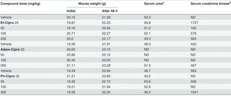
In a previous study, we showed that new prodrugs derivatives of the known antibiotic cipro-floxacin were at least 40 fold more active than the original molecule againstT.gondii tachy-zoites, the form of the parasite that causes acute disease [20]. In preparation forin vivotesting of ciprofloxacin derivatives againstT.gondiiinfection in mice, we performed an acute toxicity analysis of the test compounds using a‘single-dose assay’, where uninfected mice were admin-istered with a single oral dose of 25–200mg/kg of Cipro derivatives, and then monitored for side effects for up to 48 h. During the toxicity assay, no animal has died and no alterations in mouse behavior or body weight gain were observed 48 h after drug administration (Table 1). Biochemical assays showed that serum urea levels increased 48 hours after treatment with Cipro derivatives; however, this was also observed in vehicle-treated animals. Mice treated with higher doses of Et-Cipro and Adam-Cipro showed normal serum levels of creatinine kinase, but animals treated with 200 mg/kg of Ph-Cipro had slightly altered serum levels of
this enzyme.
Swiss mice were administrated with a single oral dose of different ciprofloxacin derivatives, and monitored for 48h.
Table 1. Toxicity of ciprofloxacin derivatives in mice.
Compound dose (mg/kg) Mouse weight (g) Serum ureaa Serum creatinine kinaseb
Initial After 48 h
Vehicle 20.15 21.28 53.0 ND
Et-Cipro25 19.87 22.25 55.8 1727
50 19.15 20.92 51.2 182
100 20.71 22.27 55.1 578
200 20.0 22.17 49.3 583
Vehicle 19.30 21.91 48.0 420
Adam-Cipro25 20.65 23.10 ND ND
50 20.80 22.12 ND ND
100 20.45 23.50 ND ND
200 21.11 23.28 51.5 467
Vehicle 19.49 22.94 46.7 563
Ph-Cipro25 21.21 23.85 49.2 ND
50 19.30 22.72 50.6 506
100 19.21 21.94 52.8 ND
200 19.93 22.20 46.2 1541
Swiss mice were administrated with a single oral dose of different ciprofloxacin derivatives, and monitored for 48h.
Serum levels of urea and creatinine kinase were measured in blood samples harvested 40h after drug administration. The following reference values (mg/kg) were used:
a18–29 mg/dL b
,1070 U/L.
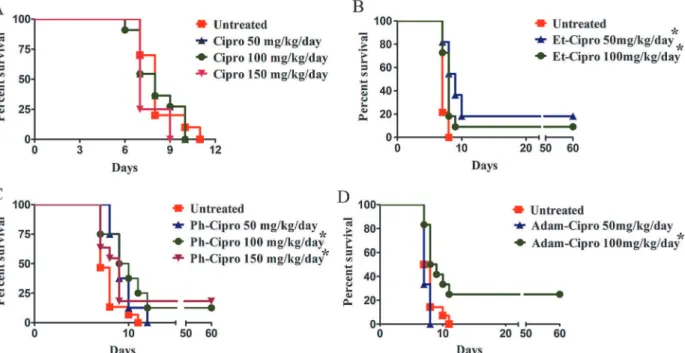
Treatment of mice infected withT.gondiitachyzoites with non-toxic doses of Cipro deriva-tives for 7 days resulted in increased mouse survival (Fig 1A–1D). All mice treated with Cipro died by day 10 post-infection (Fig 1A). In contrast, significantly increased survival ofT. gondii-infected mice was observed after treatment with 50 mg/kg/day Et-Cipro (18%;P<0.05;Fig
1B) or 100 or 150 mg/kg/day Ph-Cipro (13 and 18%, respectively;P<0.05;Fig 1C), or 100 mg/
kg/day Adam-Cipro (25%;P<0.05;Fig 1D). These mice were still alive 60 days post-infection
(Fig 1). Brain examination of 60-days survived mice did not show encysted parasites (presence of chronic infection) (data not shown).
Cipro derivatives inhibit
T
.
gondii
cell division early during infection
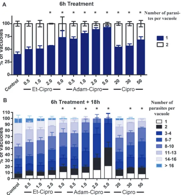
Tachyzoites of the RH strain ofT.gondiihave a doubling time of around 6–8 h inside the para-sitophorous vacuole of infected cells [25]. Treatment of infected cells with different concentra-tions of Et-Cipro, Adam-Cipro, and Cipro for just 6 h (7 h total infection) significantly decreased the number of parasitophorous vacuoles containing two parasites (as a result of cell division after invasion) compared with untreated controls, showing that the Cipro derivatives had an early effect on parasite division (Fig 2A). Adam-Cipro was the most active, as even low concentrations of this derivative (0.5μM and 1μM) significantly inhibited parasite division
compared with untreated controls. The effect of treatment with 0.5μM of Adam-Cipro was
similar to that of 50μM Cipro, with 70% and 68% of the parasitophorous vacuoles having just
one parasite, respectively, corresponding to an 100-fold increase in activity for Adam-Cipro compared with the original compound. In contrast, higher concentrations of Et-Cipro
(>2μM) were required to affect parasite division significantly (Fig 2A). Nevertheless, the effect
of 2μM of Et-Cipro on early parasite division was similar to that of 20μM of Cipro, with 57%
Fig 1. Effect of 7-day treatment with different doses of Cipro derivatives on the survival mice.Swiss Webster mice were infected i.p. with 5x10³
tachyzoites ofT.gondii(RH strain) one day prior to the start of treatment. Results were evaluated by the Kaplan-Meier product limit method, and compared using the log-rank (Mantel-Cox) test.*P<0.05 vs. untreated controls. The numbers of treated mice in each group were: untreated, 10 (A), 14 (B and D) and 15 (C); 50 and 100 mg Cipro, 11 (three groups each); 150 mg Cipro, 8 (two groups); 50 and 100 mg Et-Cipro, 11 (three groups); 50 and 100 mg Ph-Cipro, 8 (two groups); 150 mg Ph-Cipro, 11 (three groups); 50 mg Adam-Cipro, 3 (one group); 100 mg Adam-Cipro, 12 (three groups).
and 54% of the vacuoles containing just one parasite, respectively, which represents a 10-fold increase in cell division inhibition compared with the original compound.
The effect of Cipro derivatives on parasite division was sustained even after these com-pounds were removed from the culture medium and infection was allowed to proceed for an additional 18 h (Fig 2B). The number of parasites per vacuole for all Adam-Cipro treatments was significantly lower than that observed in control, untreated cells, with less than 1% of vacu-oles containing16 parasites in cells treated with 0.5μM of Adam-Cipro. After treatment with
2 or 5μM of Adam-Cipro, approximately 99% of vacuoles contained up to eight parasites only.
The presence of large numbers of vacuoles containing just one parasite 18 h after drug removal shows that some parasites were irreversibly affected by Cipro derivatives treatment.
Fig 2. Early effects of ciprofloxacin (Cipro) derivatives in tachyzoite cell division inside host cells.(A) Tachyzoites ofT.gondiiwere treated with Cipro derivatives for 6 h, from 1–7 h post-infection, and the number of parasites per vacuole was analyzed by light microscopy. (B) After 6 h, the medium containing drugs was removed and replaced with fresh medium without drugs. Tachyzoites were allowed to proliferate for an additional 18 h, and the number of parasites per vacuole was counted.*P<0.05, results were analyzed by two-way ANOVA statistical test.
The antiproliferative effect of Cipro derivatives and Cipro was also confirmed by plaque assay (S1 Fig). No growth was observed after 10 days treatment with 5μM of Adam-Cipro or
Et-Cipro, 10μM of Ph-Cipro and 20 of Cipro. In contrast, untreated cultures presented many
plaques. A significant decrease in parasite proliferation was also observed after treatment with 0.5μM Adam-Cipro and 10μM Cipro.
Treatment with ciprofloxacin derivatives prevents daughter cell budding
(but not mitosis) during tachyzoite cell division
DuringT.gondiicell division by‘endodyogeny’, two daughter cells are assembled in the interi-or of the mother cell after a single round of DNA replication, the daughter inner membrane complex (IMC) provides‘scaffolding’for the newly formed and segregating daughter cell com-ponents [26,27]. At the end of cytokinesis, the mother cell’s original IMC disassembles, and daughter cells‘bud out’incorporating the mother cell’s plasma membrane and leaving behind a residual body [26,27]. Apicomplexan mitosis is‘closed’, with the nuclear envelope remaining intact throughout the process, and being easily recognizable by its U shape [27].
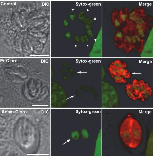
To elucidate the impact of treatment with Cipro derivatives on theT.gondiicell division process, we evaluated the effect of Cipro derivatives by immunofluorescence microscopy using antibody markers that recognize key parasite structures (Figs3and4). Untreated parasites la-beled with an antibody that recognizes SAG1, a major tachyzoite surface protein, and with Sytox-green (to label the DNA) showed a typical‘rosette’organization within host cells, with groups of parasites clustered with their apical/anterior ends projecting outwards, while their posterior ends clustered at the center of the rosette (untreated;Fig 3). In rosettes found in un-treated cells, there was one nucleus per parasite, and some parasites were in the process of nu-clear division, as evidenced by the presence of U-shaped nuclei (Fig 3, arrowheads in
untreated). Treatment with 5μM Et-Cipro and Adam-Cipro for 24 h led to the formation of
large multinucleated parasites (Fig 3, arrows), with loss of the typical rosette structure. Given that Cipro targets the apicoplast, we evaluated the effect of the new Cipro derivatives on the replication and division of this organelle, by observing dividing parasites labeled with the anti-HSP60 (Fig 4). To confirm that treatment with Cipro derivatives led to cell division ar-rest, cells were also labeled with an antibody that recognizes the specific IMC component IMC1 to improve visualization of daughter cell boundaries in dividing parasites (Fig 4). In non-dividingT.gondiitachyzoites, the IMC is a system of flattened vesicles associated to fila-mentous network found below the plasma membrane, and the combined structures of the plas-ma membrane and the IMC form the cell’s tri-laminar‘pellicle’. In dividing parasites the IMC scaffolds the new daughter cells [27]. The IMC is interrupted at the micropore region, and at apical and basal ends of the parasite only [28–30]. While untreated parasites were clearly indi-vidualized and displayed only one small and round apicoplast per cell (Fig 4A), labeling with the anti-IMC1 antibody confirmed that treatment with 1 and 5μM Et-Cipro and Adam-Cipro
and 10μM Ph-Cipro led to parasite cell division arrest, with the formation of‘tethered’
daugh-ter cells (Fig 4B–4E, arrowheads). Some enlarged parasites displayed IMC profiles indicative of the initiation of a new round of cell duplication prior to the completion of cytokinesis (Fig 4B– 4E). Thus, the multiple tetheredT.gondiicells resulted from successive rounds of cell duplica-tion combined with cytokinesis arrest. The quantificaduplica-tion of vacuoles presenting abnormal par-asite division process showed that the division arrest by Cipro derivatives was a common effect and dose dependent (Fig 4F). Treatment with 5μM of Adam-Cipro caused a drastic effect on
in parasites treated with Cipro derivatives for just 6 h and allowed to grow for additional 18 h without drugs (S2 Fig)
Parasite basal end widening (Fig 4B and 4D, asterisk) and apicoplast missegregation events (Fig 4D, large arrows) were also observed after treatment with Cipro derivatives. Additionally, some parasites treated with 5μM Et-Cipro or Adam-Cipro displayed abnormally enlarged
lobe-shaped apicoplasts (Fig 4B and 4D, arrows).
Apicoplast missegregation events were also observed by fluorescence microscopy in tachy-zoites treated for just 6 h with 2μM Et-Cipro and 1μM Adam-Cipro (Fig 4G). In these cells,
Fig 3. Cipro derivatives did not affect parasite mitosis.Parasites were labeled with anti-SAG1 (red, labeling the parasite’s cell surface) and Sytox green (to label the DNA). After 24 h of infection, untreated (control) parasites (top row) were typically arranged in‘rosettes’, and displayed normal morphology, with one nucleus per cell. Many parasites were in the process of cell division, as evidenced by the presence of U-shaped mitotic nuclei (arrowheads). In cells treated with 5μM of Et-Cipro (middle row) or Adam-Cipro (bottom row) for 24 h, tachyzoites were able to complete nuclear division, as evidenced by the
presence of individualized nuclei, but could not complete cytokinesis, which led to the formation of multinucleated cells (arrows), and loss of the typical rosette organization. Images represent maximum projections of optical slices from confocal laser scanning microscopy.
the divided apicoplast is clearly being excluded or mispositioned from one of the budding daughter cells (arrows).
The analysis of the basal IMC gap length showed that the effect of Cipro derivatives in the parasite basal end was significant after 24 h of treatment (Fig 4H). The distribution variation of the basal gap extent was higher in parasites treated with Et-Cipro, Adam-Cipro and Ph-Cipro compared to untreated control (Fig 4F). Whereas control parasites had a basal IMC length mean of 0.65 ± 0.11μm, parasites treated with Et-Cipro, Adam-Cipro or Ph-Cipro presented a
higher basal IMC length mean: 0.76 ± 0.20μm (1μM Et-Cipro), 0.80 ± 0.26 (5μM Et-Cipro),
0.75 ± 0.20 (1μM Adam-Cipro), 0.83 ± 0.34 (5μM Adam-Cipro) and 0.73 ± 0.24 (10μM
Ph-Cipro).
Ultrastructural analysis shows that Cipro derivatives affect IMC
formation, parasite budding and apicoplast positioning
To improve our understanding of the effects of Cipro derivatives on parasite cells, the ultra-structure of treated intracellular tachyzoites was examined by TEM.
In untreated control cells, observation of the parasitophorous vacuole by TEM revealed pro-files ofT.gondiitachyzoites undergoing a normal cell division process (Fig 5A). In untreated parasite with mitotic nuclei, newly formed IMCs (arrows) appeared to act as scaffolds for the assembling of daughter cell components, with daughter apicoplasts (insets) found in the appro-priate positions for the correct segregation of one apicoplast to each daughter cell (Fig 5A).
TEM analyses of cells infected withT.gondiiand treated with either 1μM Et-Cipro for 48 h
(Fig 5B) or 5μM Adam-Cipro for 24 h (Fig 5C) confirmed that these treatments led to budding
arrest, with the formation of‘tethered’parasites. Vacuoles showing large parasites (arrows) containing multiple daughter cells (arrowheads;Fig 5B) or parasites united by their basal ends (arrow;Fig 2C) were frequently observed, suggesting incomplete cytokinesis. Treatment with Cipro derivatives also affected the IMC structure, since parasites displaying wide regions of the cell surface devoid of an underlying IMC envelope were observed after 24 h treatments with 5μM Adam-Cipro (Fig 5C) or 5μM Et-Cipro (arrowheads;Fig 5D–5E).
Alterations in apicoplast inheritance during parasite cell division were also evident upon TEM analysis of infected cells treated with Cipro derivatives (Fig 5D and 5F). In non-dividing tachyzoites and in forming daughter cells, the apicoplast is always found at the anterior region of the cell (Fig 5A), which is easily recognizable by the presence of a characteristic set of cyto-skeletal components and secretory organelles (rhoptries and micronemes). Surprisingly, in in-fected cells treated with Cipro derivatives and observed by TEM, the apicoplast was often found at the posterior region of the parasite (i.e., posterior to the nucleus;Fig 5D), which is sug-gestive of apicoplast mispositioning during cell division and, consequently, of incorrect inheri-tance of this organelle by the forming daughter cells. Apicoplast missegregation was also
Fig 4. Cipro derivatives affected parasite daughter cells scission process.Immunofluorescence microscopy of LLC-MK2cells infected with tachyzoites
ofT.gondiiand treated for 24 h with Cipro derivatives. Parasites were labeled with anti-HSP60 (recognizing the apicoplast, in green) and anti-IMC1
(recognizing the inner membrane complex or IMC, in red) antibodies. (A) Untreated (control) parasites were typically organized in‘rosettes’and had apicoplasts of normal shape and size. Tachyzoites treated with (B) 5μM Et-Cipro, or (C) 1 or (D) 5μM Adam-Cipro or (E) 10μM Ph-Cipro, for 24 h, showed
enlarged and abnormally-shaped apicoplasts (arrows), signs of cell division arrest (such as‘tethered’parasites, arrowheads), abnormally-shaped basal complexes (asterisks), and also missegregated apicoplasts (thick arrows) that were left outside daughter cell boundaries (marked by the anti-IMC1 labelling). Images represent optical slices (untreated) or maximum projections of optical slices (Et-Cipro and Adam-Cipro). (F) Quantification of vacuoles (n = 120) containing parasites presenting cell division arrest after treatment with Cipro derivatives for 24h, results are the mean of two independent experiments. (G) Apicoplast segregation (arrowhead) and parasite division defects were observed as a result of Cipro derivative treatment in the first event of tachyzoite division inside host cells. (H) Comparison of the distribution of basal IMC gap length in untreated control (n = 143), 1μM Et-Cipro (n = 149), 5μM Et-Cipro
(n = 144), 1μM Adam-Cipro (n = 150), 5μM Adam-Cipro (n = 147) and 10μM Ph-Cipro (n = 143) after 24 h of treatment. All treatment groups hadP<0.05
compared to control (Kruskal-Wallis statistical test and Dunn’s Multiple Comparison Test).
evident in dividing tachyzoites treated with 5μM Et-Cipro for 24 h (Fig 5F). Apicoplast is
lo-calized posterior to budding daughter cells boundaries (arrow), which implicates the loss of this organelle after completion of the division process.
Tachyzoites treated with Cipro also shows cell division arrest, apicoplast
missegregation and IMC alteration
Although the effect of Cipro in inhibiting the apicoplast genome replication has been already shown [8,31], the phenotype induced by this drug inT.gondiihas never been studied. In order to verify and confirm that the alterations caused by Cipro derivatives are specific, we also evalu-ated the effect of Cipro (original molecule) by immunofluorescence and TEM. For that, tachy-zoites were treated with 20μM of Cipro for 24, 48 and 72h.
While the treatment of tachyzoites with Cipro for 24h has not induced any significant alter-ation, the treatments for 48h and 72h caused the same phenotype observed for Cipro deriva-tives (Fig 6). As well as seen for Cipro derivaderiva-tives, Cipro also caused cell division arrest (Fig 6A–6E). The quantification of the number of parasitophorous vacuoles containing parasites presenting cell division arrest showed that the treatment with 20μM of Cipro for 48 and 72 h
affected 24 and 26% of the vacuoles, respectively.‘Tethered’daughter cells were observed after 48 and 72 h of treatment with Cipro by immunofluorescence (arrowheads inFig 6C–6E) and TEM (Fig 6G). Enlarged parasites in division process (asterisks) and containing additional IMC profiles in the cytoplasm (inset) were observed by TEM after Cipro treatment for 48 h (Fig 6Fand inset). After 72 h of treatment the presence of multiple well-delimited daughter cells—the presence of a single individualized nucleus (N) by each daughter is evident—in the interior of a large parasite after (Fig 6G), points that Cipro also inhibitedT.gondiithe final step of cytokinesis. After Cipro treatment for 72 h, vacuoles containing large degenerated parasites showing abnormal nucleus size were also often observed (Fig 6Earrow) by immunofluorescence.
Apicoplast missegregation (arrow) and mispositioning (large arrow) events (Fig 6C and 6C’), basal end widening (square brackets inFig 6C and 6F) and parasites displaying regions of pellicle without IMC (asterisk inFig 6Dand arrows inFig 6H) were also observed after treat-ment with Cipro derivatives. InFig 6C’rosette 2, an elongated apicoplast was excluded of one forming daughter cell (arrow) during endodyogeny and large arrow points to an apicoplast lo-calized at the posterior end of the parasite. Apicoplast missegregation and mispositioning dur-ing parasite division might be the cause of apicoplast loss after Cipro treatment.
Cipro derivatives affect MORN1 localization during parasite division
Considering the crucial role of the protein MORN1 in organizingT.gondiibasal complex—cy-toskeletal structure at the basal end of the parasite [32]—and to promote daughter cell scission
Fig 5. Transmission electron microscopy analysis of tachyzoites treated with Cipro derivatives.(A) Untreated cells infected with parasites undergoing normal cell division by endodyogeny. IMC (arrowheads) scaffolds daughter cells while nucleus (N) is undergoing closed mitosis. Each daughter cell is inheriting one apicoplast (inset). (B) Tachyzoites ofT.gondiitreated with 1μM Et-Cipro for 48 h. Treatment led to parasite cell division arrest. Arrowheads
point at daughter cells delimited by the inner membrane complex and show‘tethered’daughter parasites. (C)T.gondiitachyzoites treated with 5μM
Adam-Cipro for 24 h. Treatment led to parasite cell division arrest, leading to the formation of‘tethered’daughter cells. This derivative also affected the formation of the inner-membrane complex (IMC), and one of the daughter cells is devoid of this structure (arrowhead and inset). (D) 5μM Et-Cipro for 24 h. Treatment
increased the IMC basal gap length and apicoplast positioning. Arrowhead points to the region of the parasite’s surface devoid of an IMC (gap). (E) Tachyzoites treated with 5μM Et-Cipro for 48 h. Part of the IMC envelope is missing (arrowhead) in the apical region of a daughter cell budding (arrow points
to IMC scaffolding the daughter cell bud). (F) Tachyzoites treated with 5μM Et-Cipro for 48 h. Treatment caused apicoplast (inset) to be positioned outside
the boundaries of the daughter cell IMC (arrowhead), which is likely to lead to apicoplast missegregation during parasite division. A-apicoplast; HC-host cell; M- mitochondrion; N-nucleus; PV-parasitophorous vacuole; Rp-ropthries. Scale bars: (A) 1μm, 0.2μm inset; (B) 2μm (C) 0.5μm, 0.2μm inset; (D) 1μm; (E)
1μm; (F) 1μm, 0.2μm inset.
in the division process [33–35], we evaluated the distribution of MORN1 after treatment with Cipro derivatives and Cipro by immunofluorescence using a specific antibody against this protein.
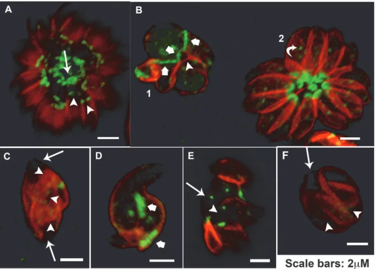
MORN1 localizes at the basal complex of the parasite (arrow) and also at the final end of the IMC in budding daughter cells during division (arrowheads) (Fig 7A). A point in the mid-dle of the cell, corresponding to the centrocone (a cone shape structure in the nuclear envelope that houses the mitotic spindle [26] was also observed (Fig 7B, curved arrow in rosette 2).
Treatment with 5μM of Et-Cipro or Adam-Cipro for 24 h and also 20μM of Cipro for 72h
led to a drastic effect in MORN1 distribution and localization, mainly in dividing parasites (Fig 7B–7F). Tethered daughter cells presenting a wide basal complex (about 2μm extension),
evi-denced by MORN1 cap, were observed after treatment with 5μM of Et-Cipro or Adam-Cipro
(Fig 7B and 7D, large arrows). However, tethered daughter cells often presented a weak locali-zation or lacked MORN1 at the end regions of IMC in mother cell (arrows) and also in budding daughter cells (arrowheads) after the treatment with 5μM of Et-Cipro or Adam-Cipro for 24 h
and 20μM of Cipro for 72 h (Fig 7B, 7C, 7E and 7F). Taking into account the importance of
MORN1 for the assembly of basal complex, and daughter cells scission [35], the lack of this protein at this region could be associated to the inhibition of daughter cells scission after Cipro and Cipro derivatives treatment.
Cipro derivatives are less active against the apicomplexan
C
.
parvum
The Apicomplexa speciesC.parvumlacks an apicoplast [36]. Thus, we hypothesized that if Cipro derivatives target the apicoplast, these compounds would be less effective at inhibitingC. parvumproliferation,. To test this hypothesis, the biological activities of Cipro derivatives were determined by measuring the inhibition ofC.parvumgrowth in MDBK cells (Table 2). Treat-ments with 50μM of Ph-Cipro and Adam-Cipro were toxic to host cells (data not shown) and50μM of Et-Cipro just inhibited 36% ofC.parvumproliferation; despite the toxicity for the
host cells, the treatment with 50μM of Ph-Cipro and Adam-Cipro did not show to decrease
the proliferation ofC.parvum, as infected cells showed a number of parasites similar to the cul-tures treated with 25μM (data not shown). Therefore, the IC50 value was only estimated for
Cipro (39.2μM after 24h;Table 2).
Discussion
Few therapy options are currently available for toxoplasmosis treatment, and effective drugs have serious toxic effects, leading to treatment interruption. The discovery of the apicoplast in the phylum Apicomplexa–which includesT.gondiiand other important human parasites, such asPlasmodiumspp.—brought new perspectives for the development of chemotherapy against these parasites, represented by the targeting of the multiple metabolic routes of pro-karyotic origin found in this organelle, and which differ significantly from those found in mammalian cells. Inhibitors of apicoplast enzymes, such as azithromycin and clindamycin
Fig 6. Treatment ofT.gondiiwith Cipro also leads to cell division arrest, apicoplast missegregation and IMC alteration.Tachyzoites ofT.gondiiwere treated with 20μM of Cipro for 24, 48 and 72 h. (A) Quantification of vacuoles (n = 120) containing parasites presenting cell division arrest after treatment with
20μM of Cipro for 24, 48 and 72 h, results are the mean of two independent experiments. (B-H) Immunofluorescence and TEM microscopy of LLC-MK2cells
infected with tachyzoites ofT.gondiiand treated for 48 and 72 h with 20μM Cipro. For immunofluorescence parasites were labeled with anti-HSP60
(recognizing the apicoplast, in green), anti-IMC1 (recognizing the inner membrane complex or IMC, in red) antibodies and DAPI (to label the DNA). While untreated tachyzoites presented normal morphology (B), parasites treated for 48 and 72 h presented cell division arrest, such as‘tethered’parasites (C-E and G), abnormally-shaped basal complexes (C and F square brackets, and inset), missegregated (C’arrow) and mispositioning apicoplasts (C’thick arrows), parasites displaying regions of pellicle without IMC (asterisk in D and arrows in H) and large degenerated parasites showing abnormal nucleus size (E arrow).
(which block prokaryotic protein synthesis), are already used in the treatment of human infec-tions caused by apicomplexan parasites [5,37]. In a previous study, we showed that derivatives of the antibiotic ciprofloxacin, a fluoroquinolone inhibitor of prokaryotic type II topoisomer-ases (DNA gyrase and topoisomerase IV), are active againstT.gondiitachyzoitesin vitro[20].
Fig 7. Cipro derivatives and Cipro affect MORN1 localization during parasite division.In parasites presenting normal division process MORN1 localizes at the basal complex of the mother cell (arrow) and also caps the final end of the IMC in budding daughter cells (arrowheads) (A), a single strong point in the middle of the cell was also observed in normal non-dividing parasites (B rosette 2 curved arrow). Tethered daughter cells resulting from the treatment with 5μM Et-Cipro (B-C) and Adam-Cipro (D-E) for 24h, and 20μM Cipro for 72h (F) showed wide MORN1 basal caps (B and D large arrows) and
lack or a weak deposition of MORN1, at the basal end of mother (arrow) and budding daughter cells (arrowheads) (B-F)
doi:10.1371/journal.pone.0125705.g007
Table 2. Effect of treatment with Cipro, Et-Cipro, Adam-Cipro and Ph-Cipro on the growth ofCryptosporidium parvumsporozoites in host cells.
Compound % inhibition of sporozoites growtha
12.5μM 25μM 50μM
Cipro 19.5±1.1 46.1±2.9 53.40±2.51
Et-Cipro 20.3±10.1 32.6±7.6 36.43±9.76
Adam-Cipro 24.7±8.4 34.2±3.8
-Ph-Cipro 33.6±11.6 42.6±14.6
-aThe % sporozoite growth inhibition was calculated by the examination of at least 1000 MDBK cells.
Here, we show that Cipro derivatives also increase the survival of mice infected withT.gondii, and affect parasite cell division by inhibiting cytokinesis and disturbing apicoplast duplication and segregation.
Thein vivostudies showed that Cipro derivative administration was well tolerated (Table 1) and did not result in serious toxicity to mice, and only mild toxicity was observed after treat-ment with high doses (200mg/kg) of Ph-Cipro (Table 1). While all mice treated with Cipro died by day 10 post-infection, some mice treated with Cipro derivatives remained alive for at least 60 days, suggesting that Cipro derivatives curedT.gondiiinfection in treated mice. Con-sidering that we used an acute model of infection, the modest rate of mice survival (13–25%) after treatment with Cipro derivatives is not irrelevant. High mice mortality was also observed in acute model of infection after the administration of drugs used for the treatment of toxoplas-mosis in the clinic [38–40]. Thus, Cipro derivatives should be considered potentially promising for the treatment of toxoplasmosis.
Fichera and Ross (1997) [8] showed that treatment ofT.gondiiwith Cipro led to progressive loss of apicoplast DNA, without affecting the first round of parasite proliferation in the host cells. However, egressed parasites that had been treated with Cipro suffered a drastic and per-manent reduction in the rate of cell duplication in the second round of proliferation in a new host cell [8]. This‘delayed effect’of drugs on parasite proliferation was also seen after treat-ment ofT.gondiiandP.falciparumwith clindamycin [41,42].
Interestingly, our results show that esterified Cipro derivatives did not trigger the same de-layed pattern of parasite death observed after treatment of infected cells with Cipro [8]. Rather, Et-Cipro and Adam-Cipro inhibited the first cell division event within the first round of para-site proliferation in host cells (Fig 2), since treatment with these Cipro derivatives for just 6 h led to a dramatic reduction in the number of parasitophorous vacuoles containing 2 parasites, 7h post-infection (Fig 2A). A possible explanation for these results is that esterified compounds act as prodrugs and this change would make them more lipophilic, favoring the diffusion across biological membranes (mainly the multiple membranes ofT.gondiiand of the apico-plast) and thus would affect apicoplast in a faster manner than the original molecule (Cipro). It is noteworthy to mention that Cipro is a mere Cipro precursor, i.e. once inside the cell, Et-Cipro ethyl ester group is hydrolysed by the action of intracellular esterases and then this deriv-ative is converted to Cipro (original molecule). Thus, the antiproliferderiv-ative effect and the mor-phological alterations caused by Et-Cipro derivative againstT.gondiican be credited to Cipro itself. An immediate effect on the proliferation ofT.gondiiwas also observed after treatment with the fluoroquinolones gatifloxacin and trovafloxacin [43,44]. So that more active com-pounds seems to subvert the delayed effect on apicoplast. The IC50values obtained for gatiflox-acin (0.52μM in HFF primary cell culture after 48h) and trovafloxacin (1.37μM in L929 cell
line) againstT.gondii[43,44] were close to those obtained for Cipro derivatives (0.42μM
Et-Cipro, 0.46μM Adam-Cipro and 1.24μM Ph-Cipro after 48h) [20]. The increased activity by
ethyl-esterified Cipro derivatives had also been previously reported inP.falciparum[45]. In-deed, Cipro had an immediate effect againstP.falciparumand did not show the delayed phe-notype [46]. Therefore, the delayed effect of Cipro againstT.gondiiis possibly due to its poor diffusion throughT.gondiimembranes.
surprising, as a moderate effect of Cipro againstC.parvumhas been reported before [47], and Cipro is possibly acting by an off target in this parasite.
Homologous genes for subunit A of the DNA gyrase—the putative target of the Cipro deriv-atives used in this study—were identified in the nuclear genome of apicomplexans, and their protein products are targeted to the apicoplast [18,19]. Thus, it is not surprising that we ob-served apicoplast alteration after treatment with Cipro derivatives and Cipro (Figs4B–4D,5D, 5Fand6C’). Our immunofluorescence and TEM data strongly suggest that the apicoplast loss observed in some cells after Cipro derivatives and Cipro treatments is likely caused by apico-plast missegregation during cell division (Figs4D–4E,5D, 5Fand6C’). Apicoplast division is concomitant with mitosis and is closely associated with the centrosome and with the nuclear spindle poles [48,49], which is likely to ensure the correct segregation of this organelle to daughter cells [48]. To date, no protein involved in the physical link between the apicoplast and the centrosome has been identified.
Immunofluorescence and TEM analyses suggest that, aside from apicoplast duplication and segregation inhibition, treatment with Cipro derivatives also affected the final step of parasite cytokinesis (cell scission), which involves constriction at the basal end of dividing parasites and cell separation. These observations suggest that parasites were able to perform mitosis (Fig 3), but were unable to complete cell scission at the end of cytokinesis, which occurs just after daughter cells bud out of the mother cell [27]. Consequently, parasites remained linked togeth-er at their basal ends, leading to the formation of groups of tethtogeth-ered parasites. This might have been the main effect of Cipro derivative treatment, since it was also visualized in parasites treat-ed with a concentration close to the IC50(1μM Et-Cipro or Adam-Cipro;Fig 4C and 4F). Cell
scission inhibition was also observed after treatment with 20μM Cipro for 48 and 72h (Fig
6A–6E). Treatment with Cipro also led to impairment ofP.falciparumcell division. After treatment with Cipro, parasites were able to initiate cell division, but failed in forming mature schizonts [50].
The immediate cell scission inhibition by Cipro derivatives is likely responsible for the strong parasite proliferation inhibition caused by Et-Cipro and Adam-Cipro treatments, which led to the formation of‘tethered’parasites, where multiple rounds of cell duplication had oc-curred in the absence of cytokinesis completion. The presence of vacuoles containing multiple tethered daughter cells even after Cipro derivatives (Et-Cipro and Adam-Cipro) removal (S2 Fig) showed that cell scission inhibition was irreversible, as daughter cells forming from new division cycles after drug removal were also unable to complete cytokinesis.
Our TEM and immunofluorescence results suggest that Cipro derivatives and Cipro treat-ments affected the integrity of IMC in mother cell (Fig 6H), as well as, the formation of this structure in daughter cells during cell division (Figs5C, 5F, and6D). The IMC supports the subpellicular microtubules [29], is important for parasite motility and host cell invasion [51], and serves as a scaffold for newly assembled structures during daughter cell formation [27,52]. During daughter cell budding, daughter IMCs is assembledde novo[53], which possibly re-quires large amounts of lipids. AlthoughT.gondiican obtain fatty acids from the host cell [54], apicoplast fatty acid synthesis is also essential for parasite survival [11,55], since the apicoplast is likely the main source of short-chain fatty acids (C14:0 and C16:0), and cooperates with the endoplasmic reticulum in the synthesis of long-chain fatty acid [55]. Thus, loss of apicoplast function by Cipro derivative treatment is expected to affect fatty acid synthesis and
IMC formation.
Morn1 forms a pair of rings around early duplicated centrioles [32,33], and caps and migrates in synchrony with the IMC during daughter cells budding [33,56]. Late in cytokinesis, the basal complex protein components constrict the basal end of newly formed parasites, eventual-ly‘resolving’cell division [32–34,56]. MORN1 showed an essential role in the assembly and maturation of basal complex, in its absence other components of the basal complex such as, Centrin2, dynein light chain and a set of basal IMC proteins could not assemble in the basal end of the parasite [32–35]. Knockout of MORN1 leads to cytokinesis and apicoplast segrega-tion defects [34,35], with the formation of‘Janus-headed’parasites [34] and also parasites with basal end wider than normal [35]. This phenotype is similar to the effect of Cipro derivatives and Cipro treatments described here (Figs4B–4G,5B, 5Cand6A–6G). By immunofluores-cence microscopy, we observed tethered tachyzoites with a wide basal end after Cipro deriva-tive treatment (Figs4B, 4D,6Cand7B and 7D), and TEM images of parasites treated with 5μM Et-Cipro and 20μM Cipro show tachyzoites with a wider basal region devoid of an
un-derlying IMC envelope (arrowheads inFig 5Dand square bracket inFig 6F, respectively). Thus, the inability of parasites treated with Cipro derivatives and Cipro to perform cell scission might be due to impaired basal complex maturation due to the lack of MORN1.
Overall, ourin vivoandin vitrostudies show that Cipro derivatives deserve to be exploited further in the search for alternative chemotherapeutic agents to treat human toxoplasmosis.
Supporting Information
S1 Fig. Plaque assay showing the antiproliferative effect of Cipro derivatives after 10 days treatment.While untreated culture (control) shows diverse plaques, treatment with just 0.5μM of Adam-Cipro or 10μM Cipro led to a drastic reduction on parasite proliferation.
In-deed, no plaques were observed in cultures treated with 5μM of Adam-Cipro or Et-Cipro,
10μM of Ph-Cipro and 20μM of Cipro.
(TIF)
S2 Fig. The effect of Cipro derivatives inT.gondiidivision is irreversible. Immunoluores-cence microscopy of tachyzoites labeled with anti-HSP60 (recognizing the apicoplast, in green) and anti-IMC1 (recognizing the inner membrane complex or IMC, in red) antibodies, and treated with Cipro derivatives for 6 h and then observed and kept in culture for a further 18h in the absence of drugs. Parasite division defects were observed (arrows) as a result of Cipro deriv-ative treatment in the first event of tachyzoite division inside host cells. Even after drug removal from the medium, cell division continued to be affected. Images represent maximum projec-tion of optical slices.
(TIF)
Acknowledgments
The authors thank Mr. Ricardo Vilela (Inmetro) and Dr. Fernando Almeida (Cenabio) for technical assistance with confocal microscopy and Mr. Helcio Evangelista da Silva for technical assistance with animal care.
Author Contributions
References
1. McLeod R, Kieffer F, Sautter M, Hosten T, Pelloux H. Why prevent, diagnose and treat congenital toxo-plasmosis? Mem Inst Oswaldo Cruz. 2009, 104: 320–44. PMID:19430661
2. Pereira-Chioccola VL, Vidal JE, Su C. Toxoplasma gondii infection and cerebral toxoplasmosis in HIV-infected patients. Future Microbiol. 2009, 4:1363–79. doi:10.2217/fmb.09.89PMID:19995194 3. Arevalo JF, Belfort R Jr, Muccioli C, Espinoza JV. Ocular toxoplasmosis in the developing world. Int
Ophthalmol Clin. 2010, 50:57–69. doi:10.1097/IIO.0b013e3181f0faeePMID:20930580
4. Holland GN. Ocular toxoplasmosis: a global reassessment. Part I: epidemiology and course of disease. Am J Ophthalmol. 2003, 136: 973–988. PMID:14644206
5. Montoya JG, Liesenfeld O. Toxoplasmosis. Lancet. 2004, 63:1965–76.
6. Iaccheri B, Fiore T, Papadaki T, Androudi S, Janjua S, Bhaila I, et al. Adverse drug reactions to treat-ments for ocular toxoplasmosis: a retrospective chart review. Clin Ther. 2008, 30:2069–74. doi:10. 1016/j.clinthera.2008.10.021PMID:19108794
7. Köhler S, Delwiche CF, Denny PW, Tilney LG, Webster P, Wilson RJ, et al. A plastid of probable green algal origin in Apicomplexan parasites. Science. 1997, 275:1485–9. PMID:9045615
8. Fichera M, Roos DS. A plastid organelle as a drug target in apicomplexan parasites. Nature. 1997, 390: 407–9. PMID:9389481
9. Goodman CD, McFadden GI. Targeting apicoplasts in malaria parasites. Expert Opin Ther Targets. 2013, 17: 167–77. doi:10.1517/14728222.2013.739158PMID:23231425
10. van Dooren GG, Striepen B. The algal past and parasite present of the apicoplast. Annu Rev Microbiol. 2013, 67: 271–89. doi:10.1146/annurev-micro-092412-155741PMID:23808340
11. Mazumdar J, Wilson EH, Masek K, Hunter CA, Striepen B. Apicoplast fatty acid synthesis is essential for organelle biogenesis and parasite survival in Toxoplasma gondii. Proc Natl Acad Sci U S A. 2006, 103: 13192–7. PMID:16920791
12. McFadden GI, Waller RF. Plastids in parasites of humans. Bioassays. 1997, 19: 1033–40. PMID: 9394626
13. Wiesner J, Reichenberg A, Heinrich S, Schlitzer M, Jomaa H. The plastid-like organelle of apicom-plexan parasites as drug target. Curr Pharm Des. 2008, 14:855–71. PMID:18473835
14. Collin F, Karkare S, Maxwell A. Exploiting bacterial DNA gyrase as a drug target: current state and per-spectives. Appl Microbiol Biotechnol. 2011, 92: 479–97. doi:10.1007/s00253-011-3557-zPMID: 21904817
15. Gouvea LR, Martins DA, Batista Dda G, Soeiro Mde N, Louro SR, Barbeira PJ, et al. Norfloxacin Zn(II)-based complexes: acid base ionization constant determination, DNA and albumin binding properties and the biological effect againstTrypanosoma cruzi. Biometals. 2013, 26: 813–25. doi:10.1007/ s10534-013-9661-zPMID:23897315
16. Hooper D. Mechanisms of action of antimicrobials: focus on fluoroquinolones. Clin Infect Dis. 2001, 32: S9–S15. PMID:11249823
17. Johannes CB, Ziyadeh N, Seeger JD, Tucker E, Reiter C, Faich G. Incidence of allergic reactions asso-ciated with antibacterial use in a large, managed care organisation. Drug Saf. 2007, 30: 705–13. PMID: 17696583
18. Dar MA, Sharma A, Mondal N, Dhar SK. Molecular cloning of apicoplast-targeted Plasmodium falcipa-rum DNA gyrase genes: unique intrinsic ATPase activity and ATP-independent dimerization of PfGyrB subunit. Eukaryot Cell. 2007, 6: 398–412. PMID:17220464
19. Raghu Ram EV, Kumar A, Biswas S, Kumar A, Chaubey S, et al. Nuclear gyrB encodes a functional subunit of thePlasmodium falciparumgyrase that is involved in apicoplast DNA replication. Mol Bio-chem Parasitol. 2007, 154: 30–39. PMID:17499371
20. Dubar F, Wintjens R, Martins-Duarte ES et al. Ester prodrugs of Ciprofloxacin as DNA-gyrase inhibitors: synthesis, antiparasitic evaluation and docking studies. Med Chem Commun. 2011, 2: 430–5. 21. Martins-Duarte EdosS, de Souza W, Vommaro RC. Itraconazole affectsToxoplasma gondii
endodyo-geny. FEMS Microbiol Lett. 2008, 282: 290–8. doi:10.1111/j.1574-6968.2008.01130.xPMID: 18371067
22. da Silva CF, Batista Dda G, Oliveira GM, de Souza EM, Hammer ER, et al. In vitro and in vivo investiga-tion of the efficacy of arylimidamide DB1831 and its mesylated salt form—DB1965—against Trypano-soma cruzi infection. PLoS One. 2012, 7:e30356. doi:10.1371/journal.pone.0030356PMID: 22291940
24. Lawton P, Hejl C, Mancassola R, Naciri M, Petavy AF. Effects of purine nucleosides on the in vitro growth of Cryptosporidium parvum. FEMS Microbiol Lett. 2003, 226:39–43. PMID:13129605
25. Roos DS, Donald RG, Morrissette NS et al. Molecular tools for genetic dissection of the protozoan
para-siteToxoplasma gondii. Methods Cell Biol. 1994, 45:27–63. PMID:7707991
26. Sheffield HG, Melton ML. The fine structure and reproduction ofToxoplasma gondii. J Parasitol. 1968, 54: 209–26. PMID:5647101
27. Hu K, Mann T, Striepen B, Beckers CJ, Roos DS, et al. (2002) Daughter cell assembly in the protozoan parasite Toxoplasma gondii. Mol Biol Cell 13: 593–606. PMID:11854415
28. Nichols BA, Chiappino ML. Cytoskeleton of Toxoplasma gondii. J Protozool. 1987, 34:217–26. PMID: 3585817
29. Morrissette NS, Murray JM, Roos DS. Subpellicular microtubules associate with an intramembranous particle lattice in the protozoan parasiteToxoplasma gondii. J Cell Sci. 1997, 110: 35–42. PMID: 9010782
30. Mann T, Beckers C. Characterization of the subpellicular network, a filamentous membrane skeletal component in the parasite Toxoplasma gondii. Mol Biochem Parasitol. 2001, 115:257–68. PMID: 11420112
31. Weissig V, VetroWidenhouse TS, Rowe TC. Topoisomerase II inhibitors induce cleavage of nuclear and 35-kb plastid DNAs in the malarial parasite Plasmodium falciparum. DNA Cell Biol. 1997, 16: 1483–92. PMID:9428797
32. Hu K. Organizational changes of the daughter basal complex during the parasite replication of Toxo-plasma gondii. PLoS Pathog. 2008, 4:e10. doi:10.1371/journal.ppat.0040010PMID:18208326 33. Gubbels MJ, Vaishnava S, Boot N, Dubremetz JF, Striepen B. A MORN-repeat protein is a dynamic
component of the Toxoplasma gondii cell division apparatus. J Cell Sci. 2006, 119: 2236–45. PMID: 16684814
34. Lorestani A, Sheiner L, Yang K, Robertson SD, Sahoo N, et al. A Toxoplasma MORN1 null mutant un-dergoes repeated divisions but is defective in basal assembly, apicoplast division and cytokinesis. PLoS One. 2010, 5: e12302. doi:10.1371/journal.pone.0012302PMID:20808817
35. Heaslip AT, Dzierszinski F, Stein B, Hu K. TgMORN1 is a key organizer for the basal complex of Toxo-plasma gondii. PLoS Pathog. 2010, 6:e1000754. doi:10.1371/journal.ppat.1000754PMID:20140195 36. Zhu G, Marchewka MJ, Keithly JS.Cryptosporidium parvumappears to lack a plastid genome.
Microbi-ology. 2000, 146:315–321. PMID:10708370
37. Fleige T, Soldati-Favre D. Targeting the transcriptional and translational machinery of the endosymbiot-ic organelle in apendosymbiot-icomplexans. Curr Drug Targets. 2008, 9:948–56. PMID:18991607
38. Martins-Duarte ES, de Souza W, Vommaro RC. Toxoplasma gondii: the effect of fluconazole combined with sulfadiazine and pyrimethamine against acute toxoplasmosis in murine model. Exp Parasitol. 2013, 133: 294–9. doi:10.1016/j.exppara.2012.12.011PMID:23270807
39. Romand S, Pudney M, Derouin F.) In vitro and in vivo activities of the hydroxynaphtoquinone atova-quone alone or combined with pyrimethamine, sulfadiazine, clarithromycin, or minocycline against Toxoplasma gondii. Antimicrob Agents Chemother. 1993, 37: 2371–8. PMID:8285620
40. Derouin F, Almadany R, Chau F, Rouveix B, Pocidalo JJ. Synergistic activity of azithromycin and pyri-methamine or sulfadiazine in acute experimental toxoplasmosis. Antimicrob Agents Chemother. 1992, 36: 997–1001. PMID:1324642
41. Fichera ME, Bhopale MK, Roos DS. In vitro assays elucidate peculiar kinetics of clindamycin action against Toxoplasma gondii. Antimicrob Agents Chemother. 1995, 39: 1530–7. PMID:7492099 42. Pfefferkorn ER, Borotz SE. Comparison of mutants of Toxoplasmagondii selected for resistance to
azi-thromycin, spiramycin, or clindamycin.Antimicrob. Agents Chemother. 1994, 38:31–37. PMID: 8141576
43. Khan A, Slifer T, Araujo F, Remington JS. Trovafloxacin is active againstToxoplasma gondii. Antimi-crob Agents Chemother. 1996, 40: 1855–9. PMID:8843293
44. Khan A, Slifer T, Araujo F, Remington JS. Activity of gatifloxacin alone or in combination with pyrimeth-amine or gamma interferon againstToxoplasma gondii. Antimicrob Agents Chemother. 2001, 45: 48– 51. PMID:11120943
45. Dubar F, Anquetin G, Pradines B et al. Enhancement of the antimalarial activity of Ciprofloxacin using a double prodrug/bioorganometallic approach. J Med Chem. 2009, 24: 7954–7. doi:10.1021/jm901357n PMID:19908867
47. Woods KM, Nesterenko MV, Upton SJ. Efficacy of 101 antimicrobials and other agents on the develop-ment of Cryptosporidium parvum in vitro. Ann Trop Med Parasitol. 1996, 90; 603–615 PMID:9039272 48. Striepen B, Crawford MJ, Shaw MK, Tilney LG, Seeber F, et al. The plastid of toxoplasma gondii is
di-vided by association with the centrosomes. J Cell Biol. 2000, 151: 1423–34. PMID:11134072 49. Nishi M, Hu K, Murray JM, Roos DS. Organellar dynamics during the cell cycle ofToxoplasma gondii. J
Cell Sci. 2008, 121:1559–68. doi:10.1242/jcs.021089PMID:18411248
50. Dahl EL, Rosenthal PJ. Multiple antibiotics exert effects against the Plasmodium falciparum apicoplast. Antimicrob Agents Chemother. 2007, 51:3485–90. PMID:17698630
51. Gaskins E, Gilk S, DeVore N et al. Identification of the membrane receptor of a class XIV myosin in Toxoplasma gondii. J Cell Biol. 2004; 165: 383–93.
52. Beck JR, Rodriguez-Fernandez IA, de Leon JC, Huynh MH, Carruthers VB, Morrissette NS, et al. A novel family of Toxoplasma IMC proteins displays a hierarchical organization and functions in coordi-nating parasite division. PLoS Pathog. 2010, 6:e1001094.
53. Ouologuem DT, Roos DS. Dynamics of the Toxoplasma gondii inner membrane complex. J Cell Sci. 2014, 127:3320–30. doi:10.1242/jcs.147736PMID:24928899
54. Charron AJ, Sibley LD. Host cells: mobilizable lipid resources for the intracellular parasiteToxoplasma
gondii. J Cell Sci. 2002, 115:3049–59. PMID:12118061
55. Ramakrishnan S, Docampo MD, Macrae JI, Pujol FM, Brooks CF, van Dooren GG, et al. Apicoplast and endoplasmic reticulum cooperate in fatty acid biosynthesis in apicomplexan parasite Toxoplasma gondii. J Biol Chem. 2012, 287:4957–71. doi:10.1074/jbc.M111.310144PMID:22179608