Pneumonic Plague
Roger D. Pechous, Vijay Sivaraman, Paul A. Price, Nikolas M. Stasulli, William E. Goldman*
Department of Microbiology and Immunology, University of North Carolina at Chapel Hill, Chapel Hill, North Carolina, United States of America
Abstract
Inhalation ofYersinia pestiscauses primary pneumonic plague, a highly lethal syndrome with mortality rates approaching 100%. Pneumonic plague progression is biphasic, with an initial pre-inflammatory phase facilitating bacterial growth in the absence of host inflammation, followed by a pro-inflammatory phase marked by extensive neutrophil influx, an inflammatory cytokine storm, and severe tissue destruction. Using a FRET-based probe to quantitate injection of effector proteins by theY. pestistype III secretion system, we show that these bacteria target alveolar macrophages early during infection of mice, followed by a switch in host cell preference to neutrophils. We also demonstrate that neutrophil influx is unable to limit bacterial growth in the lung and is ultimately responsible for the severe inflammation during the lethal pro-inflammatory phase.
Citation:Pechous RD, Sivaraman V, Price PA, Stasulli NM, Goldman WE (2013) Early Host Cell Targets ofYersinia pestisduring Primary Pneumonic Plague. PLoS Pathog 9(10): e1003679. doi:10.1371/journal.ppat.1003679
Editor:Barbara I. Kazmierczak, Yale University, United States of America
ReceivedMay 19, 2013;AcceptedAugust 20, 2013;PublishedOctober 3, 2013
Copyright:ß2013 Pechous et al. This is an open-access article distributed under the terms of the Creative Commons Attribution License, which permits unrestricted use, distribution, and reproduction in any medium, provided the original author and source are credited.
Funding:Support for this work was provided by National Institutes of Health (NIH) grant AI057157 awarded to WEG, NIH grant AI099698 awarded to WEG, and NIH National Research Service Award AI085830 awarded to RDP. The funders had no role in study design, data collection and analysis, decision to publish, or preparation of the manuscript.
Competing Interests:The authors have declared that no competing interests exist. * E-mail: [email protected]
Introduction
The historical impact ofYersinia pestis on humanity cannot be understated, as three major pandemics including the ‘‘Black Death’’ of the Middle Ages have been attributed toY. pestis[1,2]. The bacterium can be transmitted person-to-person via respira-tory droplets resulting in primary pneumonic plague, which is the deadliest manifestation ofY. pestisinfection [1,2,3]. In the event of respiratory exposure in humans, mortality rates are nearly 100% with a time to death of typically between four and seven days [1]. Its extreme lethality and history of weaponization have led to the assignment ofY. pestisas a Tier 1 Select Agent and compound fears of its intentional release as a weapon of bioterrorism [1].
Using a murine intranasal infection model, our laboratory demonstrated that the primary pneumonic plague syndrome progresses in two distinct phases [4]: an initial ‘‘pre-inflammatory phase’’ characterized by rapid bacterial replication in the lung in the absence of host innate immune responses, followed by a ‘‘pro-inflammatory’’ phase marked by extensive neutrophil influx, a massive pro-inflammatory cytokine storm, and considerable tissue destruction within the lung. In mice and humans, progression into this pro-inflammatory phase invariably results in death without immediate treatment [5,6]. Recently, our laboratory showed that
Y. pestiscreates a unique protective environment in the lungs of mice that allows for the growth of typically avirulent organisms [7], suggesting that Y. pestis suppresses host innate immune responses in the lung early during infection. The mechanism and host cell types involved in this phenomenon remain unknown.
Y. pestis utilizes a plasmid-encoded type III secretion system (T3SS) to deliver Yersinia effector proteins (Yops) directly to the cytosol of target cells. Injection of Yop effectors is essential forY.
pestisvirulence and is known to have inflammatory and anti-phagocytic effects on mammalian cells [2,8,9]. The pulmonary cells targeted byY. pestisduring primary pneumonic plague have yet to be identified. In the work presented here, we use fully virulent Y. pestis CO92 expressing a YopE-TEM b-lactamase hybrid protein to identify the host cells targeted in the lung, and to evaluate the effect of depletion of these cell types on the progression of pneumonic plague. We show that the Y. pestis
T3SS primarily targets macrophages and neutrophils early during the pre-inflammatory phase of disease. We also monitor the host cell dynamics in the lung in response toY. pestis challenge and show that neutrophils are ultimately responsible for the severe necrotizing pneumonia during the pro-inflammatory disease phase. This work is the first identification/evaluation of the host cells targeted by fully virulentY. pestisin the lung, and the results give insight into the dynamic events occurring shortly after pulmonary exposure to the highly lethal pathogenY. pestis.
Results
Injection by the T3SS is detectablein vivoduring primary pneumonic plague
CCF2-AM (Invitrogen). CCF2-CCF2-AM is a cephalosporin conjugated to the fluorophores coumarin and fluorescein that exhibit fluorescence resonance energy transfer (FRET). Uninfected mammalian cells harboring CCF2-AM can be detected as green fluorescence emission (520 nm); cells that have been targeted for Yop translocation will fluoresce blue (447 nm) due to cleavage of cytosolic CCF2-AM (and disruption of FRET) by theb-lactamase moiety of Yop-TEM. We constructed a strain ofY. pestisCO92 (Y. pestisYopE-TEM) that expresses both wild-type YopE and the N-terminal 100 amino acids of YopE fused to TEM b-lactamase. YopE is a GTPase activating protein that is highly secretedin vitro
and is known to inhibit phagocytosis through the disruption of actin microfilaments [8,9]. Incorporation of the YopE-TEM b -lactamase fusion had no effect on the virulence ofY. pestisCO92 as determined by bacterial burden, cytokine, and histopathological analysis.
To evaluate YopE-TEM injectionin vivo, C57BL/6J mice were inoculated with 106CFU ofY. pestis YopE-TEM, and single-cell suspensions were generated from infected lungs at 12 hpi for evaluation by flow cytometry [13]. Lung suspensions stained with CCF2-AM uniformly exhibited green fluorescence compared to unstained samples (Figure 1A). In contrast to mice inoculated with PBS alone, mice infected withY. pestisYopE-TEM exhibited blue fluorescence in a subset of cells (3% of total cells), indicating that YopE-TEM-mediated cleavage of CCF2-AM was detectablein vivo
by flow cytometry (Figure 1A). For a more detailed analysis, groups of mice were sacrificed at 6, 12, and 24 hpi for flow cytometry evaluation of blue fluorescence. At 6 hpi, roughly 26104injection events per mouse were detected, representing approximately 1% of total recovered cells (Figure 1B,C). This number increased over time, reaching roughly 36106, or 48% of total recovered cells by
24 hpi (Figure 1B,C). The approximately 100-fold increase in injection events coincided with a nearly 100-fold increase in bacterial burden (Figure 1D). These data demonstrate thatY. pestis
secretion of effectors occurs early during pneumonic plague, and is detectable in the lung using flow cytometry.
Y. pestistargets alveolar macrophages and neutrophils early during primary pneumonic plague
The ability of Y. pestis to create a localized protective environment in the lungs early during pneumonic plague suggests
that it deactivates host innate immune responses [7]. We hypothesized thatY. pestistargets innate immune cell populations early in the lung via type III secretion of Yop effectors. In order to examine the host cell types intoxicated early during infection, mice were sacrificed at 6, 12, and 24 hpi, and whole lung cell suspensions were incubated with CCF2-AM as well as fluores-cently labeled anti-CD45 and anti-CD3 antibodies. Cells exhib-iting blue fluorescence were gated and analyzed to distinguish between lymphocyte (CD45+
CD3+
), leukocyte (CD45+ CD32), and epithelial/endothelial (CD452CD32) cell populations. At
each of the time points examined, .96% of injection events occurred in a population of CD45+
CD32 cells, indicating that innate immune leukocyte populations were the primary targets for
Y. pestis translocation of YopE-TEM (Figure S1B). By 24 hpi roughly 40% of CD45+
CD32 cells were injected with
YopE-TEM, whereas less than 3% of the other two populations were injected (Figure S1C).
To identify the leukocyte cell populations targeted byY. pestis, lungs of mice infected withY. pestisYopE-TEM were incubated with CCF2-AM as well as a panel of fluorescently labeled antibodies to cell type-specific markers. Viable cells exhibiting blue fluorescence were gated and analyzed based on fluorescence staining patterns to identify alveolar macrophages, CD11bhigh interstitial/exudate macrophages, F4/80+
CD11bhighCD11chighmacrophages, mono-cytes, CD11bhigh and CD11blow dendritic cells (DCs), and neutrophils (Figure S2A). At 6 hpi, alveolar macrophages repre-sented roughly 55% of injected cells (Figure 1E). Neutrophils and cells that are unidentifiable based on our antibody panel (‘‘other’’) were the next most prominent target populations, each representing roughly 18% of injected cells. At 12 hpi a dramatic switch in host cell preference occurred, where greater than 70% of injected cells were neutrophils and alveolar macrophages represented less than 10% of injected cells (Figure 1E). By 24 hpi approximately 90% of injected cells were neutrophils, and more than 60% of all recovered neutrophils were positive for YopE-TEM translocation (Figure S2B). In summary,Y. pestisinitially injects effector Yops into resident alveolar macrophages during pneumonic plague, followed by a shift in host cell preference to neutrophils.
The shift inY. pestishost cell preference corresponds with an influx of neutrophils into the lung
Understanding how the preferred cellular targets of Y. pestis
change over time also demands that we assess the host cell repertoire within the infected lung. Flow cytometry analysis revealed increased numbers of neutrophils as early as 6 hpi that reached roughly 50 times those of mock-infected mice by 24 hpi (Figure 2A). Infected mice also showed elevated levels of CD11bhighDCs as well as F4/80+CD11bhighCD11chighcells that
continued to increase throughout the duration of the pre-inflammatory phase of disease (Figure 2A). Histological analysis of infected lungs showed that the influx of neutrophils resulted in minimal changes in lung pathology at 24 hpi, and small, localized areas of inflammatory infiltrate were found sporadically through-out infected lung sections (Figure 2B). These animals displayed no outward symptoms of disease at this time point. This is in contrast to what was seen at 48 hpi, where large, densely packed inflammatory foci were visible throughout a majority of infected lung sections (Figure 2B), and animals began to show disease symptoms including lethargy and decreased responses to stimuli. Thus, despite a lack of disease symptoms and a minimal level of detectable changes in lung pathology, there was an increase in bacterial burden as well as an expansion of neutrophils and other innate immune cell populations in the lung during the pre-inflammatory phase of disease.
Author Summary
Inhalation of the bacterium Yersinia pestis results in primary pneumonic plague, a severe necrotizing pneumo-nia with mortality rates approaching 100% in the absence of timely antibiotic administration. Despite the notoriety of Y. pestis as a potential biological weapon and its well-established pandemic potential, very little is known regarding early host-pathogen interactions that lead to the progression of pulmonary infection.Y. pestisharbors a type III secretion system (T3SS) for delivery of Yersinia outer protein (Yop) effectors into host cells, an early and essential step in pathogenesis. In the work presented here, we identify the host cell targets ofY. pestisYop secretion in the lung. We show that Y. pestisinitially targets alveolar macrophages, followed by a shift in host cell preference to neutrophils. Through cellular depletion studies, we dem-onstrate that Y. pestisis highly resistant to macrophage-and neutrophil-mediated clearance, macrophage-and that the accumu-lation of neutrophils in the lung is responsible for the severe necrotizing pneumonia that develops during the pro-inflammatory phase of pneumonic plague.
The abundance of neutrophils dictates levels of YopE injection
At 6 hpi, alveolar macrophages represented the primary cell population targeted byY. pestistype III secretion (Figure 1E). We
next sought to evaluate the effect of alveolar macrophage depletion on YopE injection. Clodrosome is a multilamellar liposome suspension encapsulating the drug clodronate (Encapsulum). Liposomal clodronate has been used previously to deplete alveolar Figure 1. YopE-TEM translocation in the lung during pneumonic plague.(A) Groups of mice were inoculated intranasally with 106CFU ofY.
pestisexpressing YopE-TEM or PBS alone (mock), followed by staining with CCF2-AM. Lungs were harvested at 12 hpi, and live cells were gated to evaluate blue/green fluorescence by flow cytometry. Cells showing blue fluorescence have been injected with YopE-TEM. Histograms show representative data from a single mouse; (B) Total injection events (cells exhibiting blue fluorescence) in the lungs of mice inoculated with 106CFUY.
pestisYopE-TEM; (C) The percentage of total cells in the lung injected with YopE-TEM was evaluated for samples shown in (B) at 6, 12, and 24 hpi; (D) Lung bacterial burden was evaluated in mice inoculated with 106CFUY. pestisYopE-TEM. (E) Live injected cells in lungs of mice inoculated with
106CFU Y. pestis YopE-TEM were gated for identification of alveolar macrophages (F480+
CD11blow/midCD11chigh), interstitial macrophages (F480+
CD11bhighCD11cmid/low), F480+
CD11bhighCD11chighmacrophages, monocytes (F4802CD11bhighLy-6G2), neutrophils (F4802CD11bhighLy-6G+
), and CD11b high and low dendritic cells (F4802CD11c+); All data are representative of at least three independent experiments with three to five mice
macrophages by distillation via the intranasal route [14,15,16]. In order to evaluate Y. pestis early interactions with alveolar macrophages, mice were treated with clodrosome for two days prior to inoculation with Y. pestis YopE-TEM. Intranasal administration of clodrosome resulted in a roughly 92% decrease in alveolar macrophages relative to uninoculated mice (Figure S3A,B). The targeted depletion of alveolar macrophages resulted in a roughly 1.7-fold decrease in total YopE-TEM injection events at 6 hpi (Figure 3A). Analysis of injected cells revealed that with limited numbers of alveolar macrophages available, the primary injected cell type shifted to neutrophils, which represented greater than 60% of injected cells (Figure 3C). This is in contrast to what is seen in untreated mice, where at 6 hpi alveolar macrophages represented roughly 55% of injected cells and neutrophils represented less than 3%.
We sought to determine if the abundance of neutrophils at 24 hpi was the primary factor responsible for an overall increase in
injection events. To evaluate levels of YopE-TEM injection in the absence of neutrophils, mice were injected intravenously 24 hours prior to infection with depleting antibody to the lymphocyte antigen 6 complex (Ly-6G). Ly-6G is expressed on the majority of myeloid cells in the bone marrow and peripheral granulocytes, and the use of depleting antibody clone 1A8 has been shown to specifically deplete neutrophils [17,18]. Treatment witha-Ly-6G antibody resulted in a 99% decrease in F4/802 Ly-6G+
cells (Figure S4A,B), and a 90% decrease in total F4/802Gr-1+
cells (data not shown) in the lung while having a minimal impact on other host cell populations, suggesting the efficient and specific depletion of lung neutrophils. The absence of neutrophils resulted in a .10-fold decrease in total injection events at 24 hpi (Figure 3B). Though depletion of neutrophils resulted in an increase in the representation of other cell types in the injected population, neutrophils remained the most prominent host cell target (Figure 3D). This indicates that neutrophils are the Figure 2. Lung host cell repertoire of mice inoculated intranasally withY. pestis.(A) Total cell numbers of innate immune cell populations in the lungs of mice inoculated with 106CFUY. pestisYopE-TEM or PBS (mock) at 6, 12, and 24 hpi. (B)Y. pestiscauses severe inflammation in the lungs that is visible by 48 hpi. Mice were inoculated with 106CFUY. pestisand lungs were inflated with 10% formalin at 24 and 48 hpi prior to H and
E staining. Images are representative of experiments done a minimum of three times. Data are representative of at least three independent experiments with five mice at each time point. Error bars represent SEM.
doi:10.1371/journal.ppat.1003679.g002
preferred host cell target, as their depletion resulted in an overall decrease in total injection events and failed to result in the emergence of an additional single cell type as the primary target. In summary, there is an influx of neutrophils into the lung shortly after inoculation withY. pestis, and as a result, the primary host cell target in the lung shifts from resident alveolar macrophages to neutrophils.
Depletion of neutrophils attenuates primary pneumonic plague progression in the lung
In order to determine the roles of macrophages and neutrophils in the progression of infection, we evaluated the effects of host cell depletion of each on the progression of primary pneumonic plague. Depletion of alveolar macrophage populations had little effect on bacterial survival in the lung as indicated by bacterial Figure 3. Pre-treatment of mice with clodrosome or neutrophil-depleting antibodya-Ly-6G.(A) Total YopE-TEM injection events 6 hpi in
mice inoculated with 106CFUY. pestisYopE-TEM with and without prior alveolar macrophage depletion with clodrosome. (B) Total YopE-TEM
injection events 24 hpi inY. pestis-infected mice with and without pre-treatment with neutrophil-depleting antibody. (C) Identity of injected cells 6 hpi in mice with and without prior clodrosome treatment. (D) Identity of injected cells 24 hpi with and without pre-treatment with neutrophil-depleting antibody. Data are representative of at least three independent experiments with three to five mice at each time point. Error bars represent SEM for all graphs.
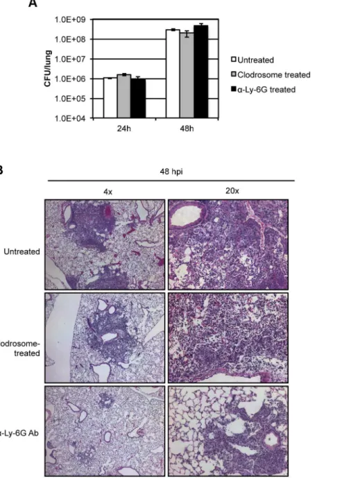
burden analysis at 24 and 48 hpi (Figure 4A). Infected mice pre-treated with clodrosome exhibited a similar time-to-death as untreated infected mice (Figure S5A), and symptoms appeared identical to untreated infected animals at 48 hpi. Histopathology of infected mouse lungs from mice pre-treated with clodrosome revealed the presence of inflammatory foci at 48 hpi that were similar to those in untreated mice (Figures 4B, S5B). These data indicate that depletion of greater than 90% of alveolar macro-phages in the lung changed the initial host cell target, but had little effect on progression of primary pneumonic plague.
As neutrophils were the primary host cell target ofY. pestistype III secretion (Figure 1E), we hypothesized that depletion of
neutrophils would hasten disease progression and enhance bacterial proliferation. Mice pre-treated with a-Ly-6G depleting antibody were infected with the minimum lethal dose of 104CFU ofY. pestisand sacrificed at 48 hpi to determine bacterial burdens in the lung. Depletion of greater than 99% of neutrophil populations had no effect on bacterial burdens the lung at 48 hpi (Figure 4A) and extended mean time-to-death of infected animals by approximately 7 hours, though this difference is not statistically significant (Figure S5A). Surprisingly, mice that were pretreated witha-Ly-6G showed no outward disease symptoms at 48 hpi, whereas untreated infected mice showed increased lethargy and a decreased response to stimuli. Histological
Figure 4. Evaluating the progression of pneumonic plague in mice pre-treated with clodrosome or neutrophil-depleting antibody a-Ly-6G.(A) Lung bacterial burden in mice with and without pre-treatment with clodrosome or neutrophil-depleting antibody and after inoculation
with 104CFUY. pestis; (B) Lung histology 48 hpi in mice inoculated with 104CFUY. pestiswith and without prior treatment with clodrosome or
neutrophil-depleting antibody. Bar graph represents experiments repeated at least three times with three to five mice. Error bars represent SEM. Histopathology shows representative images from experiments repeated at least twice.
doi:10.1371/journal.ppat.1003679.g004
evaluation of lungs of infected mice pre-treated with Ly-6G antibody revealed a dramatic decrease in the magnitude and overall area of inflammatory foci compared to that seen in untreated animals (Figures 4B, S5B). Further, whereas the foci found in wild-type infected lungs 48 hpi were densely packed with neutrophils and beginning to lose alveolar structure, lung sections froma-Ly-6G-treated mice demonstrated foci with intact alveolar architecture primarily consisting of bacteria with minimal neutrophil infiltrate (Figure 4B). These data indicate that neutrophils are the primary cell type responsible for the dramatic necrotizing pneumonia attributed to the pro-inflammatory phase of disease. In summary, depletion of neutrophils did not enhance disease, but rather limited progression into the lethal pro-inflammatory phase of pneumonic plague.
Discussion
The work presented here is the first to identify the initial host cell targets of fully virulentY. pestis during pulmonary infection. Using flow cytometry to monitor injection of a YopE-TEM fusion protein by the T3SS, we showed that Y. pestis initially targets CD11chigh alveolar macrophages and neutrophils in the lung. Previously, Bosio et al. demonstrated that the attenuated (pgm -pCD1-)Y. pestisstrain A1122 was taken up by a distinct CD11c+ CD11b2 host population in the lungs following intratracheal inoculation, confirming that Y. pestisinitially encounters alveolar macrophages early during infection [14]. By 12 hpi there is a dramatic shift in host target cell preference from macrophages to neutrophils that continues to increase by 24 hpi. This corresponds with a massive influx of neutrophils into the lung that is detected as early as 6 hpi. In addition to neutrophils, flow cytometry analysis of infected lungs revealed a 10-fold increase in numbers of CD11bhigh DCs by 24 hpi. CD11bhigh DCs are known to accumulate in airways adjacent to alveolar spaces after allergen challenge, and have been implicated in the events that define asthma as well as contributing to CD4+T-cell priming [19,20]. Infected lungs also saw an overall increase in levels of F4/ 80+
CD11b+ CD11c+
cells. These cells exhibited positivity for both interstitial (CD11b+
) and alveolar (CD11c+
) macrophage markers and were not targeted by clodrosome in uninfected mice. This population likely consists of a subset of dendritic cells expressing F4/80, activated alveolar macrophages expressing CD11b, and macrophages that have entered the lung in response to bacterial challenge and are induced to express CD11c [21]. Taken together, these results outline a highly dynamic lung environment early during infection where different host innate immune populations are expanding in the lung in an attempt to control Y. pestis
infection, albeit without much success. Despite the expansion of these cell types, macrophages and neutrophils remained the primary targets of YopE injection.
The Y. pestis specificity for injection of macrophages and neutrophils cannot be explained by abundance alone, as the mammalian respiratory tract is comprised of dozens of host cell types with direct access to the airways. We observed minimal injection of CD452CD32 epithelial/endothelial populations at
each of the time points examined, despite the fact that this includes alveolar epithelial type I cells which are known to cover roughly 95% of the surface area of the alveoli [22]. Depletion of approximately 92% of alveolar macrophages hastened the shift in target cells seen during normal infection but resulted in only slight increases in levels of injection for other cell types, even though the abundance of these populations was roughly equivalent to that of neutrophils. Conversely, the depletion of roughly 99% of neutrophils in the lung prior to infection resulted in a roughly
10-fold decrease in total injection events. This is similar to what was seen for the enteric pathogen and recentY. pestisancestorY. pseudotuberculosis, where depletion of neutrophils resulted in a significant overall decrease in total translocation events in Peyer’s patches of orogastrically inoculated mice [10]. After treatment witha-Ly-6G antibody, neutrophils remained the primary host cell target. It is known that neutrophils can regenerate rapidly, particularly in the bone marrow, which may partly explain the failed emergence of an additional preferred cell target after depletion of lung neutrophils [23]. The targeting of professional phagocytes, specifically neutrophils, has been seen in other bacterial species and may represent a common virulence mechanism for pathogens harboring a functional T3SS [10,24,25].P. aeruginosa has been shown to target macrophages and neutrophils in the lung [24], andSalmonella entericahas been shown to target neutrophils in the spleen via type III secretion to promote intracellular survival [25]. This phenomenon also occurs in other organs of the body duringYersiniainfection. In the first example of in vivo detection of Yop translocation via a Yop-b -lactamase fusion, Marketon et al. showed that the attenuatedY. pestis pgm2strain KIM D27 targeted CD11b+
, CD11c+
, and Gr-1+ cells in the spleen when delivered intravenously, suggesting targeting of macrophages, dendritic cells, and neutrophils [12]. It will be intriguing to compare the injected host cell repertoire of
Y. pestis delivered via additional routes of infection, including subcutaneous and intradermal, and in other tissues including lymph nodes and in the skin.
As alveolar macrophages and neutrophils are the primary targets ofY. pestistype III secretion, we sought to further evaluate their roles during pulmonary infection. Depletion of roughly 92% of alveolar macrophages had little or no effect on bacterial burden in the lung. This indicates that either macrophages are not involved in limitingYersiniasurvival in the lung, or thatY. pestisis able to neutralize the anti-bacterial effects of sentinel alveolar macrophages, presumably as a result of antiphagocytic/anti-inflammatory factors including the Yops, F1 capsular protein, and pH 6 antigen [2]. Also, depletion of alveolar macrophages did not significantly alter progression into the pro-inflammatory phase of disease. Infected animals demonstrated typical disease symptoms as well as the formation of inflammatory foci in infected lungs. Thus, the absence of greater than 90% of alveolar macrophages did not inhibit early host signaling that leads to the massive influx of neutrophils into the lung during pulmonaryY. pestisinfection. This indicates that either the remaining number of alveolar macrophages is sufficient, or an additional cell type is responsible for this early host response.
neutrophil-mediated killing by inhibiting phagocytosis and re-pressing reactive oxygen species production [31]. Also, gene expression profiling in neutrophils exposed to Y. pestis indicated that the T3SS suppresses the activation of a number of genes responsible for pro-inflammatory cytokine output and prolonged cell survival [32]. Surprisingly, infected animals pre-treated with neutrophil-depleting antibody did not demonstrate outward signs of disease at 48 hpi despite significant bacterial burdens, indicating a failure to fully progress into the pro-inflammatory phase of pneumonia. Upon histological examination, the small sporadic foci that were detected in the lungs of these animals were primarily made up of alveolar-residing bacteria and a relatively small number of mononuclear cells and neutrophils. These animals eventually succumbed, not from the typical lung inflammation and pathology of pneumonic plague, but as a result of the unhindered spread ofY. pestisto other tissues of the body.
The information gathered in this study supports a model of the pre-inflammatory phase of pneumonic plague where Y. pestis
initially targets resident alveolar macrophages and then neutro-phils to facilitate proliferation in the lung. As neutroneutro-phils are unable to sufficiently controlY. pestis, their continued chemotaxis results in the formation of massive inflammatory foci and causes severe damage to the alveolar architecture of the lung. Work evaluating the fate of injected macrophages and neutrophils will provide insight into the downstream signaling events that lead to progression into the pro-inflammatory phase of disease.
In summary, the work presented here is the first to identify target host cells and evaluate population dynamics in the lung during primary pneumonic plague. Though much of the previous literature in the field highlights interactions betweenY. pestisand macrophages, we show thatY. pestisprimarily targets neutrophils early after inoculation in the lung, presumably to limit host innate immune mechanisms aimed at bacterial killing and clearance. To our surprise, depletion of neutrophils, a key innate immune mechanism for controlling infection during pneumonia, attenuat-ed much of the intense inflammation seen in the lung during the pro-inflammatory phase of disease. Thus, neutrophils appear to be key players in the manifestation of what is ultimately a host-mediated necrotizing pneumonia. Understanding the early delay in innate immune response is crucial to understanding the events that ultimately lead to the progression of this highly lethal syndrome. Further, therapy targeting both bacterial growth as well as the destructive capacity of neutrophils in the lung may be key to expanding the time window during which treatment can be administered during pneumonic plague.
Materials and Methods
Ethics statement
The use of live vertebrate animals was performed in accordance with the Public Health Service (PHS) policy on Humane Care and Use of Laboratory Animals, the Amended Animal Welfare Act of 1985, and the regulations of the United States Department of Agriculture (USDA). All animal studies were approved by the University of North Carolina at Chapel Hill Office of Animal Care and Use, protocol#09-057.0.
Bacterial strains and culture conditions
The fully virulentYersinia pestisstrain CO92 was obtained from the U.S. Army, Ft. Detrick, MD.Y. pestisstrains were grown on brain-heart infusion (BHI) agar (Difco Laboratories) at 26uC for two days. For infections, liquid cultures of Y. pestis CO92 were grown in BHI broth for 6–12 h at 26uC. The cultures were then diluted to an OD620 of 0.05–0.1 in BHI supplemented with
2.5 mM CaCl2and grown 12–16 h at 37uC with constant shaking.
For construction of the YopE-TEM strain, Joan Mecsas (Tufts University) kindly provided us with plasmid pSR47s-E-TEM containing the TEM1 portion ofb-lactamase from pBR322 fused to the sequence encoding the promoter plus the first 100 amino acids of YopE [33]. Plasmid pSR47s-E-TEM was transferred into
Y. pestisCO92 fromEscherichia coliS17 harboring pSR47-E-TEM via bacterial conjugation in the presence of 10 mM MgSO4,
followed by growth on BHI agar containing kanamycin (75mg/
mL) and Polymyxin B (25mg/mL) to select forY. pestisconjugates. Single-copy integration of the plasmid results in two copies ofyopE: one copy consisting of the native YopE promoter driving the TEM reporter, followed by plasmid sequence and the intact YopE promoter present on the plasmid driving a wild-type YopE open reading frame.
Animals and animal infections
Six to eight-week-old female C57BL/6J mice were obtained from Jackson Laboratories. Mice were provided with food and water ad libitum and maintained at 25uC and 15% humidity with alternating 12 h periods of light and dark. For animal infections, groups of four to five mice were lightly anesthetized with ketamine/xylazine and inoculated intranasally with the lethal dose of 104colony forming units (CFU) suspended in 20mL PBS.
A dose of 106CFUs was necessary for analysis of YopE injection in order to assure sufficient detection in vivo at early time points. Numbers of CFUs were determined by plating serial dilutions of the inoculum on BHI. Moribund animals were euthanized with an overdose of sodium pentobarbital. For determination of bacterial burden, lungs were removed at the indicated times and homogenized in 1 mL PBS using an Omni Tissue Tearer. Serial dilutions of each organ homogenate were plated on BHI agar and reported as CFU/lung.
Depletion of innate immune cell populations
For depletion of neutrophils, mice were inoculated via tail vein injection with 20mg LEAF purified anti-mouse Ly-6G Ab clone 1A8 (Biolegend) diluted in 100ml PBS 24 hours prior to infection.
For depletion of alveolar macrophage populations, mice were anaesthetized with katamine/xylazine and clodrosome (Encapsula NanoSciences) or manufacturer-provided control liposomes were delivered intranasally in five successive 15-mL doses. This process was repeated 24 h later, and mice were typically infected 24 hours after the second treatment. For both neutrophil and alveolar macrophage depletion, mice were sacrificed at various times post-inoculation, and lung cells were analyzed by flow cytometry (see below) to evaluate the presence/absence of the appropriate cell types.
Histopathology
Groups of three mice were inoculated intranasally as described above, and lungs were inflated with 1 mL of 10% neutral buffered formalin via tracheal cannulation, then removed and incubated in 10% formalin for a minimum of 12 h. Lungs were washed once in PBS for 2 hours, immersed in 70% EtOH, and embedded in paraffin. Three five-micrometer sections 200mm apart per lung
were stained with hematoxylin/eosin for examination.
To evaluate the average area of inflammation, images were taken at 46 magnification from inflamed mouse lung sections
(three sections per mouse) from at least six mice per treatment condition (untreated, antibody-treated, or clodrosome-treated) from two independent experiments. A total of 30 sections per each treatment condition were examined and inflammatory lesions were defined and area was quantified using ImageJ software [34].
Generating single-cell suspensions from lungs
For generating a lung single-cell suspension for flow cytometry, mice were euthanized with an overdose of sodium pentobarbital, and the hepatic portal vein was cut and bled. The tracheas were cannulated with a 22G catheter, and lungs were inflated with 1 mL (5 U/mL) Dispase I (BD Biosciences) [13]. Tracheas were tied off with silk thread, and lungs were removed and incubated in Dispase I (5 U/mL) at room temperature for 25 minutes. Lungs were then placed in a petri plate with PBS-DNase I (100mg/mL, Sigma), and tissue was teased apart using forceps. At this point the trachea and surrounding connective tissue were removed. When most of the tissue was disrupted into suspension, cells were gently swirled for 1 minute, followed by passage through a 100-mm nylon cell strainer
(BD Falcon). Filtered suspensions were spun at 5006g for 5 minutes at 4uC, then resuspended in 1 mL red blood cell lysis solution (0.15 M NH4Cl, 10 mM KHCO3, 0.1 mM EDTA). After
incuba-tion at room temperature for 2 minutes, 9 mL PBS was added to neutralize osmolarity, and cells were pelleted as above for staining.
Staining of lung cell suspensions for flow cytometry After red blood cell lysis, cell suspensions from each animal were resuspended in 100mL of 2.4G2 hybridoma supernatant and
incubated at room temperature for 20 minutes to block macro-phage Fc receptors. Cell suspensions were then pelleted at 5006g for five minutes at 4uC. Cells were resuspended and incubated for 30 minutes at 4uC with the following fluorescent labeled antibodies in flow cytometry buffer (2% fetal bovine serum in PBS) for staining of cell surface markers (1:100 dilutions): CD11b-phyoerythrin (Clone M1/70.15; Invitrogen), CD11c-phycoery-thrin-Texas Red (Clone N418; Invitrogen), Ly6G-phycoerythrin-Cy7 (Clone 1A8; BD Bioscience), LIVE/DEAD fixable aqua dead cell stain (Clone L34957; Invitrogen), and F4/80-allophycocyanin (Clone BM8; Biolegend). Stained cells were analyzed based on fluorescence staining patterns to identify alveolar macro-phages (F4/80+CD11bmid/low
CD11chigh), CD11bhigh interstitial/ exudate macrophages (F4/80+CD11bhigh
CD11clow/mid), F4/ 80+
CD11bhigh CD11chigh macrophages, monocytes (F4/ 802CD11bhighCD11clowLy-6G2), CD11bhighand CD11blow den-dritic cells (F4/802CD11chighCD11bhigh or low), and neutrophils (F4/802CD11clow
CD11bhighLy-6G+) [11,12,13,14,23]. In the absence of CCF2-AM addition, 500mL PBS was added to samples, followed by centrifugation at 5006g at 4uC and fixation as described below. For addition of CCF2-AM substrate after antibody staining, we prepared 66CCF2-AM in loading solution
(Invitrogen) per manufacturer’s instructions, which was diluted into each sample to a final 16concentration. After incubation at room temperature for 30 minutes, the samples were diluted with PBS and centrifuged at 5006g at 4uC. Cells were then
resuspended in 2% formalin and incubated at room temperature for 15 minutes for fixation, followed by centrifugation and resuspension in PBS with gentamicin for removal from the Biosafety Level 3 laboratory.
Supporting Information
Figure S1 Y. pestisprimarily targets CD32CD45+ innate immune cells for injection of YopE-TEM.To discriminate between epithelial/endothelial (CD32CD452), innate immune
leukocyte (CD32CD45+
), and lymphocyte (CD3+ CD45+
) cell populations, lungs were harvested from groups of mice (n = 3) inoculated with 106CFU Y. pestis YopE-TEM, and stained with
antibodies against CD3 and CD45. Samples were analyzed by flow cytometry to discern (A) percentage of each population represented in total recovered cells, (B) the percentage of injected
cells represented by each cell type, and (C) the percentage of each cell population demonstrating blue fluorescence, and therefore injection of YopE-TEM. Data is representative of at least two independent experiments. Error bars represent SEM.
(TIFF)
Figure S2 Identifying and evaluating the host cell repertoire duringY. pestispulmonary infection.(A) For identification of host-cell types, digested lung homogenates were stained with fluorescent antibodies against F4/80, CD11b, CD11c, and Ly-6G. The gating strategy used to identify various host cell types is shown in representative histograms of data from an uninfected mouse: G1 = interstitial macrophages; G2 = CD11b+
CD11c+
cells; G3 = alveolar macrophages; G4 = CD11bHigh DCs; G5 = CD11bLow DCs; G6 = monocytes; G7 = neutrophils. (B) The percentage of each of the cell types demonstrating YopE-TEM injection (blue fluorescence) at 6, 12, and 24 hpi was evaluated by flow cytometry; Data are representative of at least three independent experiments with five mice at each time point. Error bars represent SEM.
(TIFF)
Figure S3 Depletion of alveolar macrophages with clodrosome.Cells from lungs of mice treated or untreated with clodrosome were harvested and analyzed by flow cytometry 48 h after the first of two treatments to evaluate alveolar macrophage (F4/80+
CD11blow/midCD11chigh) populations. (A) Histograms show F4/80+
populations from for a single representative mouse (B) Bar graphs show quantitation of host cell types in PBS (mock)-treated animals or animals (mock)-treated with clodrosome from a representative of experiments repeated at least twice.
(TIFF)
Figure S4 Depletion of neutrophils with a-Ly-6G anti-body.Cells from lungs of mice treated witha-Ly-6G antibody, or left untreated, were analyzed for CD11b and Ly-6G expression by flow cytometry 24 h after treatment to evaluate neutrophil (CD11b+Ly-6G+) populations. (A) Histograms show F4/ 802CD11c2populations from a single representative mouse; (B)
Bar graphs show quantitation of host cell types in PBS (mock)-treated animals or animals (mock)-treated 24 h prior witha-Ly-6G from a representative of experiments repeated at least twice.
(TIFF)
Figure S5 Evaluating infected mice treated with a -Ly-6G antibody or Clodrosome. (A) Mice (n = 10) were left untreated, or pretreated with a-Ly-6G antibody or Clodrosome followed by inoculation with 104CFU Y. pestis CO92. Infected mice were monitored for disease symptoms and were sacrificed when moribund. (B) The lungs of mice inoculated with 104CFU
Y. pestisCO92 and treated witha-Ly-6G antibody or clodrosome were harvested at 48 hpi and processed for H and E staining. Lung sections showing inflamed regions were analyzed to calculate the area occupied by inflammatory foci using ImageJ software. Bars represent the area (mm2) of inflammation per field in three sections from at least six mice. The mean inflamed area from a total of 30 fields per treatment condition are shown. Asterisks indicate a significant difference in area between sample conditions (* =,.001, ** =,.0001 by two-way ANOVA).
(TIFF)
Acknowledgments
Author Contributions
Conceived and designed the experiments: RDP VS. Performed the experiments: RDP VS NMS. Analyzed the data: RDP VS. Contributed
reagents/materials/analysis tools: RDP VS PAP. Wrote the paper: RDP WEG.
References
1. Inglesby TV, Dennis DT, Henderson DA, Bartlett JG, Ascher MS, et al. (2000) Plague as a biological weapon: medical and public health management. Working Group on Civilian Biodefense. JAMA: the journal of the American Medical Association 283: 2281–2290.
2. Perry RD, Fetherston JD (1997) Yersinia pestis–etiologic agent of plague. Clinical microbiology reviews 10: 35–66.
3. Stenseth NC, Atshabar BB, Begon M, Belmain SR, Bertherat E, et al. (2008) Plague: past, present, and future. PLoS medicine 5: e3.
4. Lathem WW, Crosby SD, Miller VL, Goldman WE (2005) Progression of primary pneumonic plague: a mouse model of infection, pathology, and bacterial transcriptional activity. Proceedings of the National Academy of Sciences of the United States of America 102: 17786–17791.
5. Krishna G, Chitkara RK (2003) Pneumonic plague. Seminars in respiratory infections 18: 159–167.
6. Lazarus AA, Decker CF (2004) Plague. Respiratory care clinics of North America 10: 83–98.
7. Price PA, Jin J, Goldman WE (2012) Pulmonary infection by Yersinia pestis rapidly establishes a permissive environment for microbial proliferation. Proceedings of the National Academy of Sciences of the United States of America 109: 3083–3088.
8. Matsumoto H, Young GM (2009) Translocated effectors of Yersinia. Current opinion in microbiology 12: 94–100.
9. Trosky JE, Liverman AD, Orth K (2008) Yersinia outer proteins: Yops. Cellular microbiology 10: 557–565.
10. Durand EA, Maldonado-Arocho FJ, Castillo C, Walsh RL, Mecsas J (2010) The presence of professional phagocytes dictates the number of host cells targeted for Yop translocation during infection. Cellular microbiology 12: 1064–1082. 11. Koberle M, Klein-Gunther A, Schutz M, Fritz M, Berchtold S, et al. (2009)
Yersinia enterocolitica targets cells of the innate and adaptive immune system by injection of Yops in a mouse infection model. PLoS pathogens 5: e1000551. 12. Marketon MM, DePaolo RW, DeBord KL, Jabri B, Schneewind O (2005)
Plague bacteria target immune cells during infection. Science 309: 1739–1741. 13. Hall JD, Woolard MD, Gunn BM, Craven RR, Taft-Benz S, et al. (2008) Infected-host-cell repertoire and cellular response in the lung following inhalation of Francisella tularensis Schu S4, LVS, or U112. Infection and immunity 76: 5843–5852.
14. Bosio CM, Goodyear AW, Dow SW (2005) Early interaction of Yersinia pestis with APCs in the lung. Journal of immunology 175: 6750–6756.
15. Buiting AM, Van Rooijen N (1994) Liposome mediated depletion of macrophages: an approach for fundamental studies. Journal of drug targeting 2: 357–362.
16. Leemans JC, Juffermans NP, Florquin S, van Rooijen N, Vervoordeldonk MJ, et al. (2001) Depletion of alveolar macrophages exerts protective effects in pulmonary tuberculosis in mice. Journal of immunology 166: 4604–4611. 17. Daley JM, Thomay AA, Connolly MD, Reichner JS, Albina JE (2008) Use of
Ly6G-specific monoclonal antibody to deplete neutrophils in mice. Journal of leukocyte biology 83: 64–70.
18. Fleming TJ, Fleming ML, Malek TR (1993) Selective expression of Ly-6G on myeloid lineage cells in mouse bone marrow. RB6-8C5 mAb to granulocyte-differentiation antigen (Gr-1) detects members of the Ly-6 family. Journal of immunology 151: 2399–2408.
19. Jakubzick C, Bogunovic M, Bonito AJ, Kuan EL, Merad M, et al. (2008) Lymph-migrating, tissue-derived dendritic cells are minor constituents within steady-state lymph nodes. The Journal of experimental medicine 205: 2839– 2850.
20. Thornton EE, Looney MR, Bose O, Sen D, Sheppard D, et al. (2012) Spatiotemporally separated antigen uptake by alveolar dendritic cells and airway presentation to T cells in the lung. The Journal of experimental medicine 209: 1183–1199.
21. Guth AM, Janssen WJ, Bosio CM, Crouch EC, Henson PM, et al. (2009) Lung environment determines unique phenotype of alveolar macrophages. American journal of physiology Lung cellular and molecular physiology 296: L936–946. 22. McElroy MC, Kasper M (2004) The use of alveolar epithelial type I cell-selective
markers to investigate lung injury and repair. The European respiratory journal: official journal of the European Society for Clinical Respiratory Physiology 24: 664–673.
23. Frazer LC, O’Connell CM, Andrews CW, Jr., Zurenski MA, Darville T (2011) Enhanced neutrophil longevity and recruitment contribute to the severity of oviduct pathology during Chlamydia muridarum infection. Infection and immunity 79: 4029–4041.
24. Diaz MH, Hauser AR (2010) Pseudomonas aeruginosa cytotoxin ExoU is injected into phagocytic cells during acute pneumonia. Infection and immunity 78: 1447–1456.
25. Geddes K, Cruz F, Heffron F (2007) Analysis of cells targeted by Salmonella type III secretion in vivo. PLoS pathogens 3: e196.
26. Craig A, Mai J, Cai S, Jeyaseelan S (2009) Neutrophil recruitment to the lungs during bacterial pneumonia. Infection and immunity 77: 568–575.
27. Garvy BA, Harmsen AG (1996) The importance of neutrophils in resistance to pneumococcal pneumonia in adult and neonatal mice. Inflammation 20: 499– 512.
28. Jeyaseelan S, Young SK, Yamamoto M, Arndt PG, Akira S, et al. (2006) Toll/ IL-1R domain-containing adaptor protein (TIRAP) is a critical mediator of antibacterial defense in the lung against Klebsiella pneumoniae but not Pseudomonas aeruginosa. Journal of immunology 177: 538–547.
29. Tateda K, Moore TA, Deng JC, Newstead MW, Zeng X, et al. (2001) Early recruitment of neutrophils determines subsequent T1/T2 host responses in a murine model of Legionella pneumophila pneumonia. Journal of immunology 166: 3355–3361.
30. Laws TR, Davey MS, Titball RW, Lukaszewski R (2010) Neutrophils are important in early control of lung infection by Yersinia pestis. Microbes and infection/Institut Pasteur 12: 331–335.
31. Spinner JL, Cundiff JA, Kobayashi SD (2008) Yersinia pestis type III secretion system-dependent inhibition of human polymorphonuclear leukocyte function. Infection and immunity 76: 3754–3760.
32. Subrahmanyam YV, Yamaga S, Prashar Y, Lee HH, Hoe NP, et al. (2001) RNA expression patterns change dramatically in human neutrophils exposed to bacteria. Blood 97: 2457–2468.
33. Harmon DE, Davis AJ, Castillo C, Mecsas J (2010) Identification and characterization of small-molecule inhibitors of Yop translocation in Yersinia pseudotuberculosis. Antimicrobial agents and chemotherapy 54: 3241–3254. 34. Schneider CA, Rasband WS, Eliceiri KW (2012) NIH Image to ImageJ: 25
years of image analysis. Nature methods 9: 671–675.