Recebido em 20.10.2000. / Received in October, 20thof 2000.
Aprovado pelo Conselho Consultivo e aceito para publicação em 10.03.2002. / Approved by the Consultive Council and accepted for publication in March, 10thof 2002. * Trabalho realizado no Ambulatório de Dermatologia do Hospital Universitário da Universidade Federal de Santa Catarina. / Work done at "Ambulatório de Dermatologia do Hospital Universitário da Universidade Federal de Santa Catarina".
1Médico dermatologista Sócio Efetivo da Sociedade Brasileira de Dermatologia / M.D. Dermatologist, Associate of the Brazilian Society of Dermatology
2Especializando do 1oano em Dermatologia do Hospital Universitário da Universidade Federal de Santa Catarina / First-year student of Dermatology specialization, University Hospital, Federal University of Santa Catarina
3Coordenador do Serviço de Dermatologia do Hospital Universitário da Universidade Federal de Santa Catarina / Coordinator of the Dermatology Service, University Hospital, Federal University of Santa Catarina
4Professor Titular do Departamento de Clínica Médica do Hospital Universitário da Universidade Federal de Santa Catarina / Titular Professor of the Dept. of Clinical Medicine, University Hospital, Federal University of Santa Catarina
5Médico Visitante do Serviço de Dermatologia do Hospital Universitário da Universidade Federal de Santa Catarina / Visiting M.D. of the Dermatology Service, University Hospital, Federal University of Santa Catarina
©2002by Anais Brasileiros de Dermatologia
Hiperqueratose palmo-plantar epidermolítica (Vörner)
Relato de caso e revisão da literatura
*Epidermolytic palmoplantar keratoderma (Vörner type)
Case report and revision of literature
*Alexandre Bortoli Machado
1Rafael Lenzi Tarnowsky
2Roberto Moreira Amorim
3Jorge José de Souza Filho
4Marcelo Rigatti
5Resumo: As queratodermias palmo-plantares familiares são doenças pouco comuns. As manifestações clínicas são variadas e exuberan-tes, atraindo a atenção dos dermatologistas. Apesar de sua maioria ser limitada à pele, algumas apresentam repercussões clínicas sistê-micas, sobretudo em derivados ectodérmicos.
Devido à variabilidade clínica, bem como a mecanismos etiopatogênicos mal compreendidos, diversas classificações têm sido propostas. Nenhuma apresenta total aceitação universal, sendo comum a discordância entre os diversos autores no que diz respeito não só às classifica-ções, mas também com relação à existência de alguns tipos de queratodermia reconhecidos ora como variantes, ora como nova entidade. Recentemente, a melhor compreensão da estrutura e dinâmica da epiderme, em especial o citoesqueleto celular, o sistema de adesão inter-celular e a ultra-estrutura da membrana basal, tem permitido elucidar de forma concreta a origem de tais processos. O citoesqueleto, sobre-tudo as citoqueratinas, tem sido alvo freqüente de essobre-tudos e identificado como responsável por muitas das queratodermias palmo-plantares. O presente caso refere-se a paciente com queratodermia palmo-plantar difusa, não transgressiva, iniciada na infância, com diversos casos familiares. A avaliação clínico-histopatológica permitiu o diagnóstico de hiperqueratose epidermolítica de Vörner. Tal caso justifica-se pela raridade e exuberância do quadro, com associação de câncer urotelial no mesmo paciente, e destaca a importância do estudo his-topatológico no diagnóstico correto das queratodermias.
Palavras-chave: Ceratodermia palmar e plantar; queratina.
Summary: The family of palmoplantar keratodermas are relatively uncommon diseases. The clinical manifestations are varied and exuberant, attracting the dermatologists' attention. Although the majority are limited to the skin, some present systemic clini-cal repercussions, above all in the ectodermal derivatives.
Due to the clinical variability, as well as to the poorly understood etiopathogenic mechanisms, several classifications have been proposed. However, none has attained total universal acceptance, such that disagreement is common between the various authors with regard not only to the classifications, but also regarding the existence of some types of keratoderma that are sometimes con-sidered as variants, while other times as new entities.
Recently, better understanding of the structure and dynamics of the epidermis, especially the cellular cytoskeleton, intercellular adhesion system and ultra-structure of the basal membrane, has enabled a concrete clarification of the origin of such processes. The cytoskeleton and above all the cytokeratins, have been the frequent subject of studies and identified as responsible for many of the palmoplantar keratodermas.
The present case refers to a patient with a non-transgressing diffuse palmoplantar keratoderma, with onset in childhood and seve-ral familial cases. The clinical-histopathological evaluation enabled the diagnosis of Vörner type epidermolytic hyperkeratosis. Such a case report is justified by the rarity and exuberance of the picture, with association of urothelial cancer in the same patient, and it underscores the importance of the histopathological evaluation for the correct diagnosis of keratodermas.
Key-words: keratoderma, palmoplantar; keratin.
INTRODUÇÃO
As queratodermias palmo-plantares constituem um grupo complexo de doenças, apresentando-se com espessa-mento epidérmico das respectivas regiões, associado ou não a outras manifestações sistêmicas, que podem constituir a principal alteração do quadro clínico, devido à possível mor-bimortalidade associada. Apresentam diversas classifica-ções, dadas a diversidade genética, morfotopográfica, as manifestações sistêmicas associadas e, mais recentemente, as alterações moleculares relacionadas. Podem ainda ser adquiridas, como manifestações de diversas doenças (Tabela 1)1ou associadas a outras genodermatoses (Tabela 2).1
RELATO DO CASO
Paciente de 49 anos, do sexo masculino, motorista, natural e proveniente de Tijucas, SC. Relatava história de lesões nas mãos e pés, com início no segundo mês de vida, evolução e estabilização do quadro na infância. Negava sin-tomas locais, como hiperidrose e aparecimento de vesículas ou erosões. Internou para tratamento de carcinoma vesical, apresentando quadro clínico de polaciúria e dor lombar. Realizou-se cistectomia radical e ureteroileostomia. Laudo
INTRODUCTION
Palmoplantar keratoderma constitutes a complex group of diseases, presenting epidermal thickening of the respective areas and may or may not be associated to other systemic manifestations, that can comprise the main alte-ration in the clinical picture, due to possible associated morbidity and mortality. They present several classifica-tions, given the diversity in the genetic, morphotopographic and associated systemic manifestations and, more recently, the related molecular alterations. They can also be acqui-red, such as manifestations of several diseases (Table 1)1
or associated to other genodermatoses (Table 2).1
CASE REPORT
Patient aged 49 years, male, motorist, natural and resident in Tijucas, Santa Catarina State. He reported a his-tory of lesions in the hands and feet, with onset in the second month of life that coursed with stabilization of the picture in childhood. He denied local symptoms, such as hyperhidrosis and appearance of vesicles or erosions. He was interned for treatment of vesicular carcinoma, presenting a clinical pic-Tabela 1: Queratodermias palmo-plantares adquiridas1
lAssociada à Aids
lQueratodermia por arsênico
lCalosidades
lQueratoderma climatericum
lEczema
lHPV
lQueratoderma blenorragicum
lLíquen plano
lEscabiose crostosa
lQueratodermia paraneoplásica
lPsoríase
lSíndrome de Reiter
lSífilis secundária
lTinha pedis
lSíndrome de Sèzary
lTuberculose verrucosa
Tabela 2: Genodermatoses associadas à queratodermia palmo-plantar1
lSíndrome do nevo basocelular
lEritrodermia ictiosiforme congênita bolhosa
lDoença de Darier-White
lEpidermodisplasia verruciforme de Lutz-Lewandowsky
lEpidermólise bolhosa simples de Dowling-Meara
lIctiose vulgar
lIctiose lamelar
lPitiríase rubra pilar
Table 2: Genodermatosis associated to keratoderma palmaris et plantaris1
lBasal cell nevus syndrome
lBullous congenital ichthyosiform erythroderma
lDarier’s disease
lLutz-Lewandowsky epidermodysplasia verruciformis
lDowling-Meara epidermolysis bullosa simplex
lIchthyosis vulgaris
lIchthyosis lamellar
lPityriasis rubra pilaris
Table 1: Acquired palmoplantar keratoderma1 lAssociated to Aids
lKeratoderma due to arsenic
lCallosities
lKeratoderma climatericum
lEczema
lHPV
lKeratoderma blennorrhagicum
lLichen planus
lScabies crustosa
lParaneoplastic keratoderma
lPsoriasis
lReiter’s syndrome
lSecondary syphilis
lTinea pedis
lSezary syndrome
ture of frequency urinary stream and lumbar pain. He underwent radical cystectomy and ureteroileostomy. Histopathology demonstrated aggressive urothelial carcino-ma. Smoker of some 30 cigarettes a day, since approximately 17 years of age. He did not present other comorbidity.
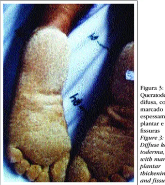
Dermatological exam (Figures 1 to 3) showed diffu-se keratoderma, with a yellowish, irregular surface and several fissures. Such alterations were limited to the palms and soles, without compromising other topographies. An erythematous halo was noticed delimiting the periphery of the lesions. The mucous membranes and cutaneous enclo-sures were unaffected.
The patient referred similar cases in the family, as can be observed in the hereditary chart below. Involvement of other generations was observed, with autosomal domi-nant inheritance. The presence of consanguinity is appa-rently coincidental.
Some of the relatives were examined (according to the hereditary chart
⇒
II 6, II 9-12, III 2, III 13-16), and a similar clinical picture, onset and course were verified.Histopathological exam of the biopsy (Figures 4 and 5) collected from the plantar surface demonstrated intense
histopatológico de carcinoma urotelial agressivo. Fumante de aproximadamente 30 cigarros por dia, há aproximada-mente 17 anos. Não apresentava demais co-morbidades.
Ao exame dermatológico (Figuras 1 a 3), o paciente mostrava hiperceratose difusa, com superfície amarelada, irregular, com presença de diversas fissuras. Tais alterações limitavam-se às palmas e plantas, não havendo comprome-timento de outras topografias. Notava-se halo eritematoso delimitando perifericamente as lesões. As mucosas e os anexos cutâneos estavam inalterados.
O paciente referiu casos semelhantes na família, como pode ser observado no heredograma anexo. Percebe-se o acometimento de outras gerações, com herança autos-sômica dominante. A presença de consangüinidade aparen-temente é ocasional.
Alguns dos parentes foram examinados (conforme heredograma ⇒II 6, II 9-12, III 2, III 13-16), verificando-se quadro clínico, história de início e evolução verificando-semelhantes. O exame histopatológico da biópsia (figuras 4 e 5) realizada na superfície plantar demonstrou intensa ortocera-tose compacta, hipergranulose e acanortocera-tose, acompanhadas de degeneração vacuolar da parte superior do estrato espi-Figura 1: Queratodermia palmar difusa, amarelada, delimitada
por halo eritematoso.
Figure 1: Diffuse palmar keratoderma, yellowish and delimited by an erythematous halo.
Figura 2: Detalhe da fotografia anterior demonstrando superfície palmar áspera e rica em fissuras.
Figure 2: Detail of the above photograph, demonstrating a rough palmar surface with many fissures.
2
Desconhecidos / Unknown Afetados / Affected Não afetados / Unaffected
I
II
III
IV
1 2 3 4 5 6 7 8 9 10 11 12 13 14 15 16 17
1 2 3
1 2 3
Figura 5: Degeneração vacuolar dos estratos granuloso e espinhoso
Figure 5: Vacuolar degeneration of the gran-ular and spiny layers nhoso e da camada granulosa. Os exames laboratoriais
esta-vam inalterados.
O paciente optou por continuar apenas em observa-ção dermatológica, não desejando nenhum tratamento local ou sistêmico.
DISCUSSÃO
As classificações das queratodermias palmo-planta-res baseiam-se principalmente nas características morfoló-gicas, topográficas e alterações sistêmicas associadas. Existem diversas classificações (Tabelas 3 e 4), o que se justifica pelas diversas características clínicas, bem como pelo fato de ainda serem doenças cujo mecanismo ainda é pouco compreendido, havendo discordância entre os auto-res. Com relação à morfologia, podem apresentar-se como difusa, focal e punctata. Quanto à topografia, podem estar restritas às superfícies palmo-plantares ou acometer regiões extensoras, como dorso das mãos e pés, bem como joelho e cotovelo (transgressivas).
Podem ainda apresentar curso estável ou agravar-se com a idade (progressivas). A pre-sença de outras alterações caracteriza as também deno-minadas displasias ectodérmi-cas palmo-plantares, com manifestações diversas,
geral-compact orthokeratosis, hypergranulosis and acanthosis, together with vacuolar degeneration of the superior part of the corneum stratum and granular layer. The laboratory exams were normal.
The patient opted to just continue under dermatolo-gical observation, without any local or systemic treatment.
DISCUSSION
Classifications of the palmoplantar keratodermas are mainly based on the morphologic and topographical characteristics and associated systemic alterations. There are several classifications (Tables 3 and 4), which is justi-fied by the diverse clinical characteristics, as well as the fact that there are still diseases whose mechanism remains little understood and with controversy between authors. Regarding the morphology, they can present as diffuse, focal and punctate. In terms of topography, they can be res-tricted to the palmoplantar surfaces or involve extensor areas, such as the back of hands and feet, as well as knees and elbows (transgres-sing). They can also present a stable course or worsen with age (progressive). The presen-ce of other alterations charac-terizes the also denominated ectodermic palmoplantar
Figura 3: Queratodermia difusa, com marcado espessamento plantar e fissuras Figure 3: Diffuse kera-toderma, with marked plantar thickening and fissures
dysplasias, with diverse manifestations, usually associated to the structures derived from the ectoderm (Table III).1
The present case concerns epidermolytic palmoplan-tar keratoderma, described initially by Vörner, in 1901.3
The manifestations begin in the first weeks of life. Most of the cases present autosomal dominant inheritan-ce;4 however, new familial cases4 (mutations) have been reported (Table 5).
The clinical picture5-6
is characterized by presenting marked palmoplantar keratoderma, with a yellowish colo-ration, well-delimited borders and often an erythematous halo outlining the lesion. The affected surface presents an aspect of snakeskin and there can be formation of blisters. The occurrence of hyperhidrosis is marked in some cases, sometimes associated to fungal infections, which is an important characteristic to be evaluated during therapeu-tics. The lesions rarely extend to the lateral surface of the fingers, as well as knuckle pad-like lesions.1,7,8
Minimal involvement of the elbows and knees can occur.1,7 Cutaneous annexes, as well as mucous surfaces, are unaf-fected. The course of the disease is stable, without progres-sion throughout the patient's life. The existence of other diseases (Table 5) in the patient with keratoderma of Vörner type, above all neoplasias,9,10
probably arises due to multiple phenomena or segregation of oncogenes.2,11,12
In the present case the patient had a prior diagnosis of urothelial carcinoma, while the cutaneous picture was considered a
mente associadas às estruturas derivadas do ectoderme (Tabela 3)1.
O presente caso diz respeito a queratodermia palmo-plantar epidermolítica, descrita inicialmente por Vorner, em 1901.3
As manifestações iniciam-se nas primeiras semanas de vida. A maioria dos casos tem padrão de herança autos-sômica dominante;4 entretanto, casos novos familiares4 (mutações) são relatados (Tabela 5).
O quadro clínico5-6caracteriza-se por marcada hiper-queratose palmo-plantar, de coloração amarelada, com bor-das bem delimitabor-das e muitas vezes com halo eritematoso contornando a lesão. A superfície afetada apresenta aspecto da "pele de cobra" (snakeskin), podendo haver formação de
bolhas. A ocorrência de hiperidrose é marcada em alguns casos, eventualmente associando-se a infecções fúngicas, característica importante a ser avaliada durante a terapêuti-ca. Raramente há extensão das lesões para superfície lateral dos dedos, bem como alterações semelhantes a coxins falan-geanos (Knuckle pad-like lesions).1,7,8 Acometimento míni-mo dos cotovelos e joelhos pode ocorrer.1,7Anexos cutâneos, bem como superfícies mucosas, apresentam-se inalterados. O curso da doença é estável, não havendo progressão duran-te a vida do pacienduran-te. A existência de outras doenças (Tabela 5) no paciente com queratodermia de Vörner, sobretudo neoplasias,9,10 provavelmente decorre de múltiplos fenôme-nos ou segregações de oncogenes.2,11,12 No presente caso o
Tabela 3: Queratodermias palmo-plantares1
/ Table 3: Palmoplantar keratodermas1
Sem manisfestações sistêmicas / without systemic manifestations
lDifusas / Diffuse
- Queratodermia palmo-plantar difusa epidermolítica (Vörner) / Diffuse epidermolytic palmoplantar keratoderma (Vörner) - Queratodermia palmo-plantar difusa não-epidermolítica (Unna-Thost) / Diffuse neoepidermolytic palmoplantar keratoderma (Unna-Thost)
- Eritroqueratodermia variabilis (Mendes da Costa) / Erythrokeratoderma variabilis (Mendes da Costa)
- Keratosis extremitaum hereditária progrediens (Greither/Sybert) / Hereditary keratosis extremitaum progrediens (Greither/Sybert)
lFocais / Focal
- Queratodermia palmo-plantar estriada (Brunauer-Fuhs-Siemens) / Striated palmoplantar keratoderma (Brunauer-Fuhs-Siemens)
lPunctatas / Punctate
- Queratodermia palmo-plantar punctata (Buschke-Fischer-Brauer) / Punctate palmoplantar keratoderma (Buschke-Fischer-Brauer) - Poroqueratose punctata palmaris et plantaris (Spiny Keratoderma) / Punctate parakeratosis palmaris et plantaris (Spiny Keratoderma)
- Acroqueratoelastoidose de Costa / - Acrokeratoelastoidosis (Costa)
Com manifestações sistêmicas (Displasias ectodérmicas palmoplantares) / with systemic manifestations (ectodermal palmoplantar dysplasias)
lClassificadas de I a XX de acordo com padrão de herança e características clínicas: alterações ungueais, capilares, dentárias e da mucosa bucal, presença de cistos epidérmicos, manifestações neurológicas, alterações pigmentárias, cardiovasculares,
paciente possuía o diagnóstico prévio de carcinoma urote-lial, sendo o quadro cutâneo achado ocasional. Contudo esse aspecto clínico não diminui a relevância da hiperqueratose nesses pacientes, pois serão eles que provavelmente se bene-ficiarão com os futuros testes de investigacão genética.
As alterações histopatológicas11 são restritas à epi-derme. Há orto-hiperceratose, hipergranulose, acantose e alterações vacuolares das camadas granulosas e parte supe-rior do estrato espinhoso. Tais alterações vacuolares carac-terizam-se pela presença de espaços claros perinucleares, acompanhada de material reticular citoplasmático, leve-mente corado, bem como grânulos querato-hialinos abun-dantes e irregulares. A derme mantém-se inalterada.
O exame histopatológico é indispensável para o diagnóstico, pois é o único que permite diferenciar a quera-todermia de Vorner da queraquera-todermia de Unna-Thost, que
apresentam alterações clínicas e padrão de herança
seme-chance finding. However this clinical aspect does not redu-ce the relevanredu-ce of keratoderma in such patients, because it is these that will probably benefit from the futures tests of genetic investigation.
The histopathological alterations11 are restricted to the epidermis. There is orthokeratoderma, hypergranulosis, acanthosis and vacuolar alterations of the granular layers and superior part of the corneum stratum. Such vacuolar alterations are characterized by the presence of clear peri-nuclear spaces, accompanied by slightly pink-colored, reti-cular cytoplasmatic material, as well as abundant and irre-gular keratohyalin granules. The dermis remains unaffected.
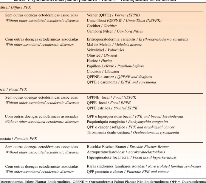
Histopathological exam is indispensable for the diagnosis, since it is the only means of differentiating kera-toderma of Vörner type from Unna-Thost type kerakera-toderma, which present similar clinical alterations and form of inhe-ritance, as well as in benign cases, or that is, those with Tabela 4: Queratodermias palmo-plantares2
/ Table IV –Palmoplantar keratoderma 2
QPP Difusa / Diffuse PPK
Sem outras doenças ectodérmicas associadas Without other associated ectodermic diseases
Vorner (QPPE) / Vörner (EPPK)
Unna-Thost (QPPNE) / Unna-Thost (NEPPK) Greither / Greither
Gamborg Nilsen / Gamborg Nilsen
Eritroqueratodermia variabilis / Erythrokeratoderma variabilis Mal de Meleda / Meleda’s disease
Vohwinkel / Vohwinkel Olmsted / Olmsted Huriez / Huriez
Papillon-Lefèvre / Papillon-Lefèvre Clouston / Clouston
QPPNE e surdez / QPPNE and deafness QPPE e carcinoma / EPPK and carcinoma
QPPNE focal / Focal NEPPK QPPE focal / Focal EPPK QPPE estriada / Striated EPPK
QPP e hiperqueratose bucal / PPK and buccal keratoderma Paquioniquia congênita / Pachyonychia congenita QPP e câncer esofágico / PPK and esophageal cancer Tirosinemia óculo-cutânea / Oculocutaneous tyrosinemia
Buschke-Fischer-Brauer / Buschke-Fischer-Brauer Acroqueratoelastoidose / Acrokeratoelastoidosis Hiperqueratose focal acral / Focal acral hyperkeratosis
Raras síndromes familiares isoladas / Rare isolated familial syndromes QPP punctata e câncer / Punctate PPK and cancer
Com outras doenças ectodérmicas associadas With other associated ectodermic diseases
Sem outras doenças ectodérmicas associadas Without other associated ectodermic diseases
Com outras doenças ectodérmicas associadas Without other associated ectodermic diseases
Sem outras doenças ectodérmicas associadas Without other associated ectodermic diseases
Com outras doenças ectodérmicas associadas With other associated ectodermic diseases QPP Focal / Focal PPK
QPP Punctata / Punctate PPK
QPPE = Queratodermia Palmo-Plantar Epidermolítica; QPPNE = Queratodermia Palmo-Plantar Não-Epidermolítico; QPP = Queratodermia Palmo-Plantar / PPK = Palmoplantar Keratoderma; EPPK = Epidermolytic Palmoplantar Keratoderma;
minimum transgression, related to keratoderma of Greither type (Sybert).12
Due to the existence of several cases diag-nosed only on the basis of the clinical pattern and without a histopathological evaluation, some authors question the real frequency of keratoderma of Vörner type.13
Several stu-dies have demonstrated its rarity; with rare cases
descri-lhantes, bem como nos casos frustros, isto é, com transgres-são mínima, relacionados à queratodermia de Greither (Sybert).12Devido à existência de diversos casos diagnosti-cados só com base no padrão clínico, estando ausente a ava-liação histopatológica, é que alguns autores questionam a real freqüência da queratodermia de Vörner.13 Diversos
Tabela 5: Casos reportados na literatura até 19914 /Table 5: Cases reported in the literature, up until 19914
Autores / Autores
Vörner Hähn
Brunsting et al. Yoshida Nagashima Klaus et al. Nagashima et al. Hussels and Davis Gómez-Orbaneja et al. Yoshida
Goette Hirai et al. Sommer et al. Moulin and Bouchet Takahashi et al. Fritsch et al. Yasaki et al. Förg
Miyanchi et al. Rabbiosi et al.
Blasik et al. Haneke Mizuno et al. Yoshiike Aso et al. Le Blainvaux Fonseca et al. Thomas et al. Camisa et al.
Chervrant-Breton et al.
Laurent et al. Puissant et al. Verdeguer et al. Blanchet-Bardon et al. Kanitakis et al. Zultak et al. Larrege et al. Fanti et al. Hamm et al. Moriwaki et al. Zarco et al. Berth-Jones et al. Requena et al.
Total 1901 1911 1962 1967 1969 1970 1970 1971 1972 1972 1974 1975 1976 1977 1977 1978 1978 1980 1980 1980 1981 1982 1982 1982 1983 1983 1984 1984 1985 1985 1985 1986 1986 1987 1987 1987 1987 1987 1988 1988 1988 1989 1991 1 1 1 3 1 2 3 1 1 1 1 2 1 1 1 1 1 1 1 1 1 1 1 1 1 1 1 1 1 1 11 1 1 1 50 1 1 2 1 1 2 1 1 1 1 1 1 14
Úlcera de perna / Leg ulcer
Esclerodermia sistêmica / Systemic scleroderma
Doença de Charcot-Marie-Tooth Charcot-Marie-Tooth disease
Granuloma anular / Granuloma annulare
Carcinomas (ovário, cólon e mama) / Carcinomas (ovary, colon and mamma)
Carcinoma (mama e ovário) / Carcinoma (mamma and ovary)
Surdez familiar /Familial deafness Anos /Year Casos familiares
Familial casesr
Casos esporádicos Sporadic cases
estudos demonstram sua raridade; com raros casos descri-tos, entretanto, por motivos acima abordados, tal doença apresenta-se provavelmente pouco notificada,13 sendo mesmo considerada por alguns autores a forma mais comum de queratodermia palmo-plantar.13 O significado real da epidermólise, entretanto, ainda é incerto, não ocor-rendo exclusivamente nessa variante, mas também em outras queratodermias, bem como demais doenças congêni-tas e adquiridas (Tabela 6).4
À microscopia eletrônica5,14há rarefação dos tonofi-lamentos nas áreas com degeneração vacuolar e seu agrupa-mento ao redor do núcleo. Os desmossomas demostram-se inalterados. Os grânulos de queratina apresentam-se aumentados e presentes desde o estrato espinhoso.
A análise genética dos casos de queratodermia palmo-plantar epidermolítica apresenta alterações no gene da citoqueratina K9, localizado no cromossomo 17.15,16Tal alteração geralmente ocorre na região central, não variável,
bed, however, for the above-mentioned reasons, such disea-se is probably little notified,13
and even considered by some authors to be the most common form of palmoplantar kera-toderma.13
The real significance of the epidermolysis, howe-ver, is still uncertain and does not occur exclusively in this variant, but also in other keratodermas, as well as other congenital and acquired diseases (Table 6).4
Electron microscopy reveals5,14
a rarefaction of the tonofilaments in the areas with vacuolar degeneration and the grouping of these around the nucleus. The desmosomes are unaffected. The keratin granules are enlarged and pre-sent as of the spinous layer.
Genetic analysis of the cases of epidermolytic pal-moplantar keratoderma presents alterations in the gene of cytokeratin K9, located in chromosome 17.15,16
Such an alte-ration generally occurs in the non variable central area of the cytokeratin and above all in segment 1A. Surprisingly alterations in areas other than the cytokeratin have been Tabela 6: Doenças com padrão histopatológico de hiperqueratose epidermolítica4
Table 6: Diseases with a histopathological pattern of epidermolytic keratoderma4
Hiperqueratose epidermolítica como principal alteração histopatológica Epidermolytic keratoderma as the principal histopathological alteration
Queratodermia palmoplantar epidermolítica / Palmoplantar epidermolytic keratoderma Eritrodermia ictiosiforme congênita bolhosa / Bullous congenital ichthyosiform erythroderma
Queratodermia palmoplantar associada à tirosinemia /Palmoplantar keratoderma associated to tyrosinemia Queratodermia palmoplantar numular / Nummular palmoplantar keratoderma
Alguns tipos de nevos epidérmicos / Some types of epidermal nevus Acantoma epidermolítico isolado /Isolated epidermolytic acanthoma
Acantoma epidermolítico disseminado / Disseminated epidermolytic acanthoma Ceratose solar epidermolítica / Epidermolytic solar keratosis
Leucoplasia epidermolítica / Epidermolytic leukoplasia
Papulose neviforme epidermolítica acrossiringea /Acrosyringeal epidermolytic nevoid papulosis
Disqueratose acantolítica focal e hiperqueratose epidermolítica combinadas / Combined focal acanthoid dyskeratosis and epidermolytic keratoderma
Hiperqueratose epidermolítica como alteração histopatológica ocasional Epidermolytic Keratoderma as an occasional histopathological alteration
Ceratose seborréica / Seborrheic keratosis Corno cutâneo / Cutaneous corneum
Carcinoma espinocelular / Spine cell carcinoma Ceratose verrucosa benigna /Benign verrucose keratosis Amiloidose liquenóide /Lichenoid amyloidosis Granuloma anular /Granuloma annulare Cisto triquilemal / Trichilemmal cyst Cisto epidermóide / Epidermoid cyst Nevo intradérmico /Intradermal nevus Cicatriz hipertrófica / Hypertrophic scar Eczema numular / Eczema nummulare
Hidradenoma de células claras /Hydradenoma of clear cells Ceratose solar / Solar keratosis
da citoqueratina, sobretudo segmento 1A. Curiosamente alterações em regiões diferentes da citoqueratina têm sido relatadas, ainda que sem diferenças fenotípicas.17
A citoqueratina K9, cujo peso molecular é de 54Kda, e o ponto isoelétrico (pHi), de 5,4, é encontrada exclusiva-mente nas superfícies palmo-plantares.18 Tal característica é a principal responsável por permitir a individualização da queratodermia de Vörner como entidade única, diferencian-do-a não só das demais queratodermias, mas também de outras doenças com padrão histopatológico semelhante. A identificação das citoqueratinas e suas respectivas funções tem permitido a melhor caracterização de diversas doenças, pelas quais são responsáveis ou estão associadas. Torna tam-bém pouco provável a hipótese de mosaicos clínicos da dermólise como causa de diversas doenças com padrão epi-dermolítico, afastando, por exemplo, a hipótese da querato-dermia de Vörner como forma localizada de eritroquerato-dermia ictiosiforme congênita bolhosa.3A citoqueratina K9faz parte dos filamentos intermediários de proteínas (FIP), moléculas cuja principal função é conferir à célula estrutura, força mecânica e adesão celular.19,20 Apresentam-se intimamente relacionados com os desmossomos e o núcleo celular. Tal denominação deve-se ao tamanho intermediário (10nm) entre os microfilamentos de actina (6nm) e os microtúbulos (25nm). Os FIPs pertencem a uma grande família de proteí-nas estruturais, divididas em seis grupos,1com base em sua seqüência de aminoácidos e especificidade tissular. Os tipos I e II são constituídos pelas citoqueratinas, predominantes em epitélios estratificados escamosos, principalmente a epi-derme. Conforme o peso molecular e ponto isoelétrico (pHi), as citoqueratinas são dispostas em dois grupos:
Tipo I - ácidas - pKi 4,5/5,5
⇒
menores em tama-nho, (40-56,5KDa), compreendendo queratinas 9 a 20 (K9 -K20). Os genes dessas queratinas localizam-se no cromos-somo 17q 12 - q21.Tipo II - básicas ou neutras - pKi 5,5/7,5
⇒
maiores em tamanho (52 - 67KDa), compreendendo queratinas 1 a 8 ( K1-K8). Os genes estão localizados no cromossomo 12q11-q13.Todas as citoqueratinas, bem como os FIPs, apresen-tam estrutura homóloga central (1A, 1B, 2A, 2B) constituída em α-hélice por aproximadamente 310 aminoácidos, sepa-radas por segmentos curtos não helicoidais (L1, L1-L2, L2) responsáveis pela conformação espacial (molecular). Apresentam também estruturas externas (head and tail) variáveis, supostamente responsáveis pela interação entre os FIPs e as demais proteínas estruturais celulares, além de polimerização celular.
As citoqueratinas polimerizam-se por meio de dis-posições não paralelas das moléculas e progressiva intera-ção entre as mesmas, formando sucessivamente protofila-mentos (2-3nm), protofibrilas (4,5nm) e fibrilas (10nm).
As citoqueratinas constituem o marco de diferencia-ção epidérmica, pilosa e ungueal. Estão presentes em propor-ção variáveis de acordo com a diferenciapropor-ção e funpropor-ção celu-lar (queratinócitos basais 10% - corneócitos 85%). O
contro-reported, although without phenotypic differences.17
Cytokeratin K9, the molecular weight of which is 54Kda and isoelectric point (pHi) of 5.4, is found exclusi-vely in the palmoplantar surfaces.18
Such a characteristic is the main aspect that enables the differentiation of Vörner type keratoderma as a singular entity, differentiating it not only from the other keratodermas, but also from other diseases with a similar histopathological pattern. The iden-tification of the cytokeratins and their respective functions has allowed a better characterization of several diseases, for which they are responsible or associated. It also ren-ders unlikely the hypothesis of clinical mosaics of molysis as the cause of several diseases with an epider-molytic pattern, discarding, for instance, the hypothesis that Vörner type keratoderma is a localized form of bullous congenital ichthyosiform erythroderma.3
Cytokeratin K9is part of the proteins of intermediate filaments (PIF), mole-cules whose main function is to provide the cell structure with mechanical strength and cellular adhesion.19,20
They are closely related to the desmosomes and cellular nucleus. Such a denomination is due to the intermediate size (10nm) between the microfilaments of actin (6nm) and the microtu-bules (25nm). PIFs belong to a large family of structural proteins, divided into six groups,1 according to their sequence of amino acids and tissular specificity. Types I and II are constituted by the cytokeratins, predominant in squamous stratified epithelia, mainly the epidermis. According to the molecular weight and isoelectric point (pHi), the cytokeratins are divided into two groups:
Type I - acidic - pKi 4.5/5.5
⇒
smaller in size, (40 -56.5KDa), comprising keratins 9 to 20 (K9 - K20). The genes of these keratins are located in chromosome 17q 12 - q21.Type II - basic or neutral - pKi 5.5/7.5
⇒
larger in size (52 - 67KDa), comprising keratins 1 to 8 (K1 - K8). The genes are located in chromosome 12q11-q13.All of the cytokeratins, as well as PIFs, present a central homologous structure (1A, 1B, 2A, 2B) constituted in
α-helix by approximately 310 amino acids, separated by short non-helical segments (L1, L1-L2, L2) responsible for the spatial conformity (molecular). They also present variable external structures (head and tail), supposedly responsible for the interaction between PIFs and other cel-lular structural proteins, besides the celcel-lular polymeriza-tion.
The cytokeratins become polymerized by means of non-parallel arrangements of molecules and progressive interaction one with another, successively forming protofi-laments (2-3nm), protofibrils (4.5nm) and fibrils (10nm).
le de regulação gênica das citoqueratinas é pouco compreen-dido, sendo realizado aparentemente nas fases iniciais de transcrição do DNA. Sabidamente os retinóides apresentam eficácia terapêutica nas queratinopatias a partir desse ponto.20,21Ressalta-se, entretanto, que outras estruturas celu-lares alteradas podem ser responsáveis pelo aparecimento das queratodermias, como alterações do envelope do corneócito (loricrina) na queratodermia mutilante de Vohwinkel,22 modi-ficações nos desmossomos na queratodermia de Brunauer-Fuhs-Siemens23 e alterações bioquímicas (enzima tirosina aminotransferase) na queratodermia de Richner-Hanhart,24 bem como defeitos ainda não esclarecidos.
Os recursos terapêuticos são escassos. O tratamento é principalmente sintomático. Queratolíticos tópicos, como ácido salicílico, bem como retinóides tópicos, podem auxiliar na redução da espessura epidérmica, porém com resultados transitórios. O uso de calcipotriol tópico tem sido descrito como alternativa terapêutica, embora também com resultados transitórios.26,27 O emprego de retinóides sistêmicos,1,4-25,28-21 antes etretinato e atualmente acitretin, demonstra redução da hiperqueratose em semanas; porém, devido ao aumento da sensibilidade táctil e dolorosa, bem como ao aumento da fra-gilidade cutânea, a terapêutica geralmente é descontinuada pelos pacientes. O emprego da terapia gênica ainda permane-ce limitado, dado o padrão autossômico da doença, o que dificulta o êxito da intervenção. Mesmo com técnicas que superem a barreira epidérmica e permitam a inserção gênica nos queratinócitos, tal alelo transformado persiste em expres-sar-se. O ideal seria o reparo do alelo transformado, mas tal técnica apresenta dificuldades extremas. Logo, possíveis pas-sos seriam a inibição da expressão dos genes transformados ou o aumento da expressão do alelo inalterado.2
q
known that in keratinopathies, the retinoids present the-rapeutic efficacy after this point.20,21 However, it is underscored that other altered cellular structures can be responsible for the onset of keratodermas, such as alterations in the envelope of the corneocyte (loricrin) in Vohwinkel's syndrome (mutilating keratoderma),22
modifications in the desmosomes in Brunauer-Fuhs-Siemens type keratoderma23
and biochemical altera-tions (tyrosine aminotransferase enzyme) in Richner-Hanhart type keratoderma,24 as well as defects which have yet to be clarified.
The therapeutic resources are scarce. Treatment is mainly symptomatic. Topical keratolytic, such as salicylic acid, as well as topical retinoids, can aid in reducing the epidermal thickness, however with transitory results. The use of topical calcipotriol has been described as a thera-peutic alternative, although again with transitory results.26,27
The use of systemic retinoids,1,4-25,28-21
etretinate and currently acitretin, has demonstrated a reduction in the keratoderma within several weeks; however, due to pain and greater tac-tile sensitivity as well as an increase in cutaneous fragility, this therapy is usually discontinued by the patients. The use of gene therapy continues limited, given the autosomal pat-tern of the disease, which hinders the success of the inter-vention. Even with techniques that overcome the epidermal barrier and allow the gene insert into the keratinocytes, the transformed allele persists in expressing itself. The ideal answer would be to repair the transformed allele, but such a technique presents extreme difficulties. Such that possible steps would be inhibition of the expression of the transfor-med genes or an increase in the expression of the unaltered allele.2
q
8. Mascaró Jr JM, Torras H, Mascaró JM. A child with unusual palms and soles. Case Report. Arch Dermatol. 1996; 132:1507-1512. 9. Chevrant-Breton J, Kerbrat P, Le Marec B et al. Kératodermie palmo-plantaire épidermolytique, autossomique dominante et adénocarcinomes familiaux (Etude de 4 génerations). Annales de Dermatologie et de Vénéréologie 1985; 112:841-844.
10. Blanchet-Bardon C, Nazarro V, Chevrant-Breton J et al. Hereditary epidermolytic palmoplantar keratoderma associated with breast and ovarian cancer in a large kindred. British Journal of Dermatology 1987; 117:363-370.
11. Ackerman AB. Histopathologic concept of epidermolytic hyperkeratosis. Arch Dermatol. 1970; 102:253-259.
12. Kansky A, Arzensek J: Is palmoplantar keratoderma of Greither's type a separate entity ? Dermatologica 1979; 158:244-248. 13. Hamm H, Happle R, Butterfass T, Traupe H. Epidermolytic palmoplantar keratoderma of Vörner: is it the most frequent type of hereditary palmoplantar keratoderma? Dermatologica 1988; 117:138-145.
14. Laurent R, Prost O, Nicollier M, Coumes S, Balzer MM, Adessi G. Composition keratohyaline granules in palmoplantar keratoderma: an ultrastructural study. Archives of Dermatological
REFERÊNCIAS / REFERENCES:
1. Stevens HP, Leigh IM: Keratoderma of palms and soles; in Fitzpatrick TB, Freeberg IM , Eisen AZ, Wolff K, Austen KF, Goldsmith LA et al: Dermatology in General Medicine, ed 5. New York, McGraw-Hill, 1999: 603-613.
2. Ratnavel RC, Griffiths WAD. The inherited palmoplantar kera-todermas. British Journal of Dermatology 1997; 137(4): 485-490. 3. Vörner H. Zur Kenntnis des Keratoma hereditarium palmare et plantare. Archiv für Dermatologie und Syphilis 1901; 56:3-31. 4. Requena L, Schoendorff C, Sanchez-Yuz E. Hereditary epider-molytic palmo-plantar keratoderma (Vörner type): report of a fam-ily and review of of the literature. Clin Exp Dermatol. 1991; 16:383-388.
5. Kanitakis J, Tsoitis G, Kanitakis C. . Hereditary epidermolytic palmo-plantar keratoderma (Vörner type). J Am Acad Dermatol. 1987; 17:414-422.
6. Moriwaki S, Toshihiro T, Horiguchi Y, Danno K, Sadao I. Epidermolytic hereditary palmoplantar keratoderma. Archives of Dermatology 1988; 124:555-559.
24. Natt E, Kida K, Odievre M, et al. Point mutations in the tyro-sine aminotransferase gene in tyrotyro-sinemia type II. Proc Natl Acad Sci USA 1992; 89: 9297-301.
25. Griffiths WAD, Leigh IM, Marks R. Disorders of keratiniza-tion. In: Champion RH, Burton JL, Ebling FJG, eds. Textbook of Dermatology. Oxford, England: Blackwell Scientific Publications; 1992: 1325-1390.
26. Yoshike T, Negi M, Manabe M, Takamori K, Ogawa H. Biochemical changes after the oral administration of retinoid in the horny layer of patients with keratinization disorders. J Dermatol 1982; 9:235-242.
27. Aso K, Toku S, Katagawa Y. Eletrophoretic pattern of preker-atins and kerpreker-atins of the soles in a case of epidermolytic palmo-plantar keratoderma successfully treated with oral retinoid. Japanese Journal of Dermatology 1983; 93:455-462.
28. Yoshiike T, Hattori M, Ogawa H. Father and daughter cases of Vörner type hereditary palmoplantar keratosis: biochemical analy-sis of stratum corneum and effect of oral retinoid therapy. Japanese Journal of Dermatology 1982; 92:751-756.
Research 1985; 277:384-394.
15. Reis A, Küster W, Eckardt R, Sperling K. Mapping of a gene for epidermolytic palmo-plantar keratoderma to the region of the acidic keratin gene cluster at 17q12-q21. Hum Genet. 1992; 90:113-116.
16. Matsumura KK, Bonifas JM, Bare JW, et al. Linkage to the type I keratin gene cluster of palmoplantar epidermolytic hyperk-eratosis. J Invest Dermatol. 1993; 100:508.
17. Coleman CM, Munro CS, Smith FJ, Uitto J, McLean WHI. Epidermolytic palmoplantar keratoderma due to a novel type of keratin mutation, a 3-bp insertion in the keratin 9 helix termination motif. Br J Dermatol 1999;140:486-490.
18. Moll R, Franke WW, Schiller DL, Geiger B, Krepler R. The catalog of human cytokeratins: patterns of expressions in normal epithelia, tumors and cultured cells. Cell 1982; 31:11-24. 19. Goldman RD, Khuon S, Chou YH, Opal P, Steinert PM. The functions of intermediate filaments in cell shape and cytoskeletal integrity. J Cell Biol 1996; 134:971-983.
20. Fuchs E, Cleveland DW. A structural scaffolding of interme-diate filaments in health and disease. Science 1998; 279:514-519. 21. Bergfeld WF, Derbes VJ, Elias PM, et al. The treatment of ker-atosis palmaris et plantaris with isotretinoin. J Am Acad Dermatol 1982; 6:727-731.
22. Maestrini E, Monaco AP, McGrath JA, et al. A molecular defect in loricrin, the major component of the cornified envelope, underlies Vohwinkel's syndrome. Nature Genet 1996; 13: 70-77. 23. Hennies H-C, Küster W, Mischke W, Reis A. Localisation of a locus for the striated form of palmoplantar keratoderma to chro-mosome 18q near the desmosomal cadherin gene cluster. Hum Mol Genet 1995; 4: 1015-20.
ENDEREÇO PARA CORRESPONDÊNCIA: / MAILINGADDRESS:
Endereço: Rafael Lenzi Tarnowsky
R. João Pio Duarte Silva, 404, Bl. Juriti - Ap. 101 Florianópolis SC 88037 001