Evaluation of the reagent test strips and microscopic examination
of urine in the diagnosis of urinary tract infection in sows
1Kelly Mazutti2,3*, Rosângela Locatelli-Dittrich3, Isabella Lunardon3, Suzana S.
Kuchiishi4,Anne C. de Lara4 , Everson Zotti5 and Geraldo C. Alberton6
ABSTRACT.- Mazutti K., Locatelli-Dittrich R., Lunardon I., Kuchiishi S.S., Lara A.C., Zotti E. & Alberton G.C. 2013. Evaluation of the reagent test strips and microscopic examination of urine in the diagnosis of urinary tract infection in sows.Pesquisa Veterinária Brasileira 33(9):1103-1108. Curso de Medicina Veterinária, Escola de Ciências Agrárias e Medicina
Ve-terinária, Pontifícia Universidade Católica do Paraná, Campus Curitiba, Rua Imaculada Con -ceição 1155, Prado Velho, Curitiba, PR 80215-901, Brazil. E-mail: kelly.mazutti@pucpr.br
The diagnosis of the urinary tract infection (UTI) in sows is usually performed by using reagent test strips, since it is a fast and practical method, and capable of being done at the farm. The microscopic examination of the urine is rarely used at the farm since it is a more time consuming and difficult technique. However, there are no studies on the accuracy of those two techniques for the UTI diagnosis on this species. This study aims to assess the accuracy of the reagent test strip and the urine microscopic examination in the diagnosis of ITU in sows, comparing them with the bacteriological examination of urine. In order to select the sows for this study, a chemical reagent test strip was carried out previously and a total of 139 sows were selected, 66 sows of which showed positivity to nitrite in the reagent test strip and 73 without nitrituria. Then, the next day, a new sample collection for performing a complete urinalysis was carried out from those 139 sows, which included physical, chemical, microscopic and microbiological examination of these urine samples. The results revealed that the nitrite test of the reagent strip showed 100% of specificity and 93% of sensitivity. The specificity of the microscopic examination for bacteriuria was 82% and the sensitivity was 100%. The UTI diagnosis by using reagent strips and/or the urine sediment test is reliable if compared to the urine bacteriological examination, which makes possible the rapid diagnosis of UTI in sows at the farm.
INDEX TERMS:Urinary tract infection, cystitis,Escherichia coli, leukocyte esterase, nitrite, urinalysis,
swine.
1 Received on May 23, 2013.
Accepted for publication on August 6, 2013.
2 Curso de Medicina Veterinária, Escola de Ciências Agrária e Medicina
Veterinária, Pontifícia Universidade Católica do Paraná (PUCPR), Campus Curitiba, Rua Imaculada Conceição 1155, Prado Velho, Curitiba, PR 80215-901, Brazil. *Corresponding author: kelly.mazutti@pucpr.br
3 Depto Medicina Veterinária, Universidade Federal do Paraná (UFPR),
Campus Curitiba, Rua dos Funcionários 1540, Curitiba, PR 80035-050.
4 Centro de Diagnóstico de Sanidade Animal (CEDISA), Rodov. BR-153
Km 110, Vila Tamanduá, Concórdia, SC 89700-000, Brazil.
5 Curso de Medicina Veterinária, Escola de Ciências Agrária e Medicina
Veterinária, PUCPR, Campus Toledo, Avenida da União 500, Jardim Coopa -gro, Toledo, PR 85902-532, Brazil.
6 Departamento de Medicina Veterinária, UFPR, Campus Palotina, Rua
Ipiranga 860, Apto 603, Palotina, PR 85950-000, Brazil.
RESUMO.- [Precisão da tira reagente e do exame mi-croscópico da urina no diagnóstico de infecções do trato urinário em porcas.]O diagnóstico de infecção do
trato urinário (ITU) em porcas geralmente é feito com o au
-xílio de tiras reagentes, por ser um método rápido, prático e passível de ser realizado na própria granja. O exame mi
-croscópico da urina raramente é utilizado em granjas por ser uma técnica mais demorada e trabalhosa. No entanto, não existem estudos sobre a precisão destas duas técni
-cas no diagnóstico de ITU nesta espécie. O objetivo deste estudo foi avaliar a precisão da tira reagente e do exame microscópico da urina no diagnóstico de ITU em porcas, comparando-os com o exame bacteriológico da urina. Para selecionar as porcas que iriam compor o estudo foi reali
-zado um exame químico prévio com tira reagente, do qual foram selecionadas 139 porcas, 66 positivas para nitrito na tira reagente e 73 negativas. No dia seguinte foi realizada uma nova coleta de urina destas 139 porcas para realiza
-mico, microscópico e microbiológico destas amostras de urina. Os resultados demonstraram que a prova de nitrito da tira reagente apresentou 100% de especificidade e 93% de sensibilidade. A especificidade do exame microscópico para bacteriúria foi de 82% e a sensibilidade de 100%. O diagnóstico de ITU com o uso de tiras reagentes e/ou com exame microscópico da urina é confiável, quando compara
-do com o exame bacteriológico da urina, o que torna possí
-vel o diagnóstico rápido de ITU em porcas na granja. TERMOS DE INDEXAÇÃO: Infecção do trato urinário, cistite, Es-cherichia coli, esterase leucocitária, nitrito, urinálise, suínos.
INTRODUCTION
Urinary tract infection (UTI) is the most important ende
-mic disease affecting sows and also one of the main cau
-ses of reproductive failures, general health complications and reduction of the life expectancy of the herd (Girotto et al. 2000, Porto et al. 2004). The microorganisms most frequently found in these infections are Escherichia coli,
Proteus mirabilis, Staphylococcus sp., Streptococcus sp., Ae-romonas hydrophila and Actinobaculum suis (Meister 2006,
Sobestiansky et al. 2007, Menin et al. 2008).
The clinical examination has a limited value in UTI diag
-nosis since in most of the cases the clinical signs are not evident (Fairbrother 2006), and it is necessary to perform
a urinalysis to achieve a conclusive diagnostic. One of the
routine practices used on farms is the collection of urine samples by spontaneous urination and the performance of the diagnosis by using reagent test strips. The preventive and/or healing procedures are carried out in accordance to the prevalence obtained through this method.
The use of the reagent strips method is widely used since it is fast, practical and can be performed at the farm. To supplement this method, a complete urinalysis, which includes the microscopic examination of the urine and the bacteriological test, may be also performed. However, the distance between the laboratories and the farms, the addi
-tional cost and the time demanded, are limiting factors for performing these tests.
Since the reagent strips used in Veterinary Medicine have been developed for diagnosing UTI in humans, they may, therefore, generate doubtful results when applied to swine. Furthermore, even on humans, there are various studies that show a wide variation in the sensitivity and specificity of the components of the strips used for diagno
-sing UTI (Kellogg et al. 1987, Bolann et al. 1989, Lachs et al. 1992, Holland et al. 1995, Sultana et al. 2001).
The aim of this study is to assess the accuracy of the rea
-gent strip and of the urine microscopic examination as me
-thods for diagnosing the urinary tract infections in sows, comparing obtained results to the bacteriological test.
MATERIALS AND METHODS
The experiment was carried out in two Piglet Production Units – PPU, one located in the State of Santa Catarina and the other in the State of Paraná, Brazil. The study was composed by 139 pregnant sows of commercial lineage, with different parity times, placed in individual cages with a channel drinker. All sows were preg
-nant, with gestational ages ranging between 50 and 70 days. The
amount and the type of ration consumed by the animals during the time of the experiment followed the routine pattern already set by the farmers, in accordance with the length of pregnancy and in compliance with the NRC (1998) recommendations for pregnant sows. The animals were provided with plenty of water during the whole period of the experiment.
In order to select the sows for this study, a chemical reagent strip test was carried out previously and a total of 139 sows were selected, 66 sows of which showed positivity to nitrite in the re
-agent strip and 73 without nitrituria. Then, the next day, a new sample collection for performing a complete urinalysis was car
-ried out from those 139 sows.
The urine samples were collected at dawn, before feeding and in sterile flasks. The collectors waited for the spontaneous urina
-tion of the sows and collected the middle urine, disregarding the first discharge. After each collection, the flasks were closed and placed behind the cages of the respective sows. After collecting the samples, the flasks were dried with tissue paper and numbe
-red according to the sows earrings. The urine samples were pla
-ced into isothermal boxes with ice and taken to the laboratory on the farm for the immediate performance of the physical, chemical and microscopic tests.
The evaluation of the urine was performed in accordance with standard methods (Strasinger et al. 1998). The chemical test was carried out with reagent test strips (Uriquest®, Labtest Diagnós
-tica S.A., Brazil). The parameters evaluated were the following: nitrite, pH, urinary specific gravity and leukocytes. The urinary specific gravity was also obtained by refractometry and pH by pH meter. The microscopic examination of the urine (sedimentosco
-py) was performed with a regular optical microscope in the 45 x objective. Leukocytes were quantified as a number per average of ten fields. The bacteria were classified in accordance with visual and subjective criteria and were recorded as absent (-), rare (R), discreet (+), moderate (++), pronounced or uncountable (+++).
The urine samples were placed in isothermal boxes with ice and were sent to the Animal Sanity Diagnosis Center – CEDISA, located in Concórdia/SC, for bacterial count and bacteriological isolation. The samples were seeded in 5% ovine blood Agar, Mac Conkey and in Tryptic Soy Agar(TSA) for colony count. Samples that showed a count equal or higher than 105 UFC/ml were con
-sidered positive for urinary tract infection (Fairbrother 2006). The bacteria were identified through Gram and biochemical tests (SIM, TSI, CIT, O/F, VM, Catalase). The bacteria identified as Gram negative were submitted to supplemental biochemical tests by using the Api 20 E commercial kit (BioMérieux®, Marcy l’Etoile,
France).
The computations for determining the sensitivity and speci
-ficity of the reagent strip and of the microscopic examination of the urine were carried out in accordance with a described me -thod (Menezes & Santos 1999). In the case of the reagent strip, the
animals positive for nitrite in the reagent strip were considered positive. In the urine microscopic examination, samples with rare or absent bacteria were considered negative, while the samples with a discreet bacterial presence (+), moderate (++), pronoun
-ced or uncountable (+++) were considered positive. The results obtained from both techniques were compared to the urine bacte
-riological test, which is considered the “gold standard” diagnostic test for UTI.
During the statistical analysis, the data obtained with conti
-nuous numerical variables of normal distribution, were submit -ted to the t test and considering the statistical differences when
the value of P<0.05. The statistical correlations were carried out
the bacterial count by seeding and by the bacteriological test was analyzed by Pearson’s test, when using crosstabulation method (Statgraphics® 1982-2010), and by Kappa’s test. The latter test was also used to establish the agreement between nitrite in the reagent test strip and the bacteriological test.
RESULTS
The UTI diagnosis with reagent test strip, considering the nitrite strip, showed 100% specificity, which means that all the positive samples for nitrite in the reagent strip (66) showed a bacterial count over 105 UFC/ml. The sensitivity
of the reagent strip was 93% because five, from 73 of the negative samples for nitrite in the strip, showed a bacterial
count of over 105 UFC/ml (Table 1). None of the samples
was positive for leukocytes in the strip.
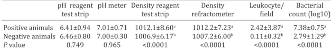
Urinary parameters of sows affected and non-affected by ITU can be seen in Table 2. There was no statistical di
-fference in the urine pH, obtained by both the reagent test
strip as obtained by pH meter, between positive and ne
-gative animals for ITU. There was a statistical correlation between the two techniques used (r=0.62, P<0.0001). The urine specific gravity was significantly higher in animals positive for UTI compared to the negative ones, in both te
-chniques (Table 2), and there was also a correlation betwe
-en urinary d-ensity obtained by the reag-ent test strip and the refractometer (r=0.86, P<0.0001).
The number of leukocytes per field was significantly hi
-gher in animals positive for UTI than in the negative ones, as well as bacterial count (Table 2). There was an agreement between the bacterial count obtained during the microsco
-pic examination and the one obtained by seeding (r=-0.27, P=0.0013). The specificity of the microscopic examination for bacteriuria was 82% and sensitivity was 100% (Table 1). According Landis & Koch (1977), the obtained values of Kappa’s test revealed an almost perfect agreement between nitrite in the reagent test strip and the bacteriological test (K=0.928, P<0.001) and a substantial agreement between
the bacterial count by seeding and by the bacteriological
test (K=0.7, P<0.001) (Table 1).
The result of the urine culture of the 71 urine samples positive for UTI (Table 3) showed that the Escherichia coli bacteria was the most frequently isolated agent (81.69%).
DISCUSSION
The nitrite proof with reagent strip showed 100% specifi
-city and 93% sensitivity for UTI diagnostic. The false-ne
-gative results may have two explanations: the first one is that not all the bacteria are capable of converting nitrate into nitrite; nevertheless, the Gram-negative, which are the main ones responsible for UTI, have this capacity (Morgan & McKenzie 1993, Strasinger 1998, Memişoğullari et al. 2010); the second explanation is that the reaction depends on the urinary stasis in the bladder for a minimum period Table 1. Results from Reagent test strip and Microscopic
examination of urine, comparing to Bacteriological test results (gold standard)
Reagent Microscopic exami-
test strip nation of urine gical test
Number of positive animals 66 84 71
Number of negative animals 73 55 68
True positives 66 72
False positives 0 12
True negatives 68 55
False negatives 5 0
Specificity 100% 82%
Sensitivity 93% 100%
Kappa’s value* 0.928 0.7
*According to the interpretation table of Kappa’s value from Landis & Koch (1977), values of Kappa’s test between 0.6 and 0.79 means a subs -tantial agreement, and values between 0.8 and 1.00 means an almost perfect agreement (P<0.001).
Table 2. Comparative results of urinary parameters of sows positive and negative for ITU
pH reagent pH meter Density reagent Density Leukocyte/ Bacterial
test strip test strip refractometer field count (log10)
Positive animals 6.41±0.94 7.01±0.71 1012.1±8.60a 1012.2±7.23a 2.42±3.87a 7.38±0.75a
Negative animals 6.46±0.80 7.00±0.30 1006.9±6.17b 1007.2±6.00b 0.11±0.32b 2.79±1.29b
P value 0.749 0.965 <0.0001 <0.0001 <0.0001 <0.0001
a,b Different superscript letters in the same column indicate statistical significance (P<0,05).
Table 3. Results of the urine culture of 71 urine samples of sows positive for UTI
Isolated bacteria Number of samples Frequency %
Escherichia coli 58 81.69
Gram-negative Coccobacillus* 4 5.63
Escherichia coli / Streptococcus sp. 4 5.63
Escherichia coli / Staphylococcus sp. 1 1.41
Escherichia coli / Proteus sp. 1 1.41
Enterobacter sp. 1 1.41
Streptococcus sp. 1 1.41
Proteus sp. 1 1.41
Total 71 100
* Positive for negative Gram bacteria: excluding the possibility of being
Enterobacter sp., Klebsiella sp., Edwardsiella sp., Salmonella sp. and Es-cherichia coli.
of time of four hours (Almond & Stevens 1995); therefore, these sows may have urinated within an interval of time
shorter than four hours.
There are several studies which evaluate the sensitivi
-ty and the specifici-ty of the positive reaction for nitrite in the reagent strip for UTI diagnosis in human beings (Bollan et al. 1989, Tincello & Richmond 1998, Sultana et al. 2001, Devillé et al. 2004, Ali et al. 2007, Ducharme et al. 2007, Taneja et al. 2010). The values found range from 33 to 57% and 78 to 99%, respectively.
None of the samples showed leukocyturia in the reagent test strip, even when significant amounts of these were pre
-sence of infection or inflammation in the urogenital system. Among the most frequent causes are bacterial infections, such as cystitis, pyelonephritis, prostatitis and urethritis (Alberton & Locatelli-Dittrich 2010). The tests for leu -kocytes, or leukocyte esterase, are based on the hydrolysis
of protein substrates esters with esterase activity. Human neutrophils produce up to 10 proteins with esterase ac
-tivity. These proteins react with the substrate to produce alcohols and acids, which then react with other substances in order to produce a change in color that is proportional to the amount of esterase in the sample (Fuller et al. 2001). González & Silva (2006) say that the proof for leukocytes in the reagent strip is based on human leukocyte esterase and do not seem to be so sensitive in animals as in humans. Therefore, this study showed that the reagent strip was not a reliable parameter and the proof of the presence of leu
-kocytes in the urine of sows must be obtained by analyzing the urinary sediment.
There was a correlation between the urinary specific gravity values obtained by reagent test strip and the refrac
-tometer (r=0.86, P<0.0001), demonstrating that the strip can be considered reliable for the assessment of urinary specific gravity. In both methodologies the values obtained were significantly higher in positive animals for UTI than in the negative ones. Urinary specific gravity is directly rela
-ted to the amount of water intake by the sow. Thus, when the amount is sufficient, insufficient, or is in a critical limit, the urinary specific gravity is less than 1008, greater than 1012, and between 1008 and 1012, respectively (Sobes
-tiansky et al. 1992). The water supply system was the same for all animals, with water ad libitum. A likely explanation for this observed difference would be that, probably, posi
-tive animals feel pain during urination caused by UTI and, thus, avoid urinating frequently, leading to a urinary stag
-nation and increasing consequently the urinary concentra
-tion. Also, because of the pain, these animals prefer to stay longer in bed, avoiding getting up to drink water.
There was also a correlation between the pH value ob
-tained by the reagent test strip and by pH meter (r=0.62, P <0.0001). The average pH values were lower in the strip when compared to pH meter results (table 2). This may have two explanations: the first is that the pH meter might be more accurate in measuring pH than the reagent test strip, and the second is that the urine was evaluated immediately by reagent test strip, while in evaluation by the pH meter there was the transport time to the laboratory, which means that the urine may have a alkalization process at the elapsed time between the two evaluations. The pH values obtained by positive and negative animals for UTI, in both techniques, were within normal limits because, according Menin et al. (2008), pH values for sows urine between 5,5 to 7,5 are con
-sidered normal. It was expected that sows positive for ITU presented a more alkaline pH. According Coles (1989), in urinary infections is expected to find alkaline urine, due to the microorganisms located in the urinary tract. When they are endowed with the urease enzyme, they can turn the urea into ammonia, causing alkalinization. According Sobes
-tiansky et al. (2007), a pH value of 8 or above, constitute an important sign of a predisposition to bacterial infections.
There was an agreement between the bacterial count carried out by sedimentoscopy and the bacterial count per
-formed by seeding (r=-0.27, P=0.0013). This result shows that the microscopic examination of the urine is reliable for diagnosing UTI, since even the five sows with false nega
-tive diagnosis for UTI through the nitrite test were detec
-ted with bacteriuria in the sediment test. Therefore, even though the microscopic examination of the urine to detect the presence of leukocytes and bacteria is more time con
-suming and harder to perform than the reagent strip test (Downs 1999), the former may be performed safely at the farm, whereas the bacteriological examination, regarded as the reference method for diagnosing UTI (Zorc et al. 2005), needs to be performed in a laboratory, has a high cost and
has the disadvantage of taking at least 48 hours for obtai-ning the results(Whiting et al. 2005).
The specificity and the sensitivity of the microsco
-pic examination of the urine in this study were 82% and 100%, respectively. Hiraoka et al. (1995) set out to as
-sess the usefulness of the microscopic examination of the urine for diagnosing UTI in human and obtained 91% sensitivity and 98% specificity for detecting bacteriuria. Memişoğullari et al. (2010) evaluated 250 human urine samples in order to compare the results obtained by the reagent strip with the microscopic examination of the uri
-ne and computed the performance characteristics of those tests. The sensitivity and the specificity of the microscopic examination of the urine were 91% and 68%, while in the reagent strip they were 80% and 60% respectively. The authors suggest that both urine analysis methods can be used for a quick diagnostic. Other authors obtained similar
results (Vangone & Russo 1985, Vickers et al. 1991, Lohr et
al. 1993, Al-Daghistani & Abdel-Dayem 2002). Taneja et al. (2010) evaluated the usefulness of the reagent strip (leu
-kocyte esterase and nitrite) and of the microscopic exami
-nation of the urine for diagnosing UTI and concluded that these techniques must be used jointly for added safety in quickly UTI diagnosing.
Escherichia coli was the bacteria most frequently iso
-lated in the urine culture of the samples positive for UTI (81.69%). Carr & Walton (1992) and Meister (2006) obtai
-ned similar results and found the E. coli bacteria as being
the most frequent agent among the urine samples evalua
-ted, with 90.38% and 70.45%, respectively. These findings
corroborate various other studies that have also found E. coli as the most frequent bacteria in UTI cases in sows (Reis et al. 1992, Carr et al. 1995, Menin et al. 2008). Similarly, in human beings, E. coli is the most common etiological agent (Anderson et al. 2004, Mysorekar & Hultgren 2006, Rosen et. al 2008).
CONCLUSION
The UTI diagnosis with reagent strips and/or the microsco
-pic examination of urine is reliable if compared with urine bacteriological examination, since the reagent test strip has shown 100% specificity and 93% sensitivity, and bacteriu
-ria in the microscopic examination of the urine has shown 82% specificity and 100% sensitivity, which makes possi
REFERENCES
Abreu P.F., Requião-Moura L.R. & Sesso R. 2007. Avaliação diagnóstica de hematúria.J. Bras. Nefrologia 29:158-163.
Alberton G.C. & Locatelli-Dittrich R. 2010. Infecções do trato urinário em porcas, p.83-135. In: Alfieri A.F., Barry A.F., Alfieri A.A., Silva C.A., Dalla -nora D., Zotti E., Alberton G.C., Rodrigues I.M.T.C., Machado I.P., Griessler K., Mores M.A.Z., Dittrich R.L. & Starkl V. (Eds), Tópicos em Sanidade e Manejo de Suínos. Curuca Consciência Ecológica, São Paulo.
Alberton G.C., Sobestiansky J. & Donin D.G. 2012. Infecção urinária em fêmeas em produção, p.179-194. In: Sobestiansky J. & Barcellos D. (Eds), Doenças dos Suínos. 2ª ed. Cânone Editorial, Goiânia.
Al-Daghistani H.I. & Abdel-Dayem M. 2002. Diagnostic value of various urine tests in the Jordanian population with urinary tract infection. Clin. Chem. Lab. Med. 40(10):1048-1051.
Ali S.H.G., Moodambail A.R., Hamrah E.K.B., Bin-Nakhi H.A. & Sadeq S.A. 2007. Reliability of rapid dipstick test in detecting urinary tract infec -tion in symptomatic children. Kuwait Med. J. 39:36-38.
Almond G.W. & Stevens J.B. 1995. Urinalysis techniques for swine practi -tioners. Compend. Contin. Educ. 17:121-129.
Anderson G.G., Dodson K.W. & Hooton T.M. 2004. Intracellular bacterial communities of urophatogenic Escherichia coli in urinary tract pathoge -nesis. Trends Microbiol. 12(9):424-430.
Bolann B.J., Sandberg S. & Digranes A. 1989. Implications of probability analysis for interpreting results of leukocyte esterase and nitrite test strips. Clin. Chem. 35(8):1663-1668.
Brito B.G. 2004. Fatores de virulência presentes em amostras de
Esche-richia coli uropatogênicas: UPEC para suínos. Ciência Rural
34(2):645-652.
Carr J. & Walton J.R. 1992. The microflora of the porcine urinary tract in cases of cystitis and pyelonephritis. Proc. 12th International Pig
Veteri-nary Society Congress, IPVS, The Hague, p.347.
Carr J., Walton J. & Done S. 1995. Cystitis and ascending pyelonephritis in the sow. In Practice 17:71-79.
Coles E.H. 1989. Pruebas de funcionamento renal, p.175-206. In: Ibid. (Ed.), Diagnóstico y Patologia em Veterinária. 4ª ed. Interamericana,
México.
Devillé W.L.J.M., Yzermans J.C., Van Duijn N.P., Bezemer P.D., Van der Windt D.A.W.M. & Bouter L.M. 2004. The urine dipstick test useful to rule out infections. A meta-analysis of the accuracy.BioMed Central Urology 4:1-14.
Downs S.M. 1999. Technical report: urinary tract infections in febrile in -fants and young children: the Urinary Tract Subcommittee of the Ame -rican Academy of Pediatrics Committee on quality improvement. Pedia -trics 103:54.
Ducharme J., Neilson S. & Ginn J. 2007. Can urine cultures and reagent test strips be used to diagnose urinary tract infection in elderly emergency department patients without focal urinary symptoms? Can. J. Emerg. Med. Care 9(2):87-92.
Fairbrother J.M. 2006. Urinary tract infection, p.671-674. In: Straw B.E., Zimmermann J.J., D´Allaire S. & Taylor D.J. (Eds), Diseases of Swine. 9th
ed. Blackwell Publ., Ames, IA.
Fuller C.E., Threatte G.A. & Henry J.B. 2001. Basic examination of the uri -ne. p.367-402. In: Henry J.B., Davey F.R., Herman C.J., McPherson R.A., Pincus M.R., Threatte G.A. & Woods G.L. (Eds), Clinical Diagnosis and Management by Laboratory Methods. 20th ed. W.B. Saunders, Philadel
-phia.
Garcia-Navarro C.E.K. 1996. Manual de Urinálise Veterinária. Varela, São Paulo. 94p.
Girotto A.F., Sobestiansky J., Dalla Costa O.A., Matos M.P.C. & Pôrto R.N.G. 2000. Avaliação econômica de alta prevalência de infecção urinária em matrizes em um sistema intensivo de produção de suínos. Comun. Téc. 59, Embrapa-CNPSA, Concórdia, SC. 4p.
González F.H.D. & Silva S.C. 2006. Introdução à Bioquímica Clínica Veteri -nária. 2ª ed. UFRGS, Porto Alegre. 37p.
Hiraoka M., Hida Y., Hori C., Tsuchida S., Kuroda M. & Sudo M. 1995. Urine
microscopy on a counting chamber for diagnosis of urinary infection. Acta Paediatrica Japonica 37:27-30.
Holland D.J., Bliss K.J., Allen C.D. & Gilbert G.L. 1995. A comparison of che -mical dipsticks read visually or by photometry in the routine screening of urine specimens in the clinical microbiology laboratory. Pathology 27:91-96.
Kellogg J.A., Manzella J.P., Shaffer S.N. & Schwartz B.B. 1987. Clinical rele-vance of culture versus screens for the detection of microbial pathogens in urine specimens. Am. J. Med. 83(4):739-745.
Lachs M.S., Nachamkin I., Edelstein P.H., Goldman J., Feinstein A.R. & Schwartz J.S. 1992. Spectrum bias in the evaluation of diagnostic tests: lessons from the rapid dipstick test for urinary tract infection. Annals Intern. Med. 117(2):135-140.
Landis J.R., Koch G.G. 1977. The measurement of observer agreement for categorical data. Biometrics 33: 159-174.
Lohr J.A., Portilla M.G., Geuder T.G., Dunn M.L. & Dudley S.M. 1993. Making a presumptive diagnosis of urinary tract infection by using a urinalysis performed in an on-site laboratory. J. Pediatrics 122:22-25.
Memişoğullari R., Yüksel H., Yildirim H.A. & Yavuz O. 2010. Performance characteristics of dipstick and microscopic urinalysis for diagnosis of urinary tract infection. Eur. J. General Med. 7(2):174-178.
Meister A.R. 2006. Efeito do cloreto de amônio, ácido cítrico e cloreto de sódio no controle de cistites em porcas. 2006. Dissertaçãode Mestrado em Ciências Veterinárias, Programa de Pós-graduação em Medicina Ve -terinária, Faculdade de Ciências Agrárias e Veterinárias, Universidade Estadual Paulista “Júlio de Mesquita Filho”, Jaboticabal, SP. 85p. Menezes A.M.B. & Santos I.S. 1999. Curso de Epidemiologia Básica para
Pneumologistas. 4. Epidemiologia clínica. J. Pneumol. 25(6):321-326. Menin A., Reck C., Capelli J.C., Ferraz S.N. & Vaz E.K. 2008. Diagnóstico de
infecção urinária em fêmeas suínas produtivas em granjas comerciais no sul do brasil. Ciênc. Anim. Bras. 9:199-206.
Morgan M.G. & Mckenzie H. 1993. Controversies in the laboratory diagno -sis of community acquired urinary tract infection. Eur. J. Clin. Microbiol. Infect. Dis. 12(7):491-504.
Mysorekar I.U. & Hultgren S.J. 2006. Mechanisms of uropathogenic Esche-richia coli persistence and eradication from the urinary tract. Proc. Natl
Acad. Sci. 103(38):14170-14175.
NRC 1998. Nutrient Requirements of Swine. 10th ed. National Research
Council, National Academy Press, Washington, DC.
Pôrto R.N.G., Sobestiansky J., Matos M.P.C. & Meirinhos M.L.G. 2004. As -pectos histopatológicos do sistema urinário de matrizes suínas descar -tadas. Ciênc. Anim. Bras. 5(2):109-112.
Reis R., Nakajima M. & Nascimento E.F. 1992. Infecções urinárias em por -cas. Arq. Bras. Med. Vet. 44(5):363-76.
Rosen D.A., Pinkner J.S., Jones J.M., Walker J.N., Clegg S. & Hultgren S.J. 2008. Utilization of an intracellular bacterial community pathway in
Klebsiella pneumoniae urinary tract infection and the effects of FimK on
Type 1 pilus expression. Infect. Immunity 76(7):3337-3345.
Sobestiansky J., Mores N. & Perestrelo R. 1992. Infecções urinárias na fê -mea suína. Circ. Téc. 11, Embrapa-CNPSA, Concórdia, p.7-49.
Sobestiansky J. 2007. Infecção urinária em fêmeas em produção, p.127-141. In: Sobestiansky J. & Barcellos D. (Eds), Doenças dos Suínos. Câ -none Editorial, Goiânia.
Statgraphics® 1982-2010. Centurion XVI ver.16.1.03. Statistical Graphic System by Statistical Graphic Corp.
Strasinger D.A. 1998. Uroanálise e Fluídos Biológicos. 3ª ed. Editorial Pre -mier Ltda, São Paulo. 233p.
Sultana R.V., Zalstein S., Cameron P. & Campbell D. 2001. Dipstick urinaly -sis and the accuracy of the clinical diagno-sis of urinary tract infection.J. Emerg. Med. 20:13-19.
Tincello D.G. & Richmond D.H. 1998. Evaluation of reagent strips in de -tecting asymptomatic bacteriuria in early pregnancy: prospective case series. Brit. Med. J. 316:435-437.
Vangone G. & Russo G. 1985. Bacteria and leukocyte count in the urine in the diagnosis of urinary tract infections. La Pediatria Medica e Chirur-gica7:125-129.
Vickers D., Ahmad T. & Coulthard, M.G. 1991. Diagnosis of urinary tract in -fection in children: fresh urine microscopy or culture? Lancet 338:767-770.
Whiting P., Westwood M., Watt I., Cooper J. & Kleijnen J. 2005. Rapid tests and urine sampling techniques for the diagnosis of urinary tract infec -tion (UTI) in children under five years: a systematic review. BioMed Central Pediatrics 4(4):1-13.
Wiles T.J., Kulesus R.R. & Mulvey M.A. 2008. Origins and virulence mecha -nisms of uropathogenic Escherichia coli. Exp. Molec. Pathol.
85(9):11-19.