This work is licensed under a Creative Commons Attribution 4.0 International License.
Comparison of Inhibitory Activities of
meta
and
para
Substituted
N
-aryl 3-Hydroxypyridin-4-one
Mannosides Towards Type 1 Fimbriated
E. coli
Vesna Petrović Peroković, Rosana Ribić, Željka Car, Srđanka Tomić*
University of Zagreb, Faculty of Science, Department of Chemistry, Horvatovac 102a, HR-10000 Zagreb, Croatia * Corresponding author’s e-mail address: [email protected]
RECEIVED: April 26, 2016 REVISED: September 9, 2016 ACCEPTED: September 9, 2016
THIS PAPER IS DEDICATED TO THE LOVING MEMORY OF IVANA WEYGAND-ĐURAŠEVIĆ (1952–2014)
Abstract: In uropathogenic Escherichia coli, mannose-specific adhesion is mediated by the FimH adhesin located at the tip of type 1 fimbriae. Novel mannosylated N-aryl substituted 3-hydroxypyridin-4ones with meta substituents on the aryl part of the molecule were prepared, and their inhibitory properties towards the adhesion of E. coli to guinea pig erythrocytes explored using the hemagglutination assay. These results were compared with inhibitory potencies of analogous para derivatives. The assays revealed greater preference of FimH towards para substi-tuted compounds in general, with p-nitro and p-methoxy substisubsti-tuted substrates being much more effective then the hydrophobic p-methyl compound. When substituents are in meta position the positive affect on the binding of compounds in the FimH binding site was observed with all compounds tested but the structure with an alkyl group was shown to be the most effective one. This study provides guidelines for the rational design of novel, more effective series of FimH antagonists.
Keywords: N-aryl 3-hydroxypyridin-4-one mannosides, FimH antagonist, hemagglutination, E. coli.
INTRODUCTION
esearch in the field of glycobiology has shown that the process of molecular recognition involving bacterial cell-surface proteins called lectins and complementary carbohydrates is of essentaial importance in many bio-logical processes.1,2 Adhesion of pathogenic organisms to host tissues is the initiation of the majority of infectious diseases and is often mediated by lectins present on the surface of infectious organisms which then combine with complementary sugars on the host surface.3 Therefore, the concept of anti-adhesion therapy of microbial diseases based on blocking or inhibiting lectins by suitable carbohydrates has gradually emerged.4–8 This concept seems to be an attractive and promising alternative to standard antibiotic therapy. Thus, the growing problem of bacterial multi-antibiotics resistance could be suppressed by using carbohydrates as anti-adhesion drugs for infectious diseases.9 One of the best characterized enterobacterial surface lectins is mannose-specific type 1
fimbriae, a common bacterial adhesin involved in receptor-ligand interactions.10,11 In uropathogenic Escherichia coli (UPEC), mannose-specific adhesion is mediated by the FimH adhesin located at the tip of type 1 fimbriae.12,13 Several studies were reported with a large number of -D
Croat. Chem. Acta 2016, 89(2), 237–242 DOI: 10.5562/cca2890 of aromatic aglycon moiety induces – stacking
interactions within the tyrosine gate enhancing the affinity of the relevant aromatic mannosides compared to alkyl mannosides.24 Aryl mannoside inhibitors of hemagglu-tination were first reported many years ago11,25 and several structure-activity relationship studies followed.26–28 Further studies also showed that elongation of aglycon alkyl chains of synthetic FimH mannoside antagonists is of importance and that it enhances their potency.21,22,24,29 Our previous reports were focused on the exploration of the influence of monovalent FimH mannoside antagonists with structurally different aglycons (lipophilic adamantane, aromatic ferrocene) on the resulting binding capacities.30–32 Aromatic ferrocene mannosides showed better inhibitory potency in preventing the adhesion of E. coli to erythrocytes.31,32 Since it is known that the potency of inhibition of hemagglutination can be further enhanced by extending the aglycon by a second aryl system26 which is also capable of reaching the hydrophobic rim formed by Tyr48, Tyr137 and Ile52, we have explored the inhibitory potential of -mannosides with N-aryl substituted 3-hydroxypyridin-4-ones (N-aryl 3,4-HPs) as aglycon parts of a molecule.[33] 3,4-HPs possess the needed structural characteristics since they are hydrophobic bicyclic compounds composed of the 3,4-HP core with an aryl extension. The 3,4-HPs, a family of heterocyclic compounds, have been extensively studied due to their broad field of application. They are used for therapeutical purposes and diagnosis, solvent extraction, and chemical analysis.34–37 They also have excellent chelating properties toward ‘hard’ metals, such as Fe3+, and represent a very promising structural pattern in the design of new chelating drugs.34 Structural modifications of the heterocyclic ring at different positions including 3-hydroxyl oxygen can influence the various properties of 3,4-HPs.37,38
Previously, we have prepared and evaluated the mannosides I–IV (Figure 1) that contain para substituted N -aryl 3,4-HP moieties in aglycon part.[33] Of all compounds we have tested so far, they have shown the greatest
potential as FimH antagonists. Therefore, we have exten-ded our research on novel N-aryl 3,4-HP derivatives.
Here we report the synthesis of meta substituted
N-aryl 3,4-HP mannosides, their evaluation and comparison with previously described para derivatives. Structure-acitivity relationship study using hemagglutination assay was performed in order to gain insight in structural patterns that influence the most binding affinities of N-aryl 3,4-HP mannosides towards FimH adhesin.
EXPERIMENTAL
Materials and Methods
Reagents and solvents for the synthesis of compounds were obtained from commercial sources (Sigma-Aldrich Corp. and J. T. Baker). When necessary solvents were further purified and/or dried using standard methods. Thin layer chromatography (solvents and ratios are given in the text) was performed on Fluka silica gel (60 F 254) plates (0.25 mm). Visualization was effected by use of UV light at 254 nm (UV Lamp Type 6, neoLab), iodine and/or charring with sulfuric acid. Column chromatography (solvents and ratios are also given in the text) was performed on Merck silica gel 60 (size 70–230 mesh ASTM). Melting points were determined in open capillaries using Büchi B-540 melting point apparatus and are uncorrected. 1H and 13C NMR spectra were recorded with Bruker Avance spectrometer at room temperature in deuterated dimethyl sulfoxide at 600 MHz and 150 MHz, respectively. Chemical shifts (δ) are given in parts per million (ppm) downfield from tetramethylsilane as internal standard. Electrospray ionization mass spectrometry (ESI-MS) was performed using Agilent 6410 MS instrument. High resolution mass spectra (HRMS) were obtained by Agilent Q-TOF iFunnel LC/MS instrument.
The purity of the final compounds (3a–3c, 92.4 %, 93.0 % and 96.3 %, respectively based on the Area%) was obtained using HPLC at 254 nm. The instrument used was Agilent 1100 HPLC equipped with quaternary pump, thermostatted column compartment, an autosampler and DAD detector. The conditions of the analysis were as follows: Waters Symmetry Shield TM RP 18 column (250 mm 4.6 mm; 5 µm, column temperature 25 °C), 1.6 mL min–1 flow, acetonitrile : ultra pure water = 55 : 45, v/v mobile phase, isocratic elution, sample concentration 2 mg mL–1, injection volume 7 µL.
General Procedure for Synthesis of
Pyridinones 1a–1c
A mixture of 3-hydroxy-2-methyl-4-pyrone (maltol, 1 g, 7.93 mmol), appropriate aromatic amine (7.93 mmol) and
p-toluenesulfonic acid (0.1500 g, 0.79 mmol) in water (20 mL) Figure 1. Previously evaluated N-aryl
DOI: 10.5562/cca2890 Croat. Chem. Acta 2016, 89(2), 237–242 was heated in a sealed thick-walled glass tube for 48 h at
150 °C. The crude product, obtained by cooling the reaction mixture to room temperature, was filtered off and recrystallized (from methanol). The purity of the products was monitored by TLC (ethyl acetate : methanol = 5 : 1, v/v).
3-HYDROXY-2-METHYL-1-(m -METHYLPHENYL)PYRIDIN-4-ONE (1a)
Yellow solid, 1.0201 g (60 %), mp 168.8–170.6 °C. 1H NMR δ / ppm: 1.97 (s, 3H, CH3), 2.38 (s, 3H, CH3-Ar), 6.20 (d, 1H, J = 7.31 Hz, H-5), 7.24 (d, 1H, J = 7.80 Hz, H-Ar), 7.27 (s, 1H, H-Ar), 7.35 (d, 1H, J = 7.61 Hz, H-Ar), 7.44 (t, 1H, J = 7.65 Hz, H-Ar), 7.53 (d, 1 H, J = 7.31 Hz, H-6). 13C NMR δ / ppm: 13.25, 20.64, 110.71, 123.88, 127.30, 129.30, 129.61, 128.50, 137.75, 139.41, 141.48, 144.94, 169.88. ESI-MS: m/z 216.2 [M+H]+.
3-HYDROXY-1-(m -METHOXYPHENYL)-2-METHYLPYRIDIN-4-ONE (1b)
Yellow solid, 0.2399 g (13 %), mp 240.0–241.7 °C. 1H NMR δ / ppm: 1.99 (s, 3H, CH3), 3.81 (s, 3H, OCH3), 6.20 (d, 1H, J = 7.30 Hz, H-5), 6.99–7.01 (m, 1H, H-Ar), 7.06 (s, 1H, H-Ar), 7.09–7.11 (m, 1H, H-Ar), 7.46 (t, 1H, J = 8.09 Hz, H-Ar), 7.54 (d, 1H, J = 7.30 Hz, H-6). 13C NMR δ / ppm: 13.16, 55.47, 110.64, 112.60, 114.91, 118.90, 130.28, 128.47, 137.69, 142.55, 144.88, 159.83, 169.51. ESI-MS: m/z 232.1 [M+H]+.
3-HYDROXY-2-METHYL-1-(m -NITROPHENYL)PYRIDIN-4-ONE(1c)
Yellow solid, 0.3418 g (18 %), mp 217.9–218.7 °C. 1H NMR δ / ppm: 1.93 (s, 3H, CH3), 6.25 (d, 1H, J = 7.37 Hz, H-5), 7.64 (d, 1H, J = 7.37 Hz, H-6), 7.83–7.88 (m, 1H, H-Ar), 7.98–8.00 (m, 1H, H-Ar), 8.38-8.40 (m, 2H, 2H-Ar). 13C NMR δ / ppm: 13.31, 111.16, 122.42, 123.43, 130.91, 133.94, 128.43, 137.81, 142.08, 144.96, 148.16, 169.88. ESI-MS: m/z 247.2 [M+H]+.
General Procedure for Synthesis of
Acetylated Mannosides 2a–2c
Prepared pyridinones 1a–1c were dissolved in dry DCM (4 mL). Collidine (1.17 equiv.) and silver trifluoromethanesulfonate (AgOTf, 1.17 equiv.) were added next and the mixtures were stirred in a dry ice bath. 2',3',4',6'-Tetra-O -acetyl-1-bromo-α-D-mannopyranose, prepared previously accordingto a known procedure[39], was dissolved in dry DCM (2 mL) and added gradually during 1 h to the reaction mixtures (2 equiv. for each reaction). After 4 h another 0.57 equivalent of AgOTf were added and the mixtures were stirred for 24 h. They were monitored by TLC (ethyl acetate : methanol = 5 : 1, v/v), filtered over a celite bed and washed with DCM. Organic layers were washed first with cold water, then with cold 3 % aqueous HCl solution and water again. They were dried over anhydrous Na2SO4, filtered and
concentrated in vacuo. The residues were purified by column chromatography on silica gel (ethyl acetate : methanol = 5 : 1, v/v) giving the corresponding mannosides
2a–2c.
3-(2',3',4',6'-TETRA-O-ACETYL--D
-MANNOPYRANOSYLOXY)-2-METHYL-1-(m-METHYLPHENYL)PYRIDIN-4-ONE (2a) Red oil, 0.1131 g (19 %). 1H NMR δ / ppm: 1.97 (s, 3H, CH3), 1.98, 2.06, 2.10, 2.17 (s, 12H, 4 × CH3, Ac), 2.40 (s, 3H, CH3 -Ar), 4.00 (m, 1H, H-6'b), 4.15 (m, 1H, H-6'a), 4.24 (br s, 1H, H-5'), 5.20 (t, 1H, J = 9.82 Hz, H-4'), 5.45–5.47 (m, 2H, H-2', 3'), 6.33 (s, 1H, 1'), 7.32–7.51 (m, 6H, 5, 6, 4 × H-Ar). 13C NMR δ / ppm: 14.62, 20.30, 20.33, 20.34, 20.47, 20.62, 61.50, 65.11, 68.00, 68.31, 68.90, 94.33, 122.42, 125.75, 129.53, 130.52, 139.71, 142.10, 144.20, 153.88, 153.90, 169.35, 169.41, 169.46, 169.80. ESI-MS: m/z 546.2 [M+H]+.
3-(2',3',4',6'-TETRA-O-ACETYL--D
-MANNOPYRANOSYLOXY)-1-(m-METHOXYPHENYL)-2-METHYLPYRIDIN-4-ONE (2b) Colourless oil, 0.2537 g (47 %). 1H NMR δ / ppm: 1.96 (s, 3H, CH3), 1.98, 2.06, 2.10, 2.17 (s, 12H, 4 × CH3, Ac), 3.82 (s, 3H, OCH3), 4.00 (dd, 1H, J = 12.12 Hz, J = 2.10 Hz, H-6'b), 4.14 (m, 1H, H-6'a), 4.23–4.27 (m, 1H, H-5'), 5.20 (app t, 1H, J = 9.99 Hz, J = 9.97 Hz, H-4'), 5.38–5.45 (m, 2H, H-2', H-3'), 6.35 (s, 1H, H-1'), 7.06–7.08 (m, 1H, H-Ar), 7.15–7.19 (m, 2H, 2 × H-Ar), 7.23 (d, 1H, J = 6.47 Hz, H-5), 7.34 (d, 1H, J = 6.47 Hz, H-6), 7.51 (t, 1H, J = 8.03 Hz, J = 7.97 Hz, H-Ar). 13C NMR δ / ppm: 14.53, 20.33, 20.36, 20.49, 55.61, 61.57, 65.13, 68.05, 68.43, 68.83, 94.12, 111.14, 112.75, 115.73, 117.35, 122.34, 130.58, 143.34, 144.21, 153.73, 158.44, 159.89, 169.40, 169.46, 169.53, 169.83. ESI-MS: m/z 562.3 [M+H]+.
3-(2',3',4',6'-TETRA-O-ACETYL--D
-MANNOPYRANOSYLOXY)-2-METHYL-1-(m-NITROPHENYL)PYRIDIN-4-ONE (2c) Yellow oil, 0.2610 g (21 %). 1H NMR δ / ppm: 1.97 (s, 3H, CH3), 1.98, 2.06, 2.10, 2.17 (s, 12H, 4 × CH3, Ac), 4.02 (dd, 1H, J = 12.28 Hz, J = 2.32 Hz, H-6'b), 4.15 (m, 1 H, H-6'a), 4.23–4.26 (m, 1H, H-5'), 5.21 (app t, 1H, J = 10.11 Hz, J = 10.09 Hz, 4'), 5.41–5.45 (m, 2H, 2', 3'), 6.37 (s, 1H, H-1'), 7.28 (d, 1H, J = 6.46 Hz, H-5), 7.42–7.43 (m, 1H, H-6), 7.91 (t, 1H, J = 8.13 Hz, H-Ar), 8.05–8.06 (m, 1H, H-Ar), 8.45– 8.47 (m, 1H, H-Ar), 8.49–8.50 (m, 1H, H-Ar). 13C NMR δ / ppm: 14.52, 20.23, 20.27, 20.39, 61.50, 65.11, 68.02, 68.35, 68.83, 94.16, 121.13, 124.68, 131.15, 132.29, 142.52, 144.26, 148.12, 154.19, 169.27, 169.34, 169.41, 169.73. ESI-MS: m/z 577.2 [M+H]+.
General Zemplén Deacetylation
Procedure
Croat. Chem. Acta 2016, 89(2), 237–242 DOI: 10.5562/cca2890 mixture. Reaction mixtures were stirred at room
temperature for 1 h and monitored by TLC (acetonitrile : H2O = 5 : 1, v/v). They were purified first by flash chromato-graphy using methanol as solvent. The organic layers were then concentrated in vacuo. The remaining residues were purified by column chromatography on silica gel (acetonitrile : H2O : methanol = 5 : 1 : 1, v/v/v) giving deacetylated mannosides 3a–3c.
3-(α-D-MANNOPYRANOSYLOXY)-2-METHYL-1-(m
-METHYLPHENYL)PYRIDIN-4-ONE (3a)
Red oil, 0.0793 g (24 %). 1H NMR δ / ppm: 2.06 (s, 3H, CH3), 2.40 (s, 3H, CH3-Ar), 3.43–3.62 (m, 4H, H-4', H-5', H-6'a, H-6'b), 3.81 (dd, 1H, J = 9.02 Hz, J = 3.21 Hz, H-3'), 3.92 (s, 1H, H-2'), 4.53 (br s, 1H, OH), 5.18 (br s, 3H, 3 × OH), 5.75 (d, 1H, J = 1.75 Hz, H-1'), 7.14 (d, 1H, J = 6.58 Hz, H-5), 7.20– 7.36 (m, 4H, H-6, 3 × H-Ar), 7.51 (app t, 1H, J = 7.82 Hz, J = 7.86 Hz, H-Ar). 13C NMR δ / ppm: 14.52, 20.59, 60.92, 66.94, 69.66, 70.46, 75.12, 98.21, 110.31, 122.48. 122.86, 125.81, 129.43, 130.26, 139.61, 141.74, 142.40, 156.27, 158.11. ESI-MS: m/z 378.2 [M+H]+. HRMS: calcd. for C19H23NO7 [M+H]+ 378.1553; found at m/z 378.1546.
3-(α-D-MANNOPYRANOSYLOXY)-1-(m
-METHOXYPHENYL)-2-METHYLPYRIDIN-4-ONE (3b)
Yellow oil, 0.0447 g (80 %). 1H NMR δ / ppm: 2.07 (s, 3H, CH3), 3.45–3.61 (m, 4H, H-4', H-5', H-6'a, H-6'b), 3.78 (s, 1H, H-3'), 3.81 (s, 3H, OCH3), 3.91 (br s, 1H, H-2'), 4.54 (br s, 1H, OH), 5.12 (br s, 3H, 3 × OH), 5.75 (s, 1H, H-1'), 7.13–7.17 (m, 4H, H-5, 3 × H-Ar), 7.31 (d, 1H, J = 6.51 Hz, H-6), 7.51 (app t, 1H,
J = 8.36 Hz, J = 8.05 Hz, H-Ar). 13C NMR δ / ppm: 14.52, 55.62, 60.93, 66.71 69.67, 70.46, 75.19, 98.30, 110.16, 111.25, 115.63, 117.50, 123.09, 130.53, 141.75, 143.41, 156.43, 157.93, 159.88. ESI-MS: m/z 416.2 [M+H]+. HRMS: calcd. for C19H23NO8 [M+H]+ 394.1502; found at m/z 394.1494.
3-(α-D-MANNOPYRANOSYLOXY)-2-METHYL-1-(m
-NITROPHENYL)PYRIDIN-4-ONE (3c)
Brown oil, 0.0202 g (69 %). 1H NMR δ / ppm: 2.07 (s, 3H, CH3), 3.35–3.65 (m, 4H, H-4', H-5', H-6'a, H-6'b), 3.77 (dd, 1H,
J = 9.04 Hz, J = 3.31 Hz, H-3'), 3.89–3.91 (m, 1H, H-2'), 4.54 (br s, 1H, OH), 5.19 (br s, 2H, 2 × OH), 5.79 (s, 1H, H-1'), 7.17 (d, 1H, J = 6.55 Hz, H-5), 7.35 (d, 1H, J = 6.55 Hz, H-6), 7.90 (t, 1H, J = 8.16 Hz, H-Ar), 8.00–8.08 (m, 1H, H-Ar), 8.44–8.48 (m, 2H, 2 × H-Ar). 13C NMR δ / ppm: 14.56, 62.75, 66.57, 69.66, 70.42, 75.19, 98.22, 110.34, 121.24. 126.32, 126.61, 127.93, 131.20, 139.61, 142.73, 148.15, 156.80, 158.22. ESI-MS: m/z 431.1 [M+Na]+. HRMS: calcd. for C18H20N2O9 [M+H]+ 409.1247; found at m/z 409.1240.
Inhibition Hemagglutination Test
A recombinant type 1 fimbriated E. coli strain, E. coli HB 101 (pPKl4), was used and cultured according to the protocol applied in our previous studies.[30–33] Guinea pig erythrocytes were isolated and used as described.[30,31,33] Mannosides 3a–3c were dissolved in distilled deionized water, and 10 μL of stock sugar solutions (diluted solutions starting from 20 mM concentration) was mixed with 10 μL of bacteria suspension in wells in V-shaped 96-well microtiter plates (Nunc). After 10 min, guinea-pig erythrocytes (10 μL) were added. Erythrocyte agglutination was read after approximately 10 min at room temperature. The lowest sugar concentration that inhibited hemagglu-tination was determined visually as inhibition titer (IT). IT values were obtained from four independent tests.RESULTS AND DISCUSSION
We report the synthesis of novel N-aryl 3,4-HP mannosides with methyl, methoxy and nitro substituents in the metaposition of the aryl part. The aim of our work was to examine the inhibitory potential of prepared α-mannosides with the N-aryl 3,4-HP aglycon toward the adhesion of
E. coli to erythrocytes and to study the influence of
substituents at the m- and p-aryl position on the FimH affinity. Target compounds 3a–3c were prepared as shown in Scheme 1.
Novel compounds 1a–1c were prepared following the previously published procedure for analogous para
derivatives.[33] Aryl parts of the pyridinones 1a–1c were
Scheme 1. Preparation of pyridinone mannosides 3a–3c: i) autoclave, p-TsOH, H2O, 150 °C, 48 h; ii) AgOTf, collidine,
DOI: 10.5562/cca2890 Croat. Chem. Acta 2016, 89(2), 237–242
meta substituted phenyls: m-methylphenyl (1a), m -methoxyphenyl (1b) and m-nitrophenyl (1c). Their structures were confirmed using NMR spectroscopy and mass spectrometry techniques. Acetylated glycoconjugates
2a–2c were synthesized by the Königs-Knorr method[2,33] using acetobromomannose as the glycosyl donor and meta
substituted N-aryl 3,4-HPs 1a–1c as acceptors in the presence of AgOTf. Because of participating acetyl group at the 2-position on the sugar molecule, Königs-Knorr reaction resulted with stereospecific formation of 1,2-trans
glycosides, acetylated α-mannosides 2a–2c in 19–47 % yields. Compounds 2a–2c were deprotected using Zemplén deacetylation.[2,11] The efficacy of deacetylation was confirmed by the absence of the characteristic signals in 1H and 13C NMR spectra belonging to acetyl groups. Target α-mannosides 3a–3c were obtained in 24–80 % yields.
In vitro structure-activity relationship study, using
hemagglutination assay (HA), was performed on para[33] and meta substituted N-aryl 3,4-HP mannosides. These compounds were tested as antagonists of FimH-mediated bacterial adhesion. The minimal concentration of a tested
meta substituted compound required to prevent E. coli
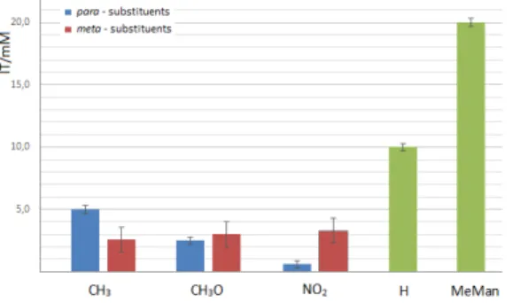
FimH from agglutinating guinea pig erythrocytes was determined (expressed as IT value) and compared to that of a reference inhibitor, methyl α-D-mannoside (MeMan) and previously described 3,4-HP mannosides (I–IV). The results are shown in Figure 2.
All tested compounds showed better inhibitory potency than the reference MeMan. Determined IT values for compounds 3a–3c were 2.6, 3.0, 3.3 mM, respectively. These results show that α-mannosides 3a–3c are 6–7 times better inhibitors of the hemagglutination than MeMan. Previously described mannoside IV showed a very
significant 32-fold improvement in potency relative to MeMan. Even more improved inhibitory potencies were also observed with mannosides that possess biaryl aglycons.[24,25] Furthermore, all derivatives with substituents in either meta or para position on the phenyl ring showed stronger activity compared to compound I
with no substituents in the aryl part of the molecule, indicating the importance of phenyl ring substitution for binding capacity. Mannosides 3b and 3c, with methoxy and nitro substituents in meta position, respectively, showed higher IT value compared to corresponding para
derivatives. This result implies that para substituent is relevant for the binding affinity. Based on obtained results and on molecular modeling study performed on para
compounds[33] which indicated the hydrogen bond between p-methoxy or p-nitro moiety and hydroxyl group of Tyr137 residue in FimH binding pocket, one can conclude that displacement of the groups from para to meta position influences unfavorably on hydrogen bonding with Tyr137 and consequently on effectiveness of binding to FimH. On the other hand, analogue 3a with methyl substituent in
meta position showed two-times better inhibitory activity than mannoside II. This suggests that hydrophobic substituent in meta position positively affects the binding of 3,4-HP mannosides in FimH binding site. Obtained results will be considered in our future studies in the design of novel 3,4-HP mannosides as FimH antagonists. Preparation and evaluation of disubstituted N-aryl 3,4-HP derivatives which have alkyl substituents in meta position and such groups in para position which can act as hydrogen bond acceptors in hydrogen bond with hydroxyl group of Tyr137 residue could be one promising route.
CONCLUSION
Novel meta substituted (methyl-, methoxy-, nitro-) N-aryl 3-hydroxypyridin-4-one mannosides were synthesized and their inhibitory potencies as FimH antagonists were evaluated and compared to previously prepared para
substituted 3,4-HP α-mannosides. The hemagglutination assays revealed greater preference of FimH towards the
para substituted N-aryl 3,4-HP mannosides, especially those with -OCH3 and -NO2 substituents. This study provides guidelines for rational design of novel, more effective FimH antagonists. In the continuation of our research on 3,4-HP derived FimH antagonists we plan further chemical transformations on aryl moiety. Based on obtained results, we conclude that the most promising route for the preparation of more efficient 3,4-HP derived
E. coli anti-adhesives is introduction of alkyl substituents in
meta position and substituents which can form hydrogen bond with Tyr137 hydroxyl group in para position of aryl part.
Figure 2. Comparison of IT values of N-aryl 3,4-HP mannosides 3a-3c towards type 1 fimbriated E. coli. The minimal concentration of a compound required to inhibit hemagglutination (IT value) was obtained from four independent tests for m-substituted compounds; 3a 2.6 mM,
Croat. Chem. Acta 2016, 89(2), 237–242 DOI: 10.5562/cca2890 Acknowledgment. We wish to thank the Croatian Science
Foundation for support of this work (project IP-2014-09-7899). We also wish to thank Vlatka Godinić from Department of Chemistry, Faculty of Science, University of Zagreb, Croatia for the preparation of the bacterial samples used in hemagglutination tests and Institute of Immunology, Department for Research and Development, Zagreb, Croatia for the donation of guinea-pig blood.
REFERENCES
[1] M. E. Taylor, K. Drickamer, Introduction to
Glycobiology, 2nd ed., Oxford University Press, New
York, 2006, p. 105.
[2] T. K. Lindhorst, Essentials of Carbohydrate Chemistry
and Biochemistry, 2nd ed., WileyVCH, Weinheim,
2003, p. 175.
[3] N. Sharon, Biochim. Biophys. Acta2006, 1760, 527. [4] D. Zopf, S. Roth, Lancet1996, 347, 1017.
[5] N. Sharon, I. Ofek, Glycoconj. J. 2000, 17, 659. [6] I. Ofek, D. L. Hasty, N. Sharon, FEMS Immunol. Med.
Microbiol. 2003, 38, 181.
[7] R. J. Pieters, Med. Res. Rev. 2007, 27, 796.
[8] M. Hartmann, T. K. Lindhorst, Eur. J. Org. Chem.
2011, 3583.
[9] S. B. Levy, Adv. Drug Delivery Rev. 2005, 57, 1446. [10] K. Lindhorst, C. Kieburg, U. Krallmann-Wenzel,
Glycoconj. J. 1998, 15, 605.
[11] T. K. Lindhorst, S. Kötter, J. Kubisch, U. Krallmann-Wenzel, S. Ehlers, V. Kren, Eur. J. Org. Chem. 1998,
15, 1669.
[12] D. Choudhury, A. Thompson, V. Stojanoff, S. Langermann, J. Pinkner, S. J. Hultgren, S. D. Knight,
Science1999, 285, 1060.
[13] P. Aprikian, V. Tchesnokova, B. Kidd, O. Yakovenko, V. Yarov Yarovoy, E. Trinchina, V. Vogel, W. Thomas, E. Sokurenko, J. Biol. Chem. 2007, 282, 23437. [14] Kötter, U. Krallmann-Wenzel, S. Ehlers, T. K.
Lindhorst, J. Chem. Soc. Perkin Trans. 1998, 1, 2193. [15] T. K. Lindhorst, C. Kieburg, U. Krallmann-Wenzel,
Glycoconj. J.1998, 15, 605.
[16] M. Touaibia, R. Roy, Mini-Rev. Med. Chem. 2007, 7, 1270.
[17] M. Almant, V. Moreau, J. Kovensky, J. Bouckaert, S. G. Gouin, Chem. Eur. J. 2011, 17, 10029.
[18] A. Schierholt, M. Hartmann, T. K. Lindhorst,
Carbohydr. Res. 2011, 346, 1519.
[19] I. Ofek, D. L. Hasty, N. Sharon, FEMS Immunol. Med.
Microbiol.2003, 38, 181.
[20] C. S. Hung, J. Bouckaert, D. Hung, J. Pinkner, C. Widberg, A. Defusco, C. G. Auguste, R. Strouse, S. Langermann, G. Waksman, S. J. Hultgren, Mol. Microbiol. 2002, 44, 903.
[21] J. Bouckaert, J. Berglund, M. Schembri, E. De Genst, L. Cools, M. Wuhrer, C. S. Hung, J. Pinkner, R. Slättegård, A. Zavialov, D. Choudhury, S. Langermann, S. J. Hultgren, L. Wyns, P. Klemm, S. Oscarson, S. D. Knight, H. De Greve, Mol. Microbiol. 2005, 55, 441.
[22] A. Wellens, C. Garofalo, H. Nguyen, N. Van Gerven, R. Slättegård, J.-P. Hernalsteens, L. Wyns, S. Oscarson, H. De Greve, S. Hultgren, J. Bouckaert,
PLoS One2008, 3, 1.
[23] S. D. Knight, J. Bouckaert, J. Top. Curr. Chem. 2009,
288, 67.
[24] Z. Han, J. S. Pinkner, B. Ford, R. Obermann, W. Nolan, S. A. Wildman, D. Hobbs, T. Ellenberger, C. K. Cusumano, S. J. Hultgren, J. W. Janetka, J. Med.
Chem.2010, 53, 4779.
[25] N. Firon, S. Ashkenazi, D. Mirelman, I. Ofek, N. Sharon, Infect. Immun. 1987, 55, 472.
[26] T. Klein, D. Abgottspon, M. Wittwer, S. Rabbani, J. Herold, X. Jiang, S. Kleeb, Christine L., M. Scharenberg, J. Bezençon, E. Gubler, L. Pang, M. Smiesko, B. Cutting, O. Schwardt, B. Ernst, J. Med. Chem. 2010, 53, 8627. [27] O. Sperling, A. Fuchs, T. K. Lindhorst, Org. Biomol.
Chem. 2006, 4, 3913.
[28] O. Sperling, A. Fuchs, T. K. Lindhorst, Infect. Immun.
1987, 55, 472.
[29] S. Rabbani, X. Jiang, O. Schwardt, B. Ernst, Anal.
Biochem.2010, 407, 188.
[30] R. Ribić, M. Kovačević, V. Petrović Peroković, I. Gruić-Sovulj, V. Rapić, S. Tomić, Croat. Chem. Acta2010,
83, 421.
[31] M. Kovačević, L. Barišić, R. Ribić, V. Petrović Peroković, S. Tomić, V. Rapić, Appl. Organometal.
Chem.2012, 26, 74.
[32] V. Kovač, R. Ribić, V. Petrović Peroković, S. Tomić, L. Barišić, Appl. Organometal. Chem.2016, 30, 524. [33] Ž. Car, T. Hrenar, V. Petrović Peroković, R. Ribić, M.
Seničar, S. Tomić, Chem. Biol. Drug Des. 2014, 84, 393. [34] M. A. Santos, S. M. Marques, S. Chaves, Coord.
Chem. Rev. 2012, 256, 240.
[35] Y. Ma, T. Zhou, X. Kong, R. C. Hider, Curr. Med. Chem.
2012, 19, 2816.
[36] C. Queiros , M. J. Amorim, A. Leite, M. Ferreira, P. Gameiro, B. De Castro, K. Biernacki, A. Magalhaes, J. Burgess, M. Rangel, Eur. J. Inorg. Chem. 2011, 131. [37] D. E. Green, M. L. Bowen, L. E. Scoot, T. Storr, M.
Merkel, K. Böhmerle, K. H. Thompson, B. O. Patrick, H. J. Schugar, C. Orvig, Dalton Trans. 2010, 39, 1604. [38] E. L. Scott, M. Telpoukhovskaia, C.
Rodriguez-Rodriguez, M. Merkel, M. L. Bowen, B. D. G. Page, D. E. Green, T. Storr, F. Thomas, D. D. Allen, P. R. Lockman, B. O. Patrick, M. J. Adam, C. Orvig, Chem. Sci.2011, 2, 642. [39] K. P. R. Kartha, H. J. Jennings, J. Carbohydr. Chem.