Abstract
Introduction: Intrathecal chemotherapy is frequently used in clinical practice for treatment and prevention of neoplastic me-ningitis. Despite its widespread use, there is little information about practical aspects such as the volume of drug to be admi-nistered or its preparation and administration.
Objective: To conduct a literature review about practical as-pects of the use of intrathecal chemotherapy.
Materials: Search in PubMed/ Medline using the terms “che-motherapy AND intrathecal”, analysis of secondary and ter-tiary information sources.
Results: The most widely used drugs in intrathecal therapy are methotrexate and cytarabine, at variable doses. One of the as-pects with higher variability among different studies is their potential combination with a glucocorticoid, the specific cor-ticoid selected and its dose. The efficacy and toxicity of the different combinations have not been compared. Regarding preparation, it is worth highlighting the recommendation to adjust pH and osmolarity to the physiological range, with the aim of improving tolerability. The volume of administration can influence distribution, and recommendated range is between 5 and 12 mL. Overall, it is recommended to extract a similar volume of cerebrospinal fluid before administration. The po-sition of the patient during and after administration can have an impact on distribution and toxicity; lateral decubitus or si-tting position is recommended in the first case, and prone and/ or supine position in the second one. Most publications don’t explain how the treatment has been prepared or administered, and the lack of standardization could affect results.
Conclusions: There is a great variability in practice when using intrathecal chemotherapy, despite being an effective therapy, accepted by all international groups. This uncertainty is not
li-Resumen
Introducción: La quimioterapia intratecal es utilizada frecuen-temente, en la práctica clínica, para el tratamiento y prevención de la meningitis neoplásica. A pesar de su uso extendido, existe poca información acerca de aspectos prácticos tales como el volumen de fármaco a administrar o la forma de preparación y administración.
Objetivo: Realizar una revisión de la literatura acerca de aspec-tos prácticos de la utilización de la quimioterapia intratecal. Material: Búsqueda en PubMed/Medline utilizando los térmi-nos “chemotherapy AND intrathecal”, análisis de fuentes de información secundarias y terciarias.
Resultados: Los fármacos más utilizados en terapia intratecal son metotrexato y citarabina, con dosis variables. La asociación o no con un glucocorticoide, el corticoide concreto seleccionado y su dosis es uno de los aspectos con mayor variabilidad entre distin-tos estudios. No se han comparado la eficacia y toxicidad de las distintas combinaciones. En la preparación destaca la recomen-dación de ajustar pH y osmolaridad al rango fisiológico, con el objetivo de mejorar la tolerancia. El volumen de administración puede influir en la distribución, oscilando las recomendaciones entre 5-12 mL. En general, se aconseja extraer previamente un volumen de líquido cefalorraquídeo similar. La posición del pa-ciente durante y tras la administración puede influir en la dis-tribución y la toxicidad; se recomienda el decúbito lateral o la sedestación, en el primer caso, y el decúbito prono y/o supino, en el segundo. La mayoría de las publicaciones no indican cómo se ha preparado o administrado el tratamiento, y la falta de es-tandarización podría afectar a los resultados.
Conclusiones: Existe gran variabilidad en la práctica a la hora de utilizar la quimioterapia intratecal, a pesar de ser una tera-pia efectiva asumida por todos los grupos internacionales. La
REVISIONES
Practical aspects of the use of intrathecal chemotherapy
Aspectos prácticos de la utilización de quimioterapia intratecal
Raquel Olmos-Jiménez
1, Alberto Espuny-Miró
1, Carlos Cárceles-Rodríguez
1and María Sacramento Díaz-Carrasco
21Pharmacology Department. Universidad de Murcia. 2Pharmacy Unit. Hospital Clínico Universitario Virgen de la Arrixaca.
Artículo bilingüe inglés/castellano
* Autor para correspondencia.
Correo electrónico: rolmosjimenez@gmail.com (Raquel Olmos Jiménez).
Recibido el 28 de julio de 2016; aceptado el 15 de octubre de 2016.
DOI: 10.7399/fh.2017.41.1.10616
Los artículos publicados en esta revista se distribuyen con la licencia:
Articles published in this journal are licensed with a: Creative Commons Attribution 4.0.
https://creativecommons.org/licenses/by-nc-nd/4.0/ La revista Farmacia Hospitalaria no cobra tasas por el envío de trabajos,
mited to the drugs and doses administered, but it also includes the manner of preparation and the administration technique. The heterogeneity in clinical practice can influence the efficacy and toxicity of this therapy.
KEYWORDS
Chemotherapy; Intrathecal; Administration; Preparation; Dosing
Farm Hosp. 2017;41(1):105-129
Introduction
The administration of intrathecal (IT) chemotherapy for the treatment and prevention of neoplastic infiltra-tion in the central nervous system (CNS) is a widely exten-ded practice that has demonstrated efficacy in different conditions. The use of such therapy for the prevention of CNS relapse in Acute Lymphoblastic Leukemia (ALL) in paediatric patients started in the 70s. The disease prog-nosis changed radically, because before the use of CNS prophylaxis, more than half of complete remissions in-duced by systemic chemotherapy would end up in CNS relapse1. IT chemotherapy has progressively displaced
radiotherapy in this indication, given its similar efficacy with a more favourable profile of adverse effects2.
Tra-ditionally, the drugs used have been methotrexate and cytarabine, alone or in combination with glucocorticoids, in the called Triple Intrathecal Therapy (TIT). The use of liposomal cytarabine has been recently introduced, as well as monoclonal antibodies such as rituximab and trastuzumab for different indications. However, even though the use of IT chemotherapy is widely accepted by the scientific community, there is a great variability in practice, in aspects such as the specific drugs and doses used, the way of preparation (volume, type of disolvent, etc.), and the administration technique. The objective of this article is to review the information available about these practical aspects.
Methods
A search was conducted in Pubmed/Medline, using the free terms “chemotherapy AND intrathecal”. The most cli-nically relevant articles were selected for this review, based on the development or description of relevant practical as-pects regarding the preparation and administration tech-nique of the IT treatment. The search was complemented with the review of quotes included in the selected articles, and the analysis of secondary information sources.
Results
Technique for intrathecal administration of drugs
Intrathecal administration consists in the direct injec-tion of the drug into the CNS3. This way of
administra-tion is occasionally necessary in order to achieve thera-peutic concentrations in the CNS, which is protected by the blood-brain barrier (BBB). The blood-brain barrier is a complex structure formed by endothelial cells from the CNS capillary network, which prevents the free exchan-ge of ions and organic molecules between the blood plasma and the nervous system4, thus protecting the
CNS from potentially neurotoxic agents; but this also makes it difficult for some drugs to reach therapeutic concentrations.
Cerebrospinal fluid (CSF) regulates the chemical environment of the CNS. The total volume of CSF in adults is 125-150 ml, with a formation and reabsorp-tion rate of approximately 20 mL/hour; therefore it is completely renewed 3 times per day. CSF secretion and reabsorption is balanced in the majority of healthy adults, in order to keep an intracranial pressure below 150 mmH20; an increase in intracranial pressure can entail severe complications. Normal CSF is clear, trans-parent and colourless. Its main physicochemical proper-ties appear in Table 1.
CSF volume varies with age. The CSF volume in new-borns is approximately 40-60 ml, and it undergoes a fast increase during the first years of life, so that the CSF volume in ≥3-year-old children is equivalent to that of adults.
Drug administration in the CNS can be conducted through two methods: into the lumbar thecal sac by lumbar puncture (LP), or directly into the lateral ventricle
incertidumbre no se limita a los fármacos y dosis administradas, sino que se extiende a la forma de preparación de las mezclas y la técnica de administración. La heterogeneidad en la práctica clínica puede influir en la efectividad y toxicidad de esta terapia.
PALABRAS CLAVE
Quimioterapia; Intratecal; Administración; Preparación; Dosificación
Farm Hosp. 2017;41(1):105-129
Table 1. Physicochemical properties of CSF
Parameter Normal cerebrospinal fluid
Aspect Crystalline
Cells 0 – 5 / mm³ (lymphocytes)
Proteins 15 – 45 mg /dL
Glucose 50 – 80 mg/dL
Chlorides 116 – 122 mEq/L
Sodium 117 – 137 mEq/L
Potassium 2.3 – 4.6 mEq/L
Osmolarity 292 – 297 mOsm/L
Density 1.0005 – 10007 g/mL
through a subcutaneous reservoir and ventricular cathe-ter (Ommaya Reservoir)3.
Lumbar Puncture
Lumbar Puncture is an invasive technique for acces-sing the subarachnoid space, with diagnostic and thera-peutic aims. The position of the patient is important for this procedure: they can be in lateral decubitus, which is the preferred position for adults, or seating position, which is the preferred position for children5.
Like any other invasive procedure, it can be painful; that is why a local anaesthetic can be used, such as sub-cutaneous mepivacaine or topical anaesthetic prepara-tions6. In paediatric patients, it could be necessary to use
pharmacological sedation. The SECIP (Spanish Society for Paediatric Intensive Care) recommends the adminis-tration of midazolam in combination with ketamine or fentanyl for this indication7.
When the LP is conducted as a diagnostic procedure, a specific amount of CSF will be extracted; it is recom-mended to restrict this to the lowest volume necessary, which is usually 6-8 mL in adults, 2-3 mL in newborns and infants, and 5-8 mL in children and adolescents. If the LP is conducted as a therapeutic procedure, the drug will be slowly introduced after extracting the relevant volume of CSF. It is recommended to conduct the admi-nistration over 3 to 5 minutes, approximately; moreover, a slower administration is recommended in patients with small brain ventricles or high intracranial pressure, in or-der to avoid headaches3,8.
There are no consistent data in scientific literature regarding the volume of drug dilution recommended for LP administration, or the CSF volume recommended for previous extraction. Overall, in order to prevent an increase in intracranial pressure when drugs are admi-nistered through the IT procedure, it is recommended not to modify the CSF volume. Therefore, it is recom-mended to extract, before IT administration, a CSF vo-lume equivalent to the vovo-lume of chemotherapy to be instilled3,8,9.
Regarding the volume of the drug to be administe-red, Pui recommends that the cytostatic agent must be dissolved in at least 6 mL of fluid, in order to achieve its adequate distribution10. The British Columbia Cancer
Agency (BCCA) recommendations also recommends 6 mL as the volume of IT drug administration for adults8.
For children, different authors have described adminis-tration volumes ranging from 6 mL for <1-year-old to 12 mL for ≤3-year-old11,12. The AHFS Drug Information
states that it is usual to extract a CSF volume similar as the one to be injected: from 5 to 15 mL13. In general, it
is considered that a CSF volume of 7 to 10 mL must be extracted in adults, and a similar volume of intrathecal chemotherapy must be instilled3.
After drug administration, it is recommended that patients should stay at least 1 hour in prone position,
in order to facilitate the penetration of the chemothera-py agent in the brain ventricles14. Other authors
recom-mend staying from 1 to 12 hours in supine position, to avoid the risk of post-puncture headache15.
Some of the complications that can appear after LP, even if conducted for diagnosis, are: development of post-puncture headache, lower back pain, nerve root irritation, infections or haemorrhages, among others16.
Ommaya Reservoir
The Ommaya Reservoir is an intraventricular device,
which is placed in the subcutaneous scalp tissue, with a catheter inserted in one of the lateral ventricles of the brain, and thus connected with CSF circulation. It is re-commended for use in those patients who require fre-quent or prolonged CNS treatments or for those cases where LP would represent a difficult technique3.
Medications are injected with a syringe in the ventri-cular reservoir. The way of administration varies depen-ding on the source. Some sources recommend the gra-dual extraction of 12 to 20 mL of CSF, at a rate <1-2 mL/ minute, with a syringe size <10 mL. After CSF extraction, the medication will be infused at a 1 mL/minute rate. Afterwards, approximately 10 mL of the previously ex-tracted CSF will be re-infused, in order to clean the Om-maya needle and catheter3. Other sources recommend
extracting a CSF volume similar to the volume of drug to be administered, and cleaning with 3-4 mL of 0.9% sodium chloride solution 0.9% after administration8.
The implantation of the Ommaya Reservoir exposes patients to a 5-10% risk of suffering complications, in-cluding haemorrhage, infection, and complications as-sociated with device malfunctioning17.
Despite the risks associated with device implantation, the main advantage of the Ommaya Reservoir over LP, is a more homogeneous drug distribution in the subara-chnoid space. The drug concentration in the ventricular CSF after LP administration is only 10% of that imme-diately achieved after the intraventricular administration of an equivalent dose of drug18. Moreover, various
stu-dies have demonstrated that the distribution in the brain ventricles of drugs administered through LP is influenced by the position after puncture; therefore, a >10 times higher reduction in concentration could occur if the pa-tient does not stay lying down during 1 hour after ad-ministration14.
General characteristics of preparations for intrathecal administration
to various authors, some factors such as pH, ionization level, or the presence of antibacterial agents during pre-paration, can be associated with the toxicity of the drug administered19. In general terms, solutions for IT
admi-nistration must be9,20: sterile, apyrogenic, limpid,
isoos-motic with CSF, with a pH close to the CSF´s ph, and without preservatives.
Taking these characteristics into account, formula-tions for intrathecal use must be prepared in laminar flow cabinets, to ensure their sterility and avoid the presence of contaminating and pyrogenic agents. Be-sides, as stated by Gil and col., it is recommended to filter the preparation through a 0.22 membrane filter, to eliminate any potential particles generated during preparation9.
Regarding osmolarity and pH, Elliott B (EB) solution was marketed in U.S.A. during some years; it was in-dicated for the IT administration of cytarabine and methotrexate, and was comparable with CSF in terms of osmolarity (288 mOsm/L), pH (6.5 to 7), electrolytic composition and glucose contents. A lower incidence of adverse effects was described, associated with the IT administration of drugs prepared in this vehicle vs. 0.9% sodium chloride solution or sterile water for injection21.
There is no solution with these characteristics currently in the market, but it is recommended that the osmolarity and pH of the preparations for intrathecal administra-tion should be close to CSF values, with the objective of improving their tolerability.
In terms of the presence of preservatives, many of the multiple-dose formulations commonly used contain preservatives such as benzyl alcohol and methylparaben or propylparaben. There have been reports of adverse events caused by the intrathecal administration of ben-zyl alcohol, such as paraparesis, nerve root demyelina-tion, and horsetail fibrosis22. There is lower experience
with parabens, and no adverse events have been des-cribed associated with their intrathecal administration; however, there are reports of anaphylactic reactions secondary to their intravenous administration, and this risk, together with the few data available about their IT administration, avoid their IT administration. Thus, it is considered in practice that all drugs for IT administration should be preservative-free23; therefore, before the
in-trathecal administration of any medication, it should be confirmed that it does not contain preservatives, taking into account that different medicinal products of the same drug can have different formulations.
Intrathecal Chemotherapy
The drugs most traditionally used for intrathecal ad-ministration in neoplastic conditions in general, and specifically for haematological conditions, have been methotrexate, cytarabine, and glucocorticoids24-27; these
drugs are often used in combination in the called TIT therapy. More recently, liposomal formulations of
cyta-rabine28 have been introduced, and even monoclonal
antibodies have been administered this way: rituximab for lymphoma29, and trastuzumab for brain metastasis in
HER2+ breast cancer30.
Methotrexate
The pharmacokinetic of methotrexate (MTX) is diffe-rent for IT administration and systemic administration. The elimination of MTX from the CSF depends mainly on the CSF flow (because there is no active transpor-tation from CSF to blood, and MTX is not metabolized in the CSF); it has an initial elimination half-life (t½) of 4.5 hours, and a final t½ of 14 hours, and is reduced to sub-therapeutical concentrations 4 days after IT admi-nistration31.
Even though IT MTX is widely used, its optimal dose has not been clearly established; but it has been de-monstrated that dose calculation based on weight or body surface is inadequate. Bleyer and col. proved that the administration of IT MTX doses based on body sur-face increased neurotoxicity in adolescent and adult patients, because very high MTX concentrations were reached in the CSF31. The reason for this, as we have
already mentioned, is that there is a fast increase in the CSF volume during the first years of life, and the volu-me in ≥3-year-old children is equivalent to the voluvolu-me in adults. Therefore, it is clearly defined that the calculation of MTX doses must be conducted according to age31,
but the IT MTX dose in different studies ranges generally from 10 to 15 mg 13,24-25,32-39, and is lower in <2-year-old
patients (Table 2).
IT administration of MTX can cause acute, sub-acute or long-term neurotoxicity. The most frequent adverse effect is aseptic meningitis or chemical arachnoiditis, which appears approximately in 10% of the patients, though incidences of up to 50% have been described. Its characteristic symptoms are headache, nausea, and neck stiffness40. The concomitant administration of IT or
oral corticosteroids can reduce the risk of its develop-ment. Geiser and col.21 observed, in a cohort of
pae-diatric patients who received IT MTX, a lower frequency in the development of chemical meningitis symptoms in those patients for whom EB solution was used as vehi-cle; this was attributed to the pH of this solution, closer to the physiological pH of CSF than those of other vehi-cles used in this study.
There have also been reports of dizziness, convul-sions, sub-acute encephalopathy or leukoencephalopa-thy40. Leukoencephalopathy can appear months or years
after the administration of methotrexate, and the risk is higher for those patients who are receiving concomitant CNS radiation and high doses of IV MTX41.
Cytarabine
intra-ventricularly a single 30 mg dose of cytarabine (ARA-C) to seven patients with leukemic meningitis in complete remission. After the injection, a maximum ARA-C con-centration >2000μmol/L was reached in the ventricular CSF, and a <1μmol/L concentration was sustained during at least 24 hours. The ARA-C by IV administration is ra-pidly eliminated from plasma through the cytidine dea-minase enzyme, which metabolizes ARA-C into uracil arabinoside; however, the concentration of the enzyme in the CSF is insignificant, and this metabolic pathway won’t practically occur43. Moreover, the value of ARA-C
clearance from the CSF (0.42 mL/minute) is similar to the CSF formation and reabsorption rate (0.35 mL/mi-nute), suggesting that ARA-C elimination from CSF is mainly due to CSF flow. The elimination of ARA-C from CSF is slower than from plasma, and clearance is eight times lower in CSF44. As MTX, there is no clearly
establi-shed ARA-C dose, and its dose must be calculated also according to age rather than to body surface24,33,34,45-47
(Table 3).
The adverse effects described with the IT administra-tion of ARA-C are: transverse myelopathy, aseptic
me-Table 2. Intrathecal methotrexate doses in monotherapy used according to different authors
Study Condition Indication Age MTX dose
Franklin and col.24 Haematological
malignancies Prophylaxis
1 to 2-year-old 2 to 3-year-old 3 to 8-year-old ≥9-year-old
8mg 10mg 12mg 15mg
Bleyer and col.31 and
AHFS Drug Information13
Haematological malignancies
Prophylaxis and Treatment
< 1-year-old 1 to 2-year-old 2 to 3-year-old ≥3-year-old
6mg 8mg 10mg 12mg
Kim et al32 Solid tumour Treatment ≥18-year-old 15mg
Kantarjian and col.33 ALL Prophylaxis ≥18-year-old 12mg
Cortes and col.34 ALL Prophylaxis ≥18-year-old 12mg
Mahoney and col.35 and
Matloub and col.25 ALL Prophylaxis
< 2-year-old 2-to-3-year-old
≥3-year-old
8mg 10mg 12mg
Siegal and col.36 Solid tumour and
lymphoma Treatment ≥18-year-old 12mg
Kiewe and col.37 Primary CNS lymphoma Treatment ≥18-year-old 15mg
Omura and col.38 ALL Prophylaxis ≥18-year-old 10mg/m²
Hill and col.39 ALL Prophylaxis
<1-year-old <2-year-old ≥3-year-old
7.5mg 10mg 12.5mg
*Abbreviations: ALL: Acute Lymphoblastic Leukemia; MTX: Methotrexate; CNS: Central Nervous System.
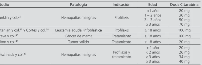
Table 3. Intrathecal cytarabine doses in monotherapy used in different studies
Study Condition Indication Age Cytarabine dose
Franklin and col.24 Haematological malignancies Prophylaxis
<1-year-old 1 to 2-year-old 2 to 3-year-old ≥3-year-old
20mg 30mg 50mg 70mg Katarjian and col.33 and
Cortes and col.34 Acute lymphoblastic leukemia Prophylaxis ≥18-year-old 100mg
Esteva and col.45 Breast cancer Treatment ≥18-year-old 100mg
Fulton and col.46 Solid tumour Treatment ≥18-year-old 20mg
Fleischhack and col.47 Haematological malignancies Prophylaxis and
Treatment
<1-year-old <2-year-old <3-year-old ≥3-year-old
ningitis, encephalopathy and convulsions; however, their development will be infrequent24,48,49.
Liposomal Cytarabine
A liposomal formulation (Depocyte®) has been
deve-loped in order to enable a lower frequency of IT ARA-C administration; this is a controlled release formulation for aqueous cytarabine, which is encapsulated in spheri-cal and multivesicular particles spheri-called DepoFoam®, which
present a longer half-life of CSF elimination. With this formulation, a two-phase elimination profile has been described, with a terminal stage t½ within 100 and 263 hours, for a dose range between 12.5 mg and 75 mg; on the other hand, standard-release cytarabine presents a terminal stage t½ of approximately 3.4 hours for 30mg doses50. After the IT injection, the liposomal particles
containing the ARA-C will break down, and the lipids will enter the normal metabolic pathways of the body. In 2007, Phuphanich and col. studied the pharmacoki-netics of Depocyte® by administering 2 intraventricular
or intralumbar doses to 8 patients, separated by a 14-day interval, and taking samples at different points up to 14 days after administration. The concentration of free and encapsulated ARA-C in the ventricular and lumbar CSF varied from 0.01 to 1500μmol/L, and was detec-table during 14 days post-dose. Systemic exposure to cytarabine was considered non-significant28.
The efficacy of IT liposomal cytarabine has been de-monstrated in two controlled and randomized clinical trials, in patients with neoplastic meningitis associated with lymphoma and solid tumours vs. conventional IT cytarabine and IT methotrexate, respectively51,52. In the
clinical trial conducted vs. conventional ARA-C, a higher response rate was observed in the liposomal cytarabine group; however, there were no statistically significant di-fferences in duration of response, progression-free survi-val and overall survisurvi-val. In the clinical trial conducted vs.
MTX, progression-free survival was also similar with the two agents.
Opposite to methotrexate and standard-release cyta-rabine, the dose of liposomal cytarabine in adults has been defined as 50mg with IT administration, in a 5 mL volume; the dosing interval varies according to the treat-ment stage. The indication approved for Depocyte® by
the Spanish Agency of Medicines and Medical Devices is the treatment of lymphomatous meningitis50.
The toxicity of liposomal cytarabine is relatively high, particularly when administered in association with other drugs which cross the BBB. In the M.D. Anderson Cen-tre for Cancer Treatment, a study was conducted on adults diagnosed with ALL and treated with the Hyper-CVAD regimen, which includes high-dose IV MTX and ARA-C. The introduction of IT liposomal cytarabine was associated with a higher toxicity rate (16%), including encephalopathy, cauda equina syndrome, convulsions,
and pseudotumor cerebri. The conclusion was that
se-vere neurotoxicity could be caused by the concomitant administration of liposomal cytarabine and drugs that can cross the BBB or radiotherapy53. In the randomized
clinical trial comparing the ARA-C liposomal and stan-dard formulations, conducted by Glantz, the majority of the adverse effects were transitory. The only effect that appeared in over 10% of treatment cycles was heada-che, which was more frequent in the liposomal cytarabi-ne group than in the traditional cytarabicytarabi-ne group (27%
vs. 2%). Chemical arachnoiditis appeared in 22% of patients treated with liposomal cytarabine vs. 13% of patients treated with the standard formulation52. In
or-der to reduce the incidence of arachnoiditis, it is recom-mended to administer systemic dexamethasone during 5 days, initiating treatment on the same day of the IT injection.
Glucocorticoids
Even though intrathecal administration of glucocor-ticoids is common, its pharmacokinetics in the CNS has not been clearly documented. Balis and col54. studied
the activity of dexamethasone and prednisolone in the CSF in a non-human primate model, after its intrave-nous and intraventricular administration. Both drugs are rapidly cleared from CSF after intraventricular adminis-tration; however, after intravenous administration, there is low penetration in the CSF due to the high binding to plasmatic proteins in both, though dexamethasone presents higher penetration due to its lower binding to plasmatic proteins (70% for dexamethasone vs. 90% for prednisolone).
IT glucocorticoids are mostly used in combination with ARA-C and MTX. This concomitant use has two ob-jectives: increasing the effectiveness of IT therapy, and reducing the incidence of meningeal irritation23. There
is wide evidence about the use of corticosteroids as part of intrathecal treatment in oncohaematology; in 1983, Muriel and col55. used IT methotrexate 12mg/m²
(maxi-mum dose = 15 mg) and dexamethasone 14mg/m², and in 1995, Gómez-Almaguer administered IT dexametha-sone 5mg/m² diluted in 5mL of 0.9% sodium chloride with MTX and ARA-C together in 8 patients with ALL and leukemic infiltration in their CNS; the cell count was reduced in CSF after the administration of combi-ned intrathecal chemotherapy in all patients56. However,
the use of corticosteroids with IT chemotherapy is not universal: its use, the one used, and its dose will vary according to the protocols of the different study groups. Overall, its use is more widespread for the treatment of paediatric patients than for adults.
The International Berlin-Frankfurt-Munster (BFM) Study Group57,58, in its protocols for ALL treatment in
Group recommends the use of prednisolone with MTX and ARA-C for prophylaxis and treatment of meningeal infiltration in ALL. This is also the corticosteroid used in the Japanese JALSG-ALL93 protocol for adults with ALL, with doses of 10mg59.
The Italian GIMEMA group has used IT MTX 12mg with methylprednisolone 40mg for ALL treatment in pa-tients from 12 to 60-year-old60. The dose of 40mg of
methylprednisolone for adult patients with ALL has also been used with MTX 15mg and ARA-C 40mg by the French LALA Group61, and in the GRAALL-2003
Proto-col62.
In U.S.A., the use of intrathecal corticosteroids for ALL treatment in adult patients has not become widespread; thus, it is not included in the CALGB-8811 study63 or in
the studies conducted by Katarjian33 and Rowe64, where
IT MTX 15 mg, 12 mg and 12.5 mg is used for CNS prophylaxis, respectively. However, for paediatric pa-tients with ALL, both the Pediatric Oncology Group and the Children’s Oncology Group use hydrocortisone with MTX and ARA-C dosed according to age25,35. The
proto-cols for treatment and prophylaxis of CNS infiltration in acute leukemia in children from the St. Jude’s Children’s Research Hospital also use hydrocortisone as part of the TIT therapy at 16 mg, 20 mg or 24 mg doses, based on age (<1 year-old, 2-3-year-old or ≥3-year-old, respecti-vely)65.
The PETHEMA Group (Program for the Study and Treatment of Haematological Malignancies) and the Spanish Society of Paediatric Haematology and Onco-logy (SEHOP) indicate the use of hydrocortisone (HC) with ARA-C and MTX in the majority of their protocols for ALL treatment, recommending this dosing: <2 year-old: 10 mg, 2-3-year-year-old: 15 mg, ≥3-year-year-old: 20 mg66.
However, other protocols from the PETHEMA Group have incorporated the use of dexamethasone. For exam-ple, Burkimab-13, widely used in Spain for the treatment of Burkitt Lymphoma in adult patients, indicates the use of MTX 15mg in combination with ARA-C 40 mg and dexamethasone 4 mg67 for prophylaxis and treatment of
CNS infiltration. PETHEMA-LAL-07OLD also uses IT dexa-methasone 4 mg with MTX and ARA-C68.
The toxicity of the intrathecal administration of glu-cocorticoids has not been studied yet by controlled stu-dies; in fact, its use in combination with IT chemothera-py is associated with a reduction of the adverse effects, primarily the development of chemical arachnoiditis. However, the development of psychiatric adverse effects caused by the use of intrathecal corticosteroids has been described in various studies; it can be ranged from depressive syndromes to psychosis69. In the study
con-ducted by Hitchins, where IT hydrocortisone was admi-nistered before the administration of IT chemotherapy, two unusual reactions were observed in two patients, who experienced headache, vomiting and confusion, in both cases after the administration using the Ommaya
reservoir. These symptoms disappeared spontaneously a few hours after administration. When both patients were re-treated with IT hydrocortisone, they suffered the same reaction again; when the administration of IT hy-drocortisone was removed from the rest of the adminis-trations of IT chemotherapy, this reaction did not appear again when chemotherapy was administered alone. The authors did not find an explanation for these reactions70.
Monoclonal Antibodies: Rituximab and Trastuzumab
There has been recent research into the administra-tion of two monoclonal antibodies; this represents an innovative targeted therapy for the treatment of lepto-meningeal carcinomatosis: trastuzumab for HER2 posi-tive breast cancer, and rituximab for the treatment of B-cell lymphoma.
Trastuzumab is highly effective in the treatment of breast cancer with HER2 overexpression; however, despite its efficacy, patients treated with trastuzumab and chemotherapy will experience an incidence of CNS metastasis ranging from 28% to 42%30. Stemmler and
col. observed that the concentration of trastuzumab in the CSF after intravenous administration was 300 or 400 times lower than its concentration in plasma71;
therefore, in order to achieve therapeutic concentra-tions of trastuzumab in the CSF, it has been considered to use IT administration. This drug has been used in series of cases, alone or in combination with intrathecal methotrexate or thiotepa, at doses between 12.5 and 25 mg administered with a frequency ranging from 3 days to 3 weeks. The most typical regimen used was 20-25 mg once a week, and all dosing regimens were well tolerated. With these regimens, there have been responses at CSF level and even an increase in survival without toxic effects30.
Rituximab is an anti-CD20 antibody; over 90% of B-Cell Non-Hodgkin lymphomas (NHL) and primary CNS lymphomas express the CD20 marker, but healthy brain tissue does not express it. Like trastuzumab, rituximab has a low penetration in the CSF; its concentration in CSF after systemic administration represents 0.1% of concentrations in blood serum. Rubenstein and col29.
conducted a Phase I clinical trial on 10 patients with B-cell NHL and primary CNS lymphoma with neoplastic meningitis, who were administered rituximab through
an Ommaya Reservoir, every week during the first week
meningitis. Each patient was administered an induction regimen including 25 mg of IT rituximab twice a week and liposomal cytarabine every 14 days, during 4 wee-ks. A maintenance stage was then conducted, inclu-ding 50 mg of liposomal cytarabine and two doses of rituximab 25 mg administered in the same week, every 4 weeks, until disease progression. After the induction regimen, 10 patients experienced partial neurological response, and received maintenance therapy. The pro-bability of survival at 6 months was 29%, and toxic effects were moderate and expected, probably due to liposomal cytarabine72.
There is lack of information about the way in which monoclonal antibodies should be prepared and admi-nistered for intrathecal administration; logically, the re-quirements for preparation for intrathecal use must be considered, always in a sterile and apyrogenic setting, and using disolvents preservatives-free20. In the case of
trastuzumab, only one of the publications explained how it had been prepared: the usual reconstitution pro-cess (150 mg in 7.2 ml water for injection; pH 6), and administration of the adequate volume (20 mg in 0.95 mL), without additional dilution73. For rituximab, one
of the studies specified that it had been prepared by diluting rituximab with 0.9% sodium chloride solution on a 1:1 ratio for 10 and 25 mg doses, and without dilution for 50mg doses, and administered over 1 to 5 minutes72.
It has not been established if intraventricular or in-tralumbar administration has any influence on the re-sults.
Intrathecal Triple Chemotherapy
The use of IT combination chemotherapy is a logical consequence, because it has been demonstrated that the use of combination systemic chemotherapy increases efficacy when compared vs. the individual administra-tion of antineoplastic agents. That is why many research groups advocate for the use of TIT chemotherapy, which consists in administering methotrexate, cytarabine and a glucocorticoid in combination26. The use of these three
agents in combination could have an additive or synergic effect for the prophylaxis and treatment of neoplastic meningitis.
In Spain, the use of TIT for the prophylaxis and treat-ment of the leukemic CNS involvetreat-ment and certain NHLs is the most widespread practice, because it is the treatment indicated in the protocols promoted by the PETHEMA Group74 and SEHOP75. The habitual use of this
strategy in adults is shown in the outcomes of the stu-dies conducted by the QUIT (Spanish Registry of Patients receiving Intrathecal Chemotherapy)74,76.
The efficacy of TIT therapy vs. MTX monotherapy was comparable in the study conducted by the Southwest Oncology Group, which included paediatric patients diagnosed with ALL and active meningeal disease
(com-plete response rate of 96% vs. 100%)27 In the
CCG-1952 clinical trial, children with standard-risk ALL were randomized to receive MTX alone (n=1018) or TIT (MTX, ARA-C and HC, n=1009) for prophylaxis of leukemic CNS involvement. Compared with IT MTX, TIT signifi-cantly reduced the risk of relapse in CNS (3.4%±1.0%
vs. 5.9%±1.2%, p=0.004); however, the survival free of disease at 6 years was equivalent between both treat-ment groups (80.7%±1.9% vs. 82.5%±1.8%, p=0.3), due to a higher proportion of bone marrow and testicu-lar relapses, which was associated with a significant re-duction in overall survival (Survival at 6 years: TIT 90.3%
vs. IT MTX 94.4%, p=0.01)25.
No direct comparisons have been published for the use of IT liposomal cytarabine vs. TIT therapy.
The adverse effects due to the use of TIT therapy are not qualitatively different from those previously described with IT MTX and ARA-C, and from the risks associated with the administration technique itself (LP or intraventricular). The most common adverse effects are headaches, nausea, vomiting and fever; more severe adverse effects will occur less frequently, such as che-mical arachnoiditis, loss of vision, and leukoencephalo-pathy24,77.
As we have mentioned previously, the use of corticos-teroids in combination with IT chemotherapy seems to have a beneficial effect on the profile of adverse events; because the risk of meningeal irritation, described with the IT administration of MTX and ARA-C in monothera-py, will be reduced with the concomitant IT administra-tion of a corticosteroid78.
As well as the IT administration of drugs in monothe-rapy, the doses of TIT to be administered are not clearly defined. However, there is consensus regarding the need to calculate the dose based on age and not on body sur-face. The MTX, ARA-C and HC doses used in different studies11,12,24,25,35,64,65,79-82 are shown in Table 4.
Other studies and work groups have used TIT with dexamethasone, prednisone or methylprednisolone, as mentioned in the section for intrathecal glucocorticoids. The TIT therapy used by the BMF Study Group was: MTX <1-year-old: 6 mg, 1-year-old, 8 mg, 2-year-old, 10 mg, ≥3-year-old: 12 mg; for ARA-C, <1-year-old: 15 mg, 1-year-old: 20 mg, 2-year-old: 25 mg, ≥3-year-old: 30 mg; for prednisolone: <1-year-old: 6 mg, 1-year-old: 8 mg, 2-year-old: 10 mg, ≥3-year-old: 12 mg57,58.
Com-binations at variable doses have also been described for adult patients61-64,67,68.
As the lack of homogeneity in dosing, the disolvent used and the administration volume are not described in almost any study; and according to various authors, these are factors that can have impact on tolerability to treatment and in the adequate distribution of the drug in the CSF12.
Of all the studies shown in Table 4, only Liu and col.12
and Lin and col.11 have described the volume used,
which varied according to age, and was in both studies: <1-year-old: 6 mL, 1-2-year-old: 8 mL, 2-3-year-old: 10 mL, and ≥3-year-old: 12 mL. Lin and col.11 stated that
they had used the dilution volume adequate to increase the efficacy of TIT therapy, though there was no argu-ment supporting this claim.
Sullivan and col., in a study comparing the efficacy and toxicity of TIT therapy vs. methotrexate and hydrocorti-sone, commented that the solvent used for preparation
was EB solution, however the dilution volume was not named27. They also described that the IT administration
of the 3 drugs was sequential: MTX was administered first, followed by HC and finally ARA-C27. This
descrip-tion of sequential administradescrip-tion is excepdescrip-tional, because in the majority of the studies it is not stated whether the administration is conducted with the total mixture in one single syringe or in separated syringes for each agent and, in this case, the order for the administration.
Currently, the most accepted trend is to mix the three components in one single syringe, to facilitate IT admi-nistration and prevent excessive handling (connecting and disconnecting the catheter), thus reducing the risk for accidental contamination during administration.
The ALL SEHOP / PETHEMA 2013 paediatric protocol describes the preparation and administration of IT treat-ment. It states that the 3 cytostatic agents must be
ad-Table 4. Dose of Intrathecal Triple Chemotherapy (methotrexate, cytarabine and hydrocortisone) based on age, in different studies
Study Age MTX dose (mg) ARA-C dose (mg) HC dose (mg)
Liu and col.12 and Lin and col.11
<1-year-old 1 to 2-year-old 2 to 3-year-old >3-year-old 6 8 10 12 12 16 20 24 6 8 10 12
Franklin and col.24
≤1 year-old 1 to 2-year-old 2 to 3-year-old 3 to 8-year-old ≥9-year-old 7.5 8 10 12 15 15 16 20 24 30 7.5 8 10 12 15
Matloub and col.25
<2-year-old 2 to 3-year-old 3 to 8-year-old >8-year-old 8 10 12 15 16 20 24 30 8 10 12 15
Mahoney and col.35
1-year-old 2-year-old ≥3 to 8-year-old
≥9-year-old 8 10 12 15 16 20 24 30 8 10 12 15
Pui and col.65
<2-year-old 2 to 3-year-old
≥3-year-old 8 10 12 24 30 36 16 20 24
Ortega and col.66
<12 month-old 12-to-23-month old 24-to-35-month old ≥35-month old 5 8 10 12 16 16 20 30 10 10 15 20
Ruggiero and col.79
<2-year-old <3-year-old ≥3-year-old 8 10 12 16 20 24 8 10 12
Tomizawa and col.80
<2-year-old <3-year-old ≥3-year-old 7.5 10 12.5 15 20 25 15 20 25 LAL SEHOP/PETHEMA 201381 12-to-23-month old 24-to-35-month old ≥35-month old 8 10 12 16 20 30 10 15 20
ministered in the same syringe, and that doubly distilled water must be used for the reconstitution of cytarabine and hydrocortisone, which will be sterile, apyrogenic and preservatives-free; the volume will then be comple-ted with 0.9% sodium chloride solution. This protocol recommends a pH and osmolarity for the preparation of 7.3 pH and a 300 mOsm/l, respectively81.
Besides the aspects considered, another important issue when using drug mixtures is to ensure the physi-cochemical stability of the final preparation. There are few stability studies for TIT preparations, and the majo-rity have been conducted with EB solution. The most re-cently published study evaluates the stability of sodium methotrexate, cytarabine and hydrocortisone sodium phosphate in a 0.9% sodium chloride solution, with pH and osmolarity adjusted to values close to the CSF ph; the conclusion was that the TIT preparations evaluated were chemically stable during at least 7 days at room temperature (RT), and under refrigeration; however, the pH of the preparations was out of the physiological va-lues of CSF at 5 days in the preparations stored with refrigeration, and at 2 days in those stored at RT84. The
characteristics of the studies conducted19,84-87 are shown
in Table 5.
There are few stability studies for TIT preparations with corticosteroids other than hydrocortisone. The Burkimab-13 protocol67, which indicates the use of TIT
with methotrexate, cytarabine and dexamethasone, re-commends administering dexamethasone in a separate syringe from methotrexate and cytarabine, due to the lack of evidence on the stability of this preparation, be-cause there are no studies on the stability of TIT mixtures with dexamethasone as a corticosteroid. The same pro-tocol recommends replacing dexamethasone by
hydro-Table 5. Summary of the stability studies for TIT
Study Drugs Conditions Disolvent Study
duration Conclusion
Cradock and col.19 MTX, ARA-C and
HCSS not mixed 22°C and 30°C EB, SCh, RL 168 hs.
MTX and ARA-C stable 7 days at 22°C and 30°C. HCSS stable 72 hs. in RL and SCh, and 24 hs. in EB. Olmos-Jiménez
and col.84
MTX, ARA-C and
HCSPh 2-8°C and 25°C SCh (with NaHCO3) 168 hs.
Chemically stable mixtures for 168 hs at 2-8°C and 25°C
Zhang and col.85 MTX, ARA-C and
HCSS not mixed 4°C and 23°C EB 48 hs.
MTX and ARA-C stable 48 hs. at 4°C and 23°C. HCSS stable 48 hs. at 4°C
and 24 hs. at 23°C
Trissel and col.86 MTX and ARA-C with
or without HCSS. 4°C and 23°C EB 48 hs.
Preparations stable 48 hs. at 4°C and 23°C
Cheungh and col.87 MTX, ARA-C and
HCSS 25°C EB, SCh, RL, Dx 24 hs.
Preparations stable in all disolvents >24 hs, except for
HCSS in EB 1.25 mg/mL.
Abbreviations: ARA-C: cytarabine; EB: Elliot B Solution; hs: hours; HCSPh: hydrocortisone sodium phosphate; HCSS: hydrocortisone sodium succinate; NaHCO3: sodium bicarbonate; MTX: methotrexate; RL: ringer’s Lactate; SCh: 0.9% sodium chloride; Dx: dextrose 5%.
cortisone, if it is preferred to administer the IT chemo-therapy in one single syringe.
In 2012, D’Hont and col. conducted a stability study for ARA-C, MTX and methylprednisolone in 0.9% so-dium chloride solution during 48 hours. The conclusion was that the preparation was stable up to 12 hours when stored at 5°C and protected from light88.
In 2014, the Ministry of Health, Social Services and Equality, together with the Spanish Society of Hospital Pharmacy, published guidelines for good practice in the preparation of medications in Hospital Pharmacy Units, where sterile preparations were classified according to a risk matrix. These guidelines classified the preparations for IT administration as high-risk preparations, and the following preparation requirements were defined: pre-paration in a laminar flow cabinet with controlled envi-ronment (clean room), and as storage requirements: a maximum storage time of 24 hours at RT and 3 days at a 2-8°C temperature89-90.
Conclusions
There is a wide variability in practice when using IT chemotherapy, despite being an effective therapy, ac-cepted by all international groups, particularly for the treatment of acute leukemia and Non-Hodgkin lympho-mas. This variability and uncertainty is not limited to the drugs and doses administered, but it also includes the way of preparation and the administration technique.
Funding
Conflict of Interests
There is no conflict of interests.
References
1. Pui CH, Thiel E. Central nervous system disease in hematologic ma-lignancies: historical perspective and practical applications. Semin Oncol 2009;36(4 Suppl 2):S2-S16.
2. Vora A, Andreano A, Pui CH, Hunger SP, Schrappe M, Moericke A, et al. Influence of Cranial Radiotherapy on Outcome in Children With Acute Lymphoblastic Leukemia Treated With Contemporary Therapy. JCO 2016;34(9):919-26.
3. Treatment of leptomeningeal metastases. Uptodate 2014 [citado 13-06-2016]. Disponible en: http://www.uptodate.com/contents/ treatment-ofleptomeningeal-metastases- carcinomatousmeningitis? source=search_result&search=treatment+of+leptomeningeal +metastases &selectedTitle=1%7E54
4. Pascual JM., Gonzalez F., Prieto R., Celdrán S., Roda JM. Blood brain barrier: development of a structure which supports the func-tional heterogeneity of the central nervous system. Rev Neurol 2004;38(6):565-81.
5. Ellenby MS, Tegtmeyer K, Lai S, Braner DA. Videos in clinical medi-cine. Lumbar puncture. N Engl J Med 2006;355(13):e12.
6. Administration of Chemotherapeutic Drugs. BC Cancer 2014 [citado 13-06-2016]. Disponible en: http://www.bccancer.bc.ca/NR/ rdonlyres/13EF6DF8-9F77-4B50-842A-0D3765B73103/73657/ C252ChemotherapeuticAgentsAdministrationOf.pdf
7. Raúl Borrego Domínguez. Sedoanalgesia para procedimientos en una UCIP. Sociedad Española de Cuidados Intensivos Pediátricos [citado 11-06-2016]. Disponible en: file:///C:/Users/Raquel/Downloads/ procedimientos%20en%20ucip.pdf
8. BCCA Protocol Summary for Solid Tumours using Intrathecal Me-thotrexate and/or Thiotepa and/or Cytarabine. BC Cancer 2015 [citado 11-06-2016]. Disponible en: http://www.bccancer.bc.ca/ NR/rdonlyres/42271735-B80E-435B-9F01-E11FB1320EF2/14239/ MOIT_1Jul05.pdf
9. Gil LG, Clemente BS, Oliveras AM, Cabanas Poy MJ, Hidalgo AE. Dosage of drugs for cerebrospinal administration. Farm Hosp 2005;29(3):185-90.
10. Pui CH. Central nervous system disease in acute lymphoblastic leu-kemia: prophylaxis and treatment. Hematology Am Soc Hematol Educ Program 2006;142-6.
11. Lin WY, Liu HC, Yeh TC, Wang LY, Liang DC. Triple intrathecal the-rapy without cranial irradiation for central nervous system preven-tive therapy in childhood acute lymphoblastic leukemia. Pediatr Blood Cancer2008;50(3):523-7.
12. Liu HC, Yeh TC, Hou JY, Chen KH, Huang TH, Chang CY, et al. Triple intrathecal therapy alone with omission of cranial radia-tion in children with acute lymphoblastic leukemia. J Clin Oncol 2014;32(17):1825- 9.
13. American Society of Health-System Pharmacists. AHFS Drug Infor-mation. Bethesda, MD: 2008.
14. Blaney SM, Poplack DG, Godwin K, McCully CL, Murphy R, Ba-lis FM. Effect of body position on ventricular CSF methotrexate concentration following intralumbar administration. J Clin Oncol 1995;13(1):177-9.
15. Giebel S, Krawczyk-Kulis M, mczyk-Cioch M, Czyz A, Lech-Ma-randa E, PiatkowskaJakubas B, et al. Prophylaxis and therapy of central nervous system involvement in adult acute lymphoblastic leukemia: recommendations of the Polish Adult Leukemia Group. Pol Arch Med Wewn 2008;118(6):356-61.
16. Sempere AP, Berenguer-Ruiz L, Lezcano-Rodas M, Mira-Berenguer F, Waez M. Lumbar puncture: its indications, contraindications, complications and technique. Rev Neurol 2007;45(7):433-6. 17. Sandberg DI, Bilsky MH, Souweidane MM, Bzdil J, Gutin PH.
Om-maya reservoirs for the treatment of leptomeningealmetastases. Neurosurgery 2000;47(1):49-54.
18. Shapiro WR, Young DF, Mehta BM. Methotrexate: distribution in cerebrospinal fluid after intravenous, ventricular and lumbar injec-tions. N Engl J Med 1975 Jul 24;293(4):161-6.
19. Cradock JC, Kleinman LM, Rahman A. Evaluation of some phar-maceutical aspects of intrathecal methotrexate sodium, cyta-rabine and hydrocortisone sodium succinate. Am J Hosp Pharm 1978;35(4):402- 6.
20. Vila Jato JL. Tecnología Farmacéutica. Madrid: Ed. Síntesis; 2001. 21. Geiser CF, Bishop Y, Jaffe N, Furman L, Traggis D, Frei E. Adverse
effects of intrathecal methotrexate in children with acute leukemia in remission. Blood 1975;45(2):189-95.
22. Golightly LK, Smolinske SS, Bennett mL, Sutherland EW, III, Ru-mack BH. Pharmaceutical excipients. Adverse effects associated with inactive ingredients in drug products (Part I). Med Toxicol Ad-verse Drug Exp 1988;3(2):128-65.
23. Hetherington NJ, Dooley MJ. Potential for patient harm from intrathecal administration of preserved solutions. Med J Aust 2000;173(3):141-3.
24. Franklin JL, Finlay J. Leukemias and lymphomas: treatment and prophylaxis of the central nervous system. Curr Treat Options Neu-rol 2006;8(4):335-45.
25. Matloub Y, Lindemulder S, Gaynon PS, Sather H, La M, Broxson E, et al. Intrathecal triple therapy decreases central nervous sys-tem relapse but fails to improve eventfree survival when compa-red with intrathecal methotrexate: results of the Children’s Cancer Group (CCG) 1952 study for standard-risk acute lymphoblastic leukemia, reported by the Children’s Oncology Group. Blood 2006;108(4):1165-73.
26. Shapiro WR, Johanson CE, Boogerd W. Treatment modalities for leptomeningeal metastases. Semin Oncol 2009;36(4 Suppl 2):S46-S54.
27. Sullivan MP, Moon TE, Trueworthy R, Vietti TJ, Humphrey GB, Komp D. Combination intrathecal therapy for meningeal leuke-mia: two versus three drugs. Blood 1977;50(3):471-9.
28. Phuphanich S, Maria B, Braeckman R, Chamberlain M. A pharma-cokinetic study of intra-CSF administered encapsulated cytarabine (DepoCyt) for the treatment of neoplastic meningitis in patients with leukemia, lymphoma, or solid tumors as part of a phase III study. J Neurooncol 2007;81(2):201-8.
29. Rubenstein JL, Fridlyand J, Abrey L, Shen A, Karch J, Wang E, et al. Phase I study of intraventricular administration of rituximab in pa-tients with recurrent CNS and intraocular lymphoma. J Clin Oncol 2007;25(11):1350-6.
30. Beauchesne P. Intrathecal chemotherapy for treatment of lepto-meningeal dissemination of metastatic tumours. Lancet Oncol 2010;11(9):871-9.
31. Bleyer WA, Coccia PF, Sather HN, Level C, Lukens J, Niebrugge DJ, et al. Reduction in central nervous system leukemia with a phar-macokinetically derived intrathecal methotrexate dosage regimen. J Clin Oncol 1983;1(5):317-25.
32. Kim DY, Lee KW, Yun T, Park SR, Jung JY, Kim DW, et al. Compa-rison of intrathecal chemotherapy for leptomeningeal carcinoma-tosis of a solid tumor: methotrexate alone versus methotrexate in combination with cytosine arabinoside and hydrocortisone. Jpn J Clin Oncol 2003;33(12):608-12.
33. Kantarjian H, Thomas D, O’Brien S, Cortes J, Giles F, Jeha S, et al. Long-term followup results of hyperfractionated cyclophospha-mide, vincristine, doxorubicin, and dexamethasone (Hyper-CVAD), a dose- intensive regimen, in adult acute lymphocytic leukemia. Cancer 2004;101(12):2788-801.
34. Cortes J, O’Brien SM, Pierce S, Keating MJ, Freireich EJ, Kantar-jian HM. The value of high-dose systemic chemotherapy and intrathecal therapy for central nervous system prophylaxis in di-fferent risk groups of adult acute lymphoblastic leukemia. Blood 1995;86(6):2091-7.
lymphoid leukemia: an association with intermediate-dose intra-venous methotrexate and intrathecal triple therapy--a Pediatric Oncology Group study. J Clin Oncol 1998;16(5):1712-22. 36. Siegal T, Lossos A, Pfeffer MR. Leptomeningeal metastases:
analy-sis of 31 patients with sustained off- therapy response following combined-modality therapy. Neurology 1994;44(8):1463-9. 37. Kiewe P, Fischer L, Martus P, Thiel E, Korfel A. Meningeal
dissemi-nation in primary CNS lymphoma: diagnosis, treatment, and survi-val in a large monocenter cohort. Neuro Oncol 2010;12(4):409-17. 38. Omura GA, Moffitt S, Vogler WR, Salter MM. Combination chemo-therapy of adult acute lymphoblastic leukemia with randomized central nervous system prophylaxis. Blood 1980;55(2):199-204 39. Hill QA, Owen RG. CNS prophylaxis in lymphoma: who to target
and what therapy to use. Blood Rev 2006;20(6):319-32.
40. Garcia-Puig M, Fons-Estupina MC, Rives-Sola S, Berrueco-Moreno R, Cruz-Martinez O, Campistol J. Neurotoxicity due to methotrexa-te in paediatric patients. Description of the clinical symptoms and neuroimaging findings. Rev Neurol2012;54(12):712-8.
41. Bhojwani D, Sabin ND, Pei D, Yang JJ, Khan RB, Panetta JC, et al. Methotrexate induced neurotoxicity and leukoencephalo-pathy in childhood acute lymphoblastic leukemia. J Clin Oncol 2014;32(9):949-59.
42. Zimm S, Collins JM, Miser J, Chatterji D, Poplack DG. Cytosine arabinoside cerebrospinal fluid kinetics. Clin Pharmacol Ther 1984;35(6):826-30.
43. Camiener GW, Smith CG. Studies of the enzymatic deamination of cytosine arabinoside. I. Enzyme distribution and species specificity. Biochem Pharmacol1965;14(10):1405-16.
44. Slevin mL, Piall EM, Aherne GW, Johnston A, Lister TA. The phar-macokinetics of cytosine arabinoside in the plasma and cerebrospi-nal fluid during conventiocerebrospi-nal and high-dose therapy. Med Pediatr Oncol 1982;10 Suppl 1:157-68.
45. Esteva FJ, Soh LT, Holmes FA, Plunkett W, Meyers CA, Forman AD, et al. Phase II trial and pharmacokinetic evaluation of cytosine ara-binoside for leptomeningeal metastases from breast cancer. Can-cerChemother Pharmacol 2000;46(5):382-6.
46. Fulton DS, Levin VA, Gutin PH, Edwards MS, Seager mL, Stewart J, et al. Intrathecal cytosine arabinoside for the treatment of me-ningeal metastases from malignant brain tumors and systemic tu-mors. Cancer Chemother Pharmacol 1982;8(3):285-91.
47. Fleischhack G, Jaehde U, Bode U. Pharmacokinetics following intraventricular administration of chemotherapy in patients with neoplastic meningitis. Clin Pharmacokinet 2005;44(1):1-31. 48. Saito T, Asai O, Dobashi N, Yano S, Osawa H, Takei Y, et al.
Peri-pheral neuropathy caused by high-dose cytosine arabinoside treat-ment in a patient with acute myeloid leukemia. J Infect Chemo-ther. 2006;12(3):148.
49. Dunton SF, Nitschke R, Spruce WE, Bodensteiner J, Krous HF. Pro-gressive ascending paralysis following administration of intrathecal and intravenous cytosine arabinoside. A Pediatric Oncology Group study. Cancer1986;57(6):1083-8.
50. Ficha técnica Depocyte. Agencia Española de Medicamentos y Pro-ductos Sanitarios 2015 [citado 14-06- 2016].Disponibleen:http:// www.ema.europa.eu/docs/es_ES/document_library/EPAR_-_Pro-duct_Information/human/000317/WC500035649.pdf
51. Glantz MJ, Jaeckle KA, Chamberlain MC, Phuphanich S, Recht L, Swinnen LJ,et al. A randomized controlled trial comparing in-trathecal sustained-release cytarabine (DepoCyt) to inin-trathecal methotrexate in patients with neoplastic meningitis from solid tu-mors. Clin Cancer Res. 1999;5(11):3394-402.
52. Glantz MJ, LaFollette S, Jaeckle KA, Shapiro W, Swinnen L, Rozen-tal JR, et al. Randomized trial of a slow- release versus a standard formulation of cytarabine for the intrathecal treatment of lympho-matous meningitis. J Clin Oncol 1999;17(10):3110-6.
53. Jabbour E, O’Brien S, Kantarjian H, Garcia-Manero G, Ferrajoli A, Ravandi F, et al. Neurologic complications associated with intra-thecal liposomal cytarabine given prophylactically in combination
with high-dose methotrexate and cytarabine to patients with acu-te lymphocytic leukemia. Blood 2007;109(8):3214-8.
54. Balis FM, Lester CM, Chrousos GP, Heideman RL, Poplack DG. Di-fferences in cerebrospinal fluid penetration of corticosteroids: pos-sible relationship to the prevention of meningeal leukemia. J Clin Oncol 1987 ;5(2):202-7.
55. Muriel FS, Svarch E, Pavlovsky S, Eppinger-Helft M, Braier J, Ver-gara B, et al. Comparison of central nervous system prophylaxis with cranial radiation and intrathecal methotrexate versus intra-thecal methotrexate alone in acute lymphoblastic leukemia. Blood 1983;62(2):241-50.
56. Gomez-Almaguer D, Gonzalez-Llano O, Montemayor J, Jaime-Pe-rez JC, Galindo C. Dexamethasone in the treatment of meningeal leukemia. Am J Hematol 1995;49(4):353-4.
57. Stary J, Zimmermann M, Campbell M, Castillo L, Dibar E, Donska S, et al. Intensive chemotherapy for childhood acute lymphoblastic leukemia: results of the randomized intercontinental trial ALL IC-BFM 2002. J Clin Oncol 2014;32(3):174-84.
58. Kamps WA, Bokkerink JP, Hakvoort-Cammel FG, Veerman AJ, Wee-ning RS, van Wering ER, et al. BFM- oriented treatment for children with acute lymphoblastic leukemia without cranial irradiation and treatment reduction for standard risk patients: results of DCLSG pro-tocol ALL-8 (1991-1996). Leukemia 2002;16(6):1099-111. 59. Takeuchi J, Kyo T, Naito K, Sao H, Takahashi M, Miyawaki S,et al.
Induction therapy by frequent administration of doxorubicin with four other drugs, followed by intensive consolidation and main-tenance therapy for adult acute lymphoblastic leukemia: the JAL-SG-ALL93 study. Leukemia. 2002;16(7):1259-66.
60. Annino L, Vegna mL, Camera A, Specchia G, Visani G, Fioritoni G, et al. Treatment of adult acute lymphoblastic leukemia (ALL): long-term follow-up of the GIMEMA ALL 0288 randomized study. Blood 2002;99(3):863-71.
61. Thomas X, Boiron JM, Huguet F, Dombret H, Bradstock K, Vey N, et al. Outcome of treatment in adults with acute lymphoblastic leukemia: analysis of the LALA-94 trial. J Clin Oncol 2004;22(20): 4075-86.
62. Huguet F, Leguay T, Raffoux E, Thomas X, Beldjord K, Delabesse E, et al. Pediatricinspired therapy in adults with Philadelphia chromo-some-negative acute lymphoblastic leukemia: the GRAALL-2003 study. J Clin Oncol 2009;27(6):911-8.
63. Larson RA, Dodge RK, Burns CP, Lee EJ, Stone RM, Schulman P, ET AL. A five-drug remission induction regimen with intensive conso-lidation for adults with acute lymphoblastic leukemia: cancer and leukemia group B study 8811. Blood. 1995;85(8):2025.
64. Rowe JM, Buck G, Burnett AK, Chopra R, Wiernik PH, Richards SM, et al. Induction therapy for adults with acute lymphoblastic leuke-mia: results of more than 1500 patients from the international ALL trial: MRC UKALL XII/ECOG E2993. Blood 2005;106(12):3760. 65. Pui CH, Campana D, Pei D, Bowman WP, Sandlund JT, Kaste SC, et
al. Treating childhood acute lymphoblastic leukemia without cra-nial irradiation. N Engl J Med. 2009;360(26):2730-41.
66. Ortega JJ, Ribera JM, Oriol A, Bastida P, González ME, Calvo C, et al. Early and delayed consolidation chemotherapy significant-ly improves the outcome of children with intermediate risk acute lymphoblastic leukemia. Final results of the prospective randomi-zed PETHEMA ALL-89 TRIAL. Haematologica. 2001;86(6):586-95. 67. Protocolo Asistencial para el Tratamiento de LLA-B madura y el
Linfoma de Burkitt (LB) en Pacientes Adultos. Sociedad Española de Hematología y Hemoterapia 2013 [citado 15-06-2016].Disponible en: http://www.sehh.es/images/stories/recursos/pethema/protocolos/ BURKIMAB/BURKIMAB-13.pdf
69. Patten SB, Neutel CI. Corticosteroid-induced adverse psychia-tric effects: incidence, diagnosis and management. Drug Saf 2000;22(2):111-22.
70. Hitchins RN, Bell DR, Woods RL, Levi JA. A prospective randomized trial of singleagent versus combination chemotherapy in menin-geal carcinomatosis. J Clin Oncol 1987;5(10):1655-62.
71. Stemmler HJ, Schmitt M, Willems A, Bernhard H, Harbeck N, Hei-nemann V. Ratio of trastuzumab levels in serum and cerebrospinal fluid is altered in HER2-positive breast cancer patients with bra-in metastases and impairment of blood-brabra-in barrier. Anticancer Drugs 2007;18(1):23-8.
72. Chamberlain MC, Johnston SK, Van HA, Glantz MJ. Recurrent lym-phomatous meningitis treated with intra-CSF rituximab and lipo-somal ara-C. J Neurooncol 2009;91(3):271-7.
73. Stemmler HJ, Mengele K, Schmitt M, Harbeck N, Laessig D, He-rrmann KA, et al. Intrathecal trastuzumab (Herceptin) and me-thotrexate for meningeal carcinomatosis in HER2-overexpres-sing metastatic breast cancer: a case report. Anticancer Drugs. 2008;19(8):832-6.
74. Sancho JM, Morgades M, Arranz R, Fernandez-Abellan P, Deben G, Alonso N, et al. Practice of central nervous system prophylaxis and treatment in acute leukemias in Spain. Prospective registry study. Med Clin (Barc ) 2008;131(11):401-5.
75. Badell I, Munoz A, Estella J, Fernandez-Delgado R, Javier G, Verdeguer A, et al. Long-term results of two consecutive trials in childhood acute lymphoblastic leukaemia performed by the Spanish Cooperative Group for Childhood Acute Lymphoblastic Leukemia Group (SHOP) from 1989 to 1998. Clin Transl Oncol 2008;10(2):117-24.
76. Sancho JM, Morgades M, Alonso N, Deben G, Fernandez de SA, Vazquez L, et al. Prospective study on the practice of central ner-vous system prophylaxis and treatment in non-Hodgkin’s lympho-ma in Spain. Med Clin (Barc ) 2008;131(12):441-6.
77. Nagpal S, Recht L. Treatment and prophylaxis of hematologic ma-lignancy in the central nervous system. Curr Treat Options Neurol 2011;13(4):400-12.
78. Vagace JM, Caceres-Marzal C, Gonzalez de MS, Gervasini G. Cen-tral nervous system chemotoxicity during treatment of pediatric acute lymphoblastic leukemia/lymphoma. Crit Rev Oncol Hematol 2012;84(2):274-86.
79. Ruggiero A, Conter V, Milani M, Biagi E, Lazzareschi I, Sparano P, et al. Intrathecal chemotherapy with antineoplastic agents in chil-dren. Paediatr Drugs 2001;3(4):237- 46.
80. Tomizawa D, Tabuchi K, Kinoshita A, Hanada R, Kigasawa H, Tsukimoto I, et al. Repetitive cycles of high- dose cytarabine are effective for childhood acute myeloid leukemia: long-term
outco-me of the children with AML treated on two consecutive trials of Tokyo Children’s Cancer Study Group. Pediatr Blood Cancer 2007 Aug;49(2):127-32.
81. Protocolo LAL SEHOP/PETHEMA 2013. Sociedad Española de He-matología y Hemoterapia 2014 [citado 11-06-2016]. Disponible en: http://www.sehh.es/images/stories/recursos/2014/noticias/LAL_ SEHOP_PETHEMA_2013.pdf
82. Ribera JM, Oriol A, Morgades M, Montesinos P, Sarrà J, Gonzá-lez-Campos J, et al. Treatment of High- Risk Philadelphia Chro-mosome–Negative Acute Lymphoblastic Leukemia in Adolescents and Adults According to Early Cytologic Response and Minimal Residual Disease After Consolidation Assessed by Flow Cytometry: Final Results of the PETHEMA ALL-AR-03 Trial. J Clin Oncol 2014 May 20;32(15):1595-604.
83. Schimmer BP, Parker KL. Adrenocorticotropic hormone; adrenocor-tical steroids and their synthetic analogs; inhibitors of the synthesis and actions of adrenocortical hormones. In: The Pharmacological Basis of Therapeutics, 11th ed, Brunton LL, Lazo JS, Parker KL (Eds), McGraw Hill, NY. p.1587.
84. Olmos-Jiménez R, Espuny-Miró A, Díaz-Carrasco MS, Fernán-dez-Varón E, Valderrey-Pulido M,Cárceles-Rodríguez C. Stability of four standardized preparations of methotrexate, cytarabine, and hydrocortisone for intrathecal use. J Oncol Pharm Pract. 2015 Aug 12. pii: 1078155215600905.
85. Zhang Y. Physical and chemical stability of methotrexate, cyta-rabine and hydrocortisone in Elliot´s B solution. Hosp Pharm 1996;31(8).
86. Trissel L.A., King K.M., Zhang Y., Wood A.M. Physical and chemical stability of methotrexate, cytarabine adn hydrocortisone in Elliot´s B solution for intrathecal use. J Oncol Pharm Pract 2002;8(1):27-32. 87. Cheung YW, Vishnuvajjala BR, Flora KP. Stability of cytarabine,
me-thotrexate sodium, and hydrocortisone sodium succinate admixtu-res. Am J Hosp Pharm 1984;41(9):1802-6.
88. D’Hondt M, Vangheluwe E, Van DS, Boonen J, Bauters T, Pelfrene B, et al. Stability of extemporaneously prepared cytarabine, metho-trexate sodium, and methylprednisolone sodium succinate. Am J Health Syst Pharm 2012;69(3):232-40.
89. Guía de buenas prácticas de preparación de medicamentos en ser-vicios de farmacia hospitalaria. Ministerio de Sanidad, Serser-vicios So-ciales e Igualdad [citado 12-11-2016]. Disponible en: http://www. msssi.gob.es/profesionales/farmacia/pdf/GuiaBPP3.pdf
90. Usarralde-Pérez A, Toro-Chico P, Pérez-Encinas M. Actualización de la estabilidad de los medicamentos citostáticos y otras mez-clas intravenosas aplicando la metodología de la matriz de ries-go para la elaboración de medicamentos estériles. Farm Hosp 2016;40(4):260-271.
Introducción
La administración de quimioterapia intratecal (IT), para el tratamiento y prevención de la infiltración neoplásica en el sistema nervioso central (SNC), es una práctica amplia-mente extendida que ha demostrado ser eficaz en distin-tas patologías. La utilización de dicha terapia para profi-laxis de recaída en SNC, en leucemia aguda linfoblástica en pacientes pediátricos, comenzó en los años 70. El pro-nóstico de la enfermedad cambió de manera radical, ya que antes del uso de profilaxis en el SNC más de la mitad de las remisiones completas inducidas por quimioterapia sistémica terminaban en recaída en el SNC1. La
quimiote-rapia vía IT ha desplazado progresivamente a la radiote-rapia en esta indicación, dada su eficacia similar con un
perfil de efectos adversos más favorable2. Los fármacos