Article
Recent Advances in Diagnostic Strategies for Diabetic
Peripheral Neuropathy
Jong Chul Won1, Tae Sun Park2
1
Department of Internal Medicine, Cardiovascular and Metabolic Disease Center, Inje University Sanggye Paik Hospital, Inje University College of Medicine, Seoul; 2Department of Internal Medicine, Research Institute of Clinical Medicine, Chonbuk
National University Hospital, Chonbuk National University Medical School, Jeonju, Korea
Diabetes is an increasing epidemic in Korea, and associated diabetic peripheral neuropathy (DPN) is its most common and dis-abling complication. DPN has an insidious onset and heterogeneous clinical manifestations, making it difficult to detect high-risk patients of DPN. Early diagnosis is recommended and is the key factor for a better prognosis and preventing diabetic foot ulcers, amputation, or disability. However, diagnostic tests for DPN are not clearly established because of the various pathophysiology developing from the nerve injury to clinical manifestations, differences in mechanisms according to the type of diabetes, comor-bidities, and the unclear natural history of DPN. Therefore, DPN remains a challenge for physicians to screen, diagnose, follow up, and evaluate for treatment response. In this review, diagnosing DPN using various methods to assess clinical symptoms and/ or signs, sensorineural impairment, and nerve conduction studies will be discussed. Clinicians should rely on established modali-ties and utilize current available testing as complementary to specific clinical situations.
Keywords: Diabetic neuropathies; Early diagnosis; Evaluation studies as topic; Electrophysiology
INTRODUCTION
Diabetes is an important health problem for the Korean popula-tion, as it affected 11.0% of adults ≥30 years of age in 2013 [1]. Patients with diabetes have higher rates of premature death, functional disability, and coexisting illnesses compared with those of healthy subjects. Among the serious and deleterious complications of diabetes, diabetic foot ulcers and amputations are the most critical complications, even though most cases are preventable [2,3]. Diabetic peripheral neuropathy (DPN) can be generally separated into distal symmetric peripheral neurop-athies and asymmetric (focal and multifocal) neuropneurop-athies
(e.g., multiple mononeuropathy, lumbosacral, thoracic, and cervical radiculoplexus neuropathies) [4]. Progressive loss of nerve fibers and resulting instability produce a wide range of clinical manifestations, which differ in their symptoms accord-ing to the type of diabetes, population, co-morbidities, and clinical course [5]. Despite the development of diagnostic methods and therapeutic modalities, DPN is not diagnosed or managed properly in some patients. In addition, no reliable es-timates are available for the frequency of DPN in different populations, because clear diagnostic guidelines are not existed [2]. Unfortunately, gold standard methods from clinical trials are not useful in the clinical setting, because they are time
con-Received: 26 April 2016, Revised: 12 May 2016, Accepted: 16 May 2016 Corresponding author: Jong Chul Won
Department of Internal Medicine, Cardiovascular and Metabolic Disease Center, Inje University Sanggye Paik Hospital, Inje University College of Medicine, 1342 Dongil-ro, Nowon-gu, Seoul 01757, Korea
Tel: +82-2-950-8860, Fax: +82-2-2091-1464, E-mail: [email protected]
Copyright © 2016 Korean Endocrine Society
suming and require trained specialists and special devices [6]. Moreover, the sensitivity and specificity of different DPN diag-nostic methods, as well as their threshold values, have not been determined uniformly.
In this review, the clinical implications of an early diagnosis of DPN and the importance of assessing the signs and symp-toms of DPN in patients with diabetes are discussed, as neuro-logical examinations are often omitted in this group of patients. Strategies for diagnosing DPN recommended by expert groups including the Korean Diabetes Association (KDA) that are helpful to clinicians in clinical practice to diagnose DPN are discussed.
DIABETES AND DPN
DPN is “the presence of symptoms and/or signs of peripheral nerve dysfunction in patients with diabetes after excluding oth-er causes” [7]. Thus, DPN should be diagnosed by clinical evaluation, because the absence of symptoms does not always imply the absence of signs. Other causes associated with symp-toms or signs mimicking DPN should always be excluded, such as neurotoxins and heavy metal poisoning, alcohol abuse, vitamin B12 deficiency, hypothyroidism, renal disease, chronic inflammatory demyelinating polyradiculoneuropathy, inherited neuropathies, and vasculitis [7].
DPN occurs in both patients with type 1 diabetes mellitus (T1DM) and type 2 diabetes mellitus (T2DM) because of chronic hyperglycemia. Its onset is usually insidious, so many patients remain asymptomatic for a long period of time, and patient complaints or a routine screening examination often
raise suspicion. Peripheral nerve fibers are classified into large myelinated Aβ-fibers (proprioception, vibration, and pressure) and small thinly myelinated Aδ-fibers and unmyelinated C-fi -bers (warm and cold input and noxious input with high thresh-old). DPN involves either or both the small and large nerve fi-bers in limbs in a length-dependent pattern (Table 1) [8,9]. Small nerve fibers injuries occur earlier [10] than do large ones [11]. Although the pathophysiology of DPN remains unclear, peripheral mechanisms have been suggested, such as neural ion channels (Na+
and Ca2+
), abnormal glycemic flux-related dam-age to the spinal cord, and central mechanisms, such as im-paired central pain processing secondary to functional and structural brain remodeling [12].
DPN is the most common and earliest complication of diabe-tes that was found to affect 33.5% of patients with T2DM at multiple hospitals across Korea (n=3,999), compared with
reti-nopathy (21.0%) and nephropathy (15.7%) [2]. Painful DPN was reported among 14.4% of that population and was associ-ated with decreased quality of life (QOL) and sleep disturbance [3].
Increased vibration and thermal thresholds occur early dur-ing the course of the disease in both patients with T1DM and T2DM [11]. However, DPN symptoms may develop much ear-lier in the course of T1DM than in T2DM [13], suggesting a difference in the natural history or different mechanisms of nerve injury in DPN between diabetes types: predominant in-volvement of small nerve fibers in patients with T2DM versus large myelinated fibers in patients with T1DM, respectively, even in those with a similar severity of neuropathy [14]. These differences were supported by large interventional studies that
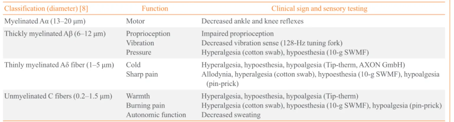
Table 1. Peripheral Nerve Fibers and Function
Classification (diameter) [8] Function Clinical sign and sensory testing
Myelinated Aα (13–20 μm) Motor Decreased ankle and knee reflexes
Thickly myelinated Aβ (6–12 μm) Proprioception Vibration Pressure
Impaired proprioception
Decreased vibration sense (128-Hz tuning fork) Hyperalgesia (cotton swab), hypoesthesia (10-g SWMF)
Thinly myelinated Aδ fiber (1–5 μm) Cold Sharp pain
Hyperalgesia, hypoesthesia, hypoalgesia (Tip-therm, AXON GmbH)
Allodynia, hyperalgesia (cotton swab), hypoesthesia (10-g SWMF), hypoalgesia (pin-prick)
Unmyelinated C fibers (0.2–1.5 μm) Warmth Burning pain Autonomic function
Hyperalgesia, hypoesthesia, hypoalgesia (Tip-therm)
Hyperalgesia (cotton swab), hypoesthesia (10-g SWMF), hypoalgesia (pin-prick) Decreased sweating
Hyperalgesia, increased pain sensitivity of the skin to heat, cold, pin-prick stimuli, or blunt pressure; allodynia, pain in response to non-nociceptive stimuli; hypoesthesia, decreased sensitivity for non-painful stimuli; hypoalgesia, decreased sensitivity to painful stimuli [35].
explored the effects of intensive glucose control on microvas-cular outcomes; patients with T1DM and T2DM responded differently to enhanced glucose control [15]. More intensive treatment of hyperglycemia in patients with T1DM leads to a substantially lower incidence of neuropathy [16], whereas it has a minimal effect on preventing neuropathy in patients with T2DM [17]. The large effect of glucose control in patients with T1DM suggests that hyperglycemia is the primary driver of nerve injury, whereas the lack of this effect in patients with T2DM suggests that factors other than hyperglycemia are im-portant, including obesity, hypertension, low high density lipo-protein concentrations, and hypertriglyceridemia, which possi-bly contribute to nerve injury and often cluster together with diabetes. However, the precise temporal sequence should be clarified in further prospective studies.
IMPORTANCE OF EARLY DETECTION
Half of the patients with DPN are asymptomatic. Typical symptoms of DPN are symmetric numbness, paresthesia, or pain in the distal lower limbs involving more than a single nerve distribution, which progresses in a centripetal direction [6]. Symmetrical sensory loss (“stocking or glove sensory loss”) in the feet, above the ankles, and in the hands is evident on clinical examination. The ankle and Achilles reflexes are usually reduced or absent, which can result in foot abnormali-ties. These symptoms collectively result in disturbed proprio-ception and abnormal muscle sensory function. However, many patients with DPN have trouble describing their symp-toms accurately, which confounds the results of clinical trials and comparisons of drug efficacy. Therefore, validated and quantified measures applicable to Korean patients must be de-veloped to assess the nature and extent of DPN at the early stage.
Painful DPN has a negative impact on physical and mental QOL compared with non-painful DPN. Patients with painful DPN have significantly decreased QOL scores because of pain and impaired balance and mobility. Pain has a large effect on QOL, including quality of sleep, mood, energy, and mobility. Therefore, an early diagnosis of DPN is critical for a good prognosis, and timely comprehensive care can help prevent falls and reduce the negative impacts on patients’ QOL [3].
No clinical test is available to identify or predict the develop-ment or worsening of symptoms in patients at risk of or with DPN. In addition, no consensus exists for the precise algorithm of medications to treat DPN, and the only known effective
dis-ease modifying treatment for DPN is enhanced glucose control [15]. Therefore, identifying patients during the early course will provide a window to identify targeted therapy to modify the course of DPN.
An observational study in Korea indicated that only 12.6% patients with DPN are aware of their disease as a complication of diabetes, and a greater proportion of patients with DPN are not receiving treatment even though they are more likely to de-velop foot ulcers [2]. Painful DPN is under-recognized and un-dertreated, indicating that an early diagnosis provides an op-portunity for improved patient care.
SCREENING AND DIAGNOSTIC
MODALITIES FOR DPN
Assessments of pressure sensation, vibration, thermal, and pain thresholds are used as screening tools for patients “at risk” for foot ulcerations [6]. Although there is a lack of uniform guide-lines on diagnosis and interpretation of the results from a neu-rological examination, it is generally accepted that DPN should be diagnosed based on more than one diagnostic test rather than on one symptom, sign, or test alone [7].
Assessment of risk factors for DPN
DPN develops with chronic hyperglycemia and subsequent po-tential mechanisms induced by chronic hyperglycemia, includ-ing oxidative injury, activation of the polyol glucose metabolic pathway, deposition of advanced glycosylation end-products within nerves, and vascular insufficiency. A number of studies have reported that DPN is related to diabetes duration, glycemia, current cigarette smoking, dyslipidemia, and hyper-tension [18,19]. In agreement with these observations, the re-sults from a nationwide survey conducted by the Diabetic Neu-ropathy Study Group of the KDA in 2010 showed that older age, female sex, long duration of diabetes, presence of retinop-athy, hypertension, or dyslipidemia, hyperglycemic status (i.e., being treated with insulin), and history of cerebrovascular dis-ease or foot ulcers are independently associated with DPN [2].
Assessment of patients’ symptoms and/or signs using scoring systems
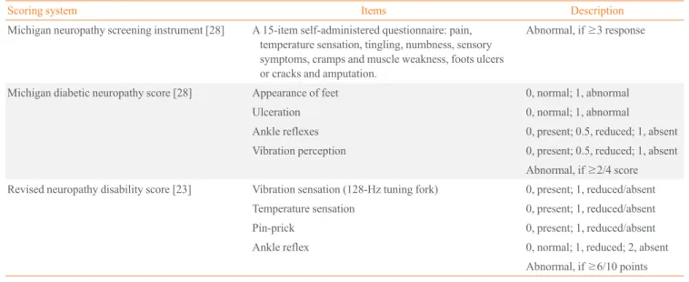
the diabetic neuropathy symptom score [22], the modified NDS, which uses a sensory score (vibration perception thresh-old using a 128-Hz tuning fork and temperature perception, pin-prick) and reflex score (Achilles reflex) [23], neuropathy impairment score (NIS) [24], NIS of the lower limbs [25], a di-abetic neuropathy examination [26], and the Toronto clinical neuropathy score are now available [27]. The Michigan neu-ropathy screening instrument (MNSI) for outpatients question-naire has high specificity of 92%, and the Korean version has been validated [2,28]. The Michigan diabetic neuropathy score (MDNS) along with the MNSI score includes assessments of foot deformities and clinical sensory nerve tests to score DPN [28]. The Neuropathy Study Group of the KDA recommends the MNSI, MDNS, and modified NDS to evaluate patients with diabetes (Table 2). These scoring systems enhance diagnostic accuracy by combining the results of individual examinations. The severity of neuropathic pain and response to treatment in patients with painful DPN can be assessed using a visual ana-log scale or valid scales and questionnaires, such as the Korean version of the Brief Pain Inventory [3].
Assessment of sensorineural impairment
Clinical sensory nerve tests conducted at bedside use various devices to generate specific physical vibratory (128-Hz tuning fork) [29], pressure (10-g Semmes-Weinstein monofilament) [30], noxious (pin-prick) [31], or thermal stimuli (Tip-therm, AXON GmbH, Dusseldorf, Germany) [32], which deliver electrical signals along the sensory pathway (Table 1, Fig. 1)
[33-35].
Quantitative sensory testing (QST) is a quantitative method usually graded using a continuous numerical scale to detect the threshold of thermal perception (cold or warm), vibration per-ception, current perper-ception, pressure pain, and sudomotor func-tion (Table 1) [28,29,32,36,37]. Vibrafunc-tion thresholds are partic-ularly sensitive to detect mild or subclinical neuropathy and correlate well with other QST measures [4]. The current per-ception threshold (CPT) to 2,000-Hz stimulation is correlated best with vibratory thresholds, and the CPT to 5-Hz stimulation is correlated with thermal perception [37]. Sweating abnormal-ities may be an early manifestation of DPN. Sudoscan is a nov-el method to detect nov-electrochemical skin conductance, which is proportional to the number of functional sweat glands [36].
While a sensory stimulus is an objective physical event, the response is highly subjective and depends on the examiner’s experience, patient’s cooperation, and confounding factors (age, sex, obesity, and smoking or alcohol consumption). This differs from an electrophysiological study of nerve conduction velocity (NCV) in which the stimulus generates evoked stimuli independent of the subjective response [38]. The QST is proba-bly effective for documenting sensory abnormalities and changes in sensory thresholds during a longitudinal evaluation of patients with DPN. The Rochester Diabetic Neuropathy Study reported that “the QST should not be used as the sole criterion for diagnosing DPN but should be accompanied by at least one other defined abnormality before the diagnosis of DPN can be made”; therefore, the QST should be
complemen-Table 2. Methods of Sensorimotor Neural Test for Diagnosis of Diabetic Peripheral Neuropathy
Scoring system Items Description
Michigan neuropathy screening instrument [28] A 15-item self-administered questionnaire: pain, temperature sensation, tingling, numbness, sensory symptoms, cramps and muscle weakness, foots ulcers or cracks and amputation.
Abnormal, if ≥3 response
Michigan diabetic neuropathy score [28] Appearance of feet 0, normal; 1, abnormal
Ulceration 0, normal; 1, abnormal
Ankle reflexes 0, present; 0.5, reduced; 1, absent
Vibration perception 0, present; 0.5, reduced; 1, absent Abnormal, if ≥2/4 score
Revised neuropathy disability score [23] Vibration sensation (128-Hz tuning fork) 0, present; 1, reduced/absent
Temperature sensation 0, present; 1, reduced/absent
Pin-prick 0, present; 1, reduced/absent
Ankle reflex 0, normal; 1, reduced; 2, absent
tary to a thorough clinical assessment [39]. Although the QST has been used in clinical practice and clinical trials, it is not ex-tensively used in clinical practice in Korea. Further studies are needed to develop the standardized test procedure, QST algo-rithms, and reference values from healthy test subjects.
Role of electrophysiological studies
The nerve conduction study (NCS) is a reliable and objective
diagnostic method to evaluate the DPN treatment response [40]. The pathological findings of DPN are axonal loss, axonal re-generation, and demyelination in some patients [41,42]. The NCV is used to detect slowing of nerve conduction in nerve ax-ons resulting from segmental demyelination and to measure the speed of both motor and sensory conduction, amplitude, distal latency, distance, F wave latency, and other factors [41]. The American Academy of Neurology (AAN) in conjunction with
Fig. 1. Bedside neurological and sensory nerve testing. (A) Vibration. Patients are notified when they cannot feel the vibrations from a 128-Hz tuning fork (first interphalangeal joint of the great toe) when the toes are extended, and the investigator feels the vibration and measures the time when the feeling disappeared. A time difference ≥10 seconds between the investigator and the patient is considered abnormal [33]. (B) Pressure: 10-g monofilaments are pressed on 10 points on the sole and dorsum of the feet until the monofilament be-gins to bend (100 mN). If the patient has sensation in fewer than seven points, the results is considered abnormal [33]. Four sites per foot, such as the hallux and metatarsal heads 1, 3, and 5, should be screened [4]. (C) Noxious stimuli and (D) light touch. The patient is touched on the foot using a sterile pin, toothpick, and cotton wisp and asked to identify a “sharp or dull” or “light touch” with their eyes closed [33]. (E) Warm/cold. Tip-therm (temperature discriminator; AXON GmbH) is a pen-like device with a plastic cylinder on one end and a metal cylinder on the other end, which is applied to the dorsum of each foot at irregular intervals so patients can identify the sensa-tion as cold or not with their eyes closed [32]. (F) Sudomotor funcsensa-tion. Indicator tests (Neuropad, miro Verbandstoffe) are applied to both soles at the level of the first and second metatarsal heads. The time to color change from blue to pink is more than 10 seconds; the result is considered abnormal [34].
A B C
the American Association of Electrodiagnostic Medicine and the American Academy of Physical Medicine and Rehabilita-tion has a recommended protocol for NCS [40], which includes unilateral studies of sural, ulnar, and medial sensory nerves and peroneal, tibial, medial, and ulnar motor nerves with F waves with a minimum case detection criterion to confirm distal sym-metric polyneuropathy for clinical research; “an abnormality (≥
99th or ≤1st percentile) of any nerve conduction attribute in two separate nerves, one of which must be the sural nerve.” The Nerve Conduction Criteria Study indicated subclinical distal symmetric polyneuropathy to be clinically acceptable if “≥1 among abnormal attributes in ≥2 separate nerves” for clinical practice [43]. Although a NCS is regarded as the gold standard in clinical research, it is not useful in clinical practice because it is time consuming, requires special devices and trained examin-ers, and has no general consensus regarding its criteria, even af-ter multiple investigations [40]. In addition, a NCS is sensitive enough to detect abnormalities in large nerve fibers but is not sensitive enough to detect to small nerve neuropathy, which is the earliest detectable sign of DPN [44].
Challenging modalities for diagnosing DPN
A nerve biopsy, typically of the sural nerve, is rarely used in clinical practice, due to its invasiveness [6]. However, a skin biopsy assessment of intraepidermal nerve fiber density (un-myelinated C fibers and small (un-myelinated fibers) has been pro-posed as a valid method to detect early small nerve neuropathy, even when signs of DPN are minimal or absent and when my-elinated nerve fiber morphology is still normal [45]. A nerve biopsy may detect pathological changes in small nerve fibers and correlate well with the structural pathology of axons. How-ever, this procedure is invasive to patients and normal reference value in Korean subjects must be established.
Corneal confocal microscopy is used to assess the pathology of the corneal subbasal plexus of nerve fibers originating from the ophthalmic division of the trigeminal nerve, particularly corneal nerve fiber length, which is highly reproducible [46]. This noninvasive technique is sensitive to detect corneal nerve fiber damage during the earlier stages of DPN, and the extent of corneal nerve damage and repair correlates with peripheral nerve function. The nerve fiber regeneration response to thera-peutic intervention enables documentation of the natural his-tory of DPN in patients at follow-up [47]. This method, how-ever, needs expensive device and expert examiner for exami-nation.
EVOLVING STRATEGIES FOR
DIAGNOSING DPN
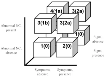
The consensus regarding the diagnosis of DPN has changed since 2004. The AAN recommends a NCV with both neuro-pathic symptoms and signs as confirmation of DPN [40]. Then, the European Federation of Neurological Societies proposed a skin biopsy and intraepidermal nerve fiber density as sensitive measures to detect small nerve neuropathy [48]. Lastly, the To-ronto consensus panel defined “possible” neuropathy as having symptoms, signs, or abnormal reflexes, “probable” as having any two or more of symptoms, signs, or abnormal reflexes, “confirmed” as having either symptoms or signs and NCS re-sults or skin biopsy, and “subclinical” as having neither symp-toms nor signs but rather abnormal nerve conduction or a vali-dated measure of small nerve neuropathy. In addition, they rec-ommended severity assessments using a staged approach based on the nerve conduction abnormality (Fig. 2) [5,6,43].
The KDA guidelines recommend screening for DPN regular-ly at patient’s visits, after a diagnosis of diabetes. In agreement with the American Diabetes Association’s recommendation [49], all patients should be screened for DPN at the time of the
Fig. 2. Definition and severity assessment of diabetic peripheral neuropathy (DPN) proposed by the Toronto Diabetic Neuropathy Expert Group. Numbers in each column refer to the definitions of the minimal criteria for DPN, and the number in parentheses is the stage of severity: 1 (“possible”), 2 (“probable”), or 3 (“confirmed”) for clinical practice and 3 or 4 (“subclinical”) for research studies [6]. Severity is staged based on the symptoms, signs, and nerve conduction (NC) abnormalities: stage 0, no NC abnormality; 1a, subclinical but without symptoms or signs; 1b, subclinical with signs but no symptoms; 2a, subclinical with symptoms regardless of signs; and 2b (not shown here), subclinical with unequivocal weakness of ankle dorsiflexion [5].
Abnormal NC, present
Signs, absence
Abnormal NC,
absence Signs,
presence
Symptoms, absence
T2DM diagnosis and 5 years after a T1DM diagnosis and at least annually thereafter. The KDA also recommends that phy-sicians perform a foot examination at each visit to inspect the feet for deformities, cracks, ulcerations, and wounds in addi-tion to sensaaddi-tion. DPN should be screened by surveys for neu-ropathic symptoms and signs, including the MNSI, clinical sensory nerve tests, the QST, and ankle and Achilles reflexes. Specifically, the KDA recommends screening via the 10-g monofilament, vibration perception clinical bedside tests using a 128-Hz tuning fork, and assessment of ankle reflexes (Fig. 3) [7]. An assessment of distal pulses is also recommended, and the ankle brachial index should be measured if peripheral arte-rial disease needs to be evaluated.
CONCLUSIONS
Early diagnosis of DPN is critical for successful management of diabetes and preventing DPN-related patient and social dis-ease burdens. Hyperglycemia and other risk factors should be controlled in patients during the early stages of DPN. In addi-tion, symptoms and/or signs must be assessed, and composite scoring systems, the QST, as well as a NCS are complementary to diagnose DPN in patients with diabetes. Further research is needed to investigate the role of the QST and NCS for early detection and predicting DPN in Korean patients with T2DM. Although the diagnosis is clearly improving with current diag-nostic tools, longitudinal investigations are needed to better un-derstand the significance of abnormalities detected by each modality, including emerging new methods and their cost-ef-fectiveness.
CONFLICTS OF INTEREST
No potential conflict of interest relevant to this article was re-ported.
ACKNOWLEDGMENTS
This work was supported by Priority Research Centers Pro-gram through the National Research Foundation of Korea (NRF) funded by the Ministry of Education, Science and Tech-nology (2010-0020224).
ORCID
Tae Sun Park http://orcid.org/0000-0001-7216-8468
REFERENCES
1. Ha KH, Kim DJ. Trends in the diabetes epidemic in Korea.
Endocrinol Metab (Seoul) 2015;30:142-6.
2. Won JC, Kwon HS, Kim CH, Lee JH, Park TS, Ko KS, et
al. Prevalence and clinical characteristics of diabetic pe-ripheral neuropathy in hospital patients with type 2 diabetes in Korea. Diabet Med 2012;29:e290-6.
3. Kim SS, Won JC, Kwon HS, Kim CH, Lee JH, Park TS, et
al. Prevalence and clinical implications of painful diabetic peripheral neuropathy in type 2 diabetes: results from a na-tionwide hospital-based study of diabetic neuropathy in Korea. Diabetes Res Clin Pract 2014;103:522-9.
4. Boulton AJ, Malik RA, Arezzo JC, Sosenko JM. Diabetic
somatic neuropathies. Diabetes Care 2004;27:1458-86. 5. Dyck PJ, Albers JW, Andersen H, Arezzo JC, Biessels GJ,
Bril V, et al. Diabetic polyneuropathies: update on research definition, diagnostic criteria and estimation of severity. Diabetes Metab Res Rev 2011;27:620-8.
6. Tesfaye S, Boulton AJ, Dyck PJ, Freeman R, Horowitz M,
Kempler P, et al. Diabetic neuropathies: update on defini-tions, diagnostic criteria, estimation of severity, and treat-ments. Diabetes Care 2010;33:2285-93.
7. Korean Diabetes Association. Treatment guideline for
dia-betes. J Korean Diabetes 2015;12:S109-12.
8. Gasser HS. The classification of nerve fibers. Ohio J Sci
1941:41;145-59.
9. Vinik AI, Mehrabyan A. Diabetic neuropathies. Med Clin
North Am 2004;88:947-99.
10. Malik RA, Tesfaye S, Newrick PG, Walker D, Rajbhandari
Fig. 3. Diagnostic approach to diabetic peripheral neuropathy (DPN) proposed by the Korean Diabetes Association [7].
Typical symptoms±signs for DPN (distal, symmetric, sensorimotor)
Diagnosis of DPN (possible or probable) Excluding other causes
of neuropathy
Signs and neurologic examination • 10-g monofilament • 128-Hz tuning fork • Ankle reflex
Symptoms Decreased sensation, asleep, numbness, prick-ling or stabbing, burning
SM, Siddique I, et al. Sural nerve pathology in diabetic pa-tients with minimal but progressive neuropathy. Diabetolo-gia 2005;48:578-85.
11. Ziegler D, Mayer P, Muhlen H, Gries FA. The natural
his-tory of somatosensory and autonomic nerve dysfunction in relation to glycaemic control during the first 5 years after diagnosis of type 1 (insulin-dependent) diabetes mellitus. Diabetologia 1991;34:822-9.
12. Selvarajah D, Wilkinson ID, Maxwell M, Davies J, Sankar
A, Boland E, et al. Magnetic resonance neuroimaging study of brain structural differences in diabetic peripheral neu-ropathy. Diabetes Care 2014;37:1681-8.
13. Partanen J, Niskanen L, Lehtinen J, Mervaala E, Siitonen O,
Uusitupa M. Natural history of peripheral neuropathy in patients with non-insulin-dependent diabetes mellitus. N Engl J Med 1995;333:89-94.
14. Myers MI, Peltier AC. Uses of skin biopsy for sensory and
autonomic nerve assessment. Curr Neurol Neurosci Rep 2013;13:323.
15. Callaghan BC, Little AA, Feldman EL, Hughes RA.
En-hanced glucose control for preventing and treating diabetic neuropathy. Cochrane Database Syst Rev 2012;6:CD007543. 16. The Diabetes Control and Complications Trial Research
Group. The effect of intensive treatment of diabetes on the development and progression of long-term complications in insulin-dependent diabetes mellitus. N Engl J Med 1993; 329:977-86.
17. Ismail-Beigi F, Craven T, Banerji MA, Basile J, Calles J,
Cohen RM, et al. Effect of intensive treatment of hypergly-caemia on microvascular outcomes in type 2 diabetes: an analysis of the ACCORD randomised trial. Lancet 2010; 376:419-30.
18. Franklin GM, Shetterly SM, Cohen JA, Baxter J, Hamman
RF. Risk factors for distal symmetric neuropathy in NID-DM: the San Luis Valley Diabetes Study. Diabetes Care 1994;17:1172-7.
19. Maser RE, Steenkiste AR, Dorman JS, Nielsen VK, Bass
EB, Manjoo Q, et al. Epidemiological correlates of diabetic neuropathy. Report from Pittsburgh Epidemiology of Dia-betes Complications Study. DiaDia-betes 1989;38:1456-61. 20. Dyck PJ, Sherman WR, Hallcher LM, Service FJ, O’Brien
PC, Grina LA, et al. Human diabetic endoneurial sorbitol, fructose, and myo-inositol related to sural nerve morphom-etry. Ann Neurol 1980;8:590-6.
21. Dyck PJ, Karnes J, O’Brien PC, Swanson CJ. Neuropathy
symptom profile in health, motor neuron disease, diabetic
neuropathy, and amyloidosis. Neurology 1986;36:1300-8. 22. Meijer JW, Smit AJ, Sonderen EV, Groothoff JW, Eisma
WH, Links TP. Symptom scoring systems to diagnose dis-tal polyneuropathy in diabetes: the Diabetic Neuropathy Symptom score. Diabet Med 2002;19:962-5.
23. Abbott CA, Vileikyte L, Williamson S, Carrington AL,
Boulton AJ. Multicenter study of the incidence of and pre-dictive risk factors for diabetic neuropathic foot ulceration. Diabetes Care 1998;21:1071-5.
24. Dyck PJ, Litchy WJ, Lehman KA, Hokanson JL, Low PA,
O’Brien PC. Variables influencing neuropathic endpoints: the Rochester Diabetic Neuropathy Study of Healthy Sub-jects. Neurology 1995;45:1115-21.
25. Singleton JR, Bixby B, Russell JW, Feldman EL, Peltier A,
Goldstein J, et al. The Utah Early Neuropathy Scale: a sen-sitive clinical scale for early sensory predominant neuropa-thy. J Peripher Nerv Syst 2008;13:218-27.
26. Meijer JW, van Sonderen E, Blaauwwiekel EE, Smit AJ,
Groothoff JW, Eisma WH, et al. Diabetic neuropathy ex-amination: a hierarchical scoring system to diagnose distal polyneuropathy in diabetes. Diabetes Care 2000;23:750-3. 27. Davies M, Brophy S, Williams R, Taylor A. The prevalence,
severity, and impact of painful diabetic peripheral neurop-athy in type 2 diabetes. Diabetes Care 2006;29:1518-22. 28. Feldman EL, Stevens MJ, Thomas PK, Brown MB, Canal N,
Greene DA. A practical two-step quantitative clinical and electrophysiological assessment for the diagnosis and stag-ing of diabetic neuropathy. Diabetes Care 1994;17:1281-9. 29. Dyck PJ, Dyck PJ, Larson TS, O’Brien PC, Velosa JA.
Pat-terns of quantitative sensation testing of hypoesthesia and hyperalgesia are predictive of diabetic polyneuropathy: a study of three cohorts. Nerve growth factor study group. Diabetes Care 2000;23:510-7.
30. Cheng WY, Jiang YD, Chuang LM, Huang CN, Heng LT,
Wu HP, et al. Quantitative sensory testing and risk factors of diabetic sensory neuropathy. J Neurol 1999;246:394-8. 31. Ziegler D, Siekierka-Kleiser E, Meyer B, Schweers M.
Vali-dation of a novel screening device (NeuroQuick) for quanti-tative assessment of small nerve fiber dysfunction as an ear-ly feature of diabetic poear-lyneuropathy. Diabetes Care 2005; 28:1169-74.
32. Viswanathan V, Snehalatha C, Seena R, Ramachandran A.
Early recognition of diabetic neuropathy: evaluation of a simple outpatient procedure using thermal perception. Post-grad Med J 2002;78:541-2.
Associa-tion. Management of diabetic neuropathy. Seoul: Korean Diabetes Association; 2011. p. 16-34.
34. Papanas N, Papatheodorou K, Papazoglou D, Monastiriotis
C, Christakidis D, Maltezos E. A comparison of the new in-dicator test for sudomotor function (Neuropad) with the vi-bration perception threshold and the clinical examination in the diagnosis of peripheral neuropathy in subjects with type 2 diabetes. Exp Clin Endocrinol Diabetes 2008;116:135-8. 35. Loeser JD, Treede RD. The Kyoto protocol of IASP Basic
Pain Terminology. Pain 2008;137:473-7.
36. Casellini CM, Parson HK, Richardson MS, Nevoret ML,
Vinik AI. Sudoscan, a noninvasive tool for detecting dia-betic small fiber neuropathy and autonomic dysfunction. Diabetes Technol Ther 2013;15:948-53.
37. Masson EA, Veves A, Fernando D, Boulton AJ. Current
perception thresholds: a new, quick, and reproducible meth-od for the assessment of peripheral neuropathy in diabetes mellitus. Diabetologia 1989;32:724-8.
38. Dyck PJ, Thomas PK. Peripheral neuropathy. 4th ed.
Phila-delphia: Saunders; 2005. Chapter 35, Nerve conduction and needle electromyography; p. 899-969.
39. Dyck PJ, Davies JL, Litchy WJ, O’Brien PC. Longitudinal
assessment of diabetic polyneuropathy using a composite score in the Rochester Diabetic Neuropathy Study cohort. Neurology 1997;49:229-39.
40. England JD, Gronseth GS, Franklin G, Miller RG, Asbury
AK, Carter GT, et al. Distal symmetric polyneuropathy: a definition for clinical research: report of the American Acad-emy of Neurology, the American Association of Electrodi-agnostic Medicine, and the American Academy of Physical Medicine and Rehabilitation. Neurology 2005;64:199-207. 41. Dyck PJ, Lais A, Karnes JL, O’Brien P, Rizza R. Fiber loss
is primary and multifocal in sural nerves in diabetic
poly-neuropathy. Ann Neurol 1986;19:425-39.
42. Behse F, Buchthal F, Carlsen F. Nerve biopsy and
conduc-tion studies in diabetic neuropathy. J Neurol Neurosurg Psychiatry 1977;40:1072-82.
43. Dyck PJ, Carter RE, Litchy WJ. Modeling nerve
conduc-tion criteria for diagnosis of diabetic polyneuropathy. Mus-cle Nerve 2011;44:340-5.
44. Breiner A, Lovblom LE, Perkins BA, Bril V. Does the
pre-vailing hypothesis that small-fiber dysfunction precedes large-fiber dysfunction apply to type 1 diabetic patients? Diabetes Care 2014;37:1418-24.
45. Sumner CJ, Sheth S, Griffin JW, Cornblath DR, Polydefkis
M. The spectrum of neuropathy in diabetes and impaired glucose tolerance. Neurology 2003;60:108-11.
46. Hertz P, Bril V, Orszag A, Ahmed A, Ng E, Nwe P, et al.
Reproducibility of in vivo corneal confocal microscopy as a novel screening test for early diabetic sensorimotor poly-neuropathy. Diabet Med 2011;28:1253-60.
47. Brines M, Dunne AN, van Velzen M, Proto PL, Ostenson
CG, Kirk RI, et al. ARA 290, a nonerythropoietic peptide engineered from erythropoietin, improves metabolic con-trol and neuropathic symptoms in patients with type 2 dia-betes. Mol Med 2015;20:658-66.
48. Lauria G, Hsieh ST, Johansson O, Kennedy WR, Leger JM,
Mellgren SI, et al. European Federation of Neurological Societies/Peripheral Nerve Society Guideline on the use of skin biopsy in the diagnosis of small fiber neuropathy. Re-port of a joint task force of the European Federation of Neurological Societies and the Peripheral Nerve Society. Eur J Neurol 2010;17:903-12, e44-9.
49. American Diabetes Association. Standards of medical care