online | memorias.ioc.fiocruz.br
Brycon hilarii (Valenciennes, 1950) (Syn. Brycon microlepis Perugia, 1897) is an endemic characid spe-cies of the upper Paraguay Basin (Resende 2003), popu-larly known as piraputanga. It is highly appreciated in central Brazil (especially in the states of Mato Grosso and Mato Grosso do Sul) (Resende 2003) and is a very valuable species, fetching high market prices. B. hilarii has recently been introduced into aquaculture in several regions of Brazil, with a production of 633,000 kg on fish farms in 2006 (IBAMA 2008), demonstrating the considerable potential of this species to this activity.
Myxosporean parasites are among the most impor-tant fish pathogens (Schmahl et al. 1989) and more than 2,300 species have been reported to infect fish (marine and/or freshwater fish) in either their natural environ-ment or on farms (Adriano et al. 2006, 2009a, b, Feist & Longshaw 2006, Eiras et al. 2008, Azevedo et al. 2009). The Myxobolus Butschli, 1882, is the genus with the greatest number of species (approximately 790 valid
spe-Financial support: FAPESP (06/59075-6), CEPTA/ICMBio, CAPES (to TM), FAPESP (to MMC)
+ Corresponding author: edapadriano@hotmail.com Received 15 December 2009
Accepted 13 July 2010
cies) (Eiras et al. 2005, Lom & Dyková 2006) and many of these species are reported as pathogenic to fish (Kent et al. 2001, Feist & Longshaw 2006). In South America, 30 Myxobolus species have been reported (Azevedo et al. 2010, Eiras et al. 2010), but none have been reported to infect fish from thegenus Brycon.
As part of ongoing research on the characteristics of myxosporea parasites of freshwater fish in Brazil, the present paper describes a new species of Myxobolus found to infect wild specimens of piraputangain the Brazilian Pantanal wetland using morphological, histological, ultra-structural and molecular phylogeny analyses.
MATERIALS AND METHODS
A total of 126 specimens of wild B. hilarii caught in the Pantanal Mato-Grossense in central Brazil and 90 farmed specimens from three fish farms in the state of São Paulo (SP) were examined. The wild fish were examined in two seasons: spring (flood period) of 2001-2004 (n = 45) and autumn (drought period) of 2003-2005 and 2008 (n = 81). The fish were captured at three loca-tions: in the Aquidauana, Miranda and Paraguay Rivers in the southern region of the Pantanal (n = 34), in rivers and lakes of the Pantanal National Park in the central region (n = 60) and in the Cuiabá and Manso Rivers in the municipality of Nobres in the northern region (n = 32). The fish from the Miranda, Aquidauana and Para-guay Rivers were caught in the spring of 2001 and 2002 as well as the autumn of 2003, whereas those from the
Phylogeny, ultrastructure, histopathology and prevalence of
Myxobolus
oliveirai
sp. nov., a parasite of
Brycon hilarii
(Characidae)
in the Pantanal wetland, Brazil
Tiago Milanin1, Jorge C Eiras2, Sarah Arana3,Antônio AM Maia4, Anderson L Alves5,
Márcia RM Silva4, Mateus M Carriero4, Paulo S Ceccarelli6, Edson A Adriano7/+
1Departamento de Biologia Animal 3Departamento de Histologia e Embriologia, Universidade Estadual de Campinas, Campinas, SP, Brasil 2Departamento de Biologia e Centro Interdisciplinar de Investigação Marinha e Ambiental, Faculdade de Ciências, Universidade do Porto, Porto, Porto, Portugal 4Departamento de Ciências Básicas, Faculdade de Zootecnia e Engenharia de Alimentos, Universidade de São Paulo,
Pirassununga, SP, Brasil 5Departamento de Biologia, Universidade Estadual Paulista Julio de Mesquita Filho, Rio Claro, SP, Brasil 6Centro Nacional de Pesquisa e Conservação de Peixes Continentais, Instituto Chico Mendes de Conservação da Biodiversidade,
Pirassununga, SP, Brasil 7Departamento de Ciências Biológicas, Universidade Federal de São Paulo, Rua Prof. Artur Riedel 275, 09972-270 Jardim Eldorado, Diadema, SP, Brasil
This paper presents the morphological, histological and ultrastructural characteristics of Myxobolus oliveirai sp. nov., a parasite of the gill filaments in Brycon hilarii from the Brazilian Pantanal. Out of 216 B. hilarii specimens examined (126 wild and 90 cultivated), 38.1% of wild specimens (n = 48) were infected. The parasites form elongated plasmodia primarily in the tip of gill filaments, reaching about 3 mm in length. A thorough comparison with all the Myxobolus species described from South American hosts, as well as nearly all the Myxobolus species described so far is provided. Partial sequencing of the 18S rDNA gene revealed a total of 1,527 bp. The Myxobolus species para-site of B. hilarii did not match any of the Myxozoa available in GenBank. In the phylogenetic analysis, M. oliveirai sp. nov. composed a monophyletic group with eight other species: five species of Myxobolus parasites of mugilid fishes, two parasites of pangasiid and one of centrarchid. Infection prevalence values of the parasite revealed no significant differences between wet and dry seasons or between males and females. The importance of the infection to the farming of the host species is emphasized.
histological analysis, fragments of infected organs were fixed in 10% buffered formalin, embedded in paraffin, cut into serial sections (4 µm in thickness) and stained with haematoxylin/eosin and Sirius red. For transmis-sion electron microscopy, plasmodia were fixed in 2.5% glutaraldehyde in 0.1 M sodium cacodylate buffer (pH 7.4) for 12 h, washed in glucose-saline solution for 2 h and post-fixed in OsO4, all at 4ºC. After dehydration in an acetone series, the material was embedded in EMbed 812 resin. Ultrathin sections, double stained with uranyl acetate and lead citrate, were examined in an LEO 906 electron microscope operated at 60 kV.
For molecular analysis, plasmodia were removed from the host tissue and fixed in ethanol. After ruptur-ing the plasmodia with the aid of a needle, the contents were collected in a 1.5 mL microcentrifuge tube. DNA was extracted using the Wizard® Genomic DNA Purifi-cation kit (Promega, USA), following the manufacturer’s instructions. DNA content was determined using the NanoDrop 2000 spectrophotometer (Thermo Scientific) at 260 nm. Polymerase chain reaction (PCR) was carried out in a final volume of 25 µL, which contained 10-50 ng of extracted DNA, 1x Taq DNA polymerase buffer (Invitrogen), 0.2 mmol of dNTP (Invitrogen), 1.5 mmol of MgCl2, 0.2 pmol of each primer (Invitrogen), 0.25 µL (1.25 U) of Taq DNA polymerase (Invitrogen) and ultra pure (MilliQ) water. The Eppendorf AG 22331 Hamburg Thermocycler was used. An ~1600 bp 5’ fragment of the SSU rDNA gene was amplified using the primers Mx5-Mx3 (Andree et al. 1999)in the following manner: an initial denaturation step at 95ºC for 5 min followed by 35 cycles of denaturation (95ºC for 60 s), annealing (62ºC for 60 s) and extension (72ºC for 120 s), finished with an extended elongation step at 72ºC for 5 min. PCR prod-ucts were submitted to electrophoresis on 1% agarose gel (BioAmerica) in a Tris-Borate-EDTA buffer (0.045 MTris-borate, 0.001 M EDTA pH 8.0), stained with ethid-ium bromide and analyzed in a FLA-3000 (Fugi) scan-ner. The size of the amplified fragments was estimated by comparisons with the 1 kb DNA Ladder (Invitrogen).
Purified PCR products were cloned in pCR®4-TOPO® vectors from the TOPO-TA Cloning® kit for sequencing (Invitrogen). A single clone was sequenced using Mx5-Mx3 and MC5-MC3 (Eszterbauer 2004) primer pairs with the BigDye® Terminator v3.1 Cycle Sequencing Kit (Applied Biosystems™) in an ABI 3730 DNA sequenc-ing analyzer (Applied Biosystems™).
in the analysis. Bootstrap analysis (1,000 replicates) was employed to assess the relative robustness of the branch-es of the NJ, MP and ML trebranch-es using the MEGA 4.0 and 5.0 programmes. The distance analyses were performed using the K2P model conducted using the MEGA 4.0 programme (Tamura et al. 2007). The species Cerato-myxa seriolae and Ceratomyxa shasta were used as out-groups in the phylogenetic analyses.
The effects of season, study location and sex of the host on the prevalence of the parasite were assessed using
the χ2 test, with the level of significance set at p< 0.05. RESULTS
Among the 126 wild and 90 cultivated specimens of piraputanga (a total of 216) examined in the present study, 22.2% (n = 48) had plasmodia of an unknown parasite from the genus Myxobolus. The prevalence was of 38.1% in the wild specimens and 0% in the cultivated specimens. In the wild specimens, the prevalence of the parasite varied significantly with regard to study location
(χ2
2 = 28.79; p = 0.00), with the lowest prevalence found in southern region of the Pantanal (2.9%). No signifi-cant difference was found between the central (43.3%)
and northern regions (65.6%) (χ2
1 = 4.15; p = 0.04). The parasite was found in both seasons studied (prevalence of 33.3% in spring and 40.7% in autumn) and the
varia-tion between seasons was not significant (χ2
1 = 0.63; p = 0.41). Regarding the sex of the host, the prevalence was 32.2% for female specimens and 16.2% for males; this
difference was also not significant (χ2
1 = 3.07; p = 0.07). Sex was not defined in seven infected specimens.
Myxobolus oliveirai sp. nov. (Figs 1-5)
The plasmodia exhibited development with different sporogenic stages, such as generative cells and disporo-blastic pansporoblasts (Fig. 3C) along the periphery of the plasmodia (occurring in a thin layer of not more than 40 µm), followed by a layer containing immature spores and mature spores occurring in the internal region (Figs 2-4). The spores were pear-shaped in the frontal view (Figs 1B, 5A), measuring 11.2 ± 0.4 µm in length, 7.4 ± 0.5 µm in width and 4.6 ± 0.6 µm in thickness. The polar capsules were elongated in shape and equal in size, measuring 5.6 ± 0.2 µm in length and 2.3 ± 0.2 µm in width. The anterior ends of polar capsules were close to one another and the polar filaments exhibited 6-8 turns arranged perpendicular to the longitudinal axis of the capsules (Figs 4C, 5A). A few, small sporoplasmosomes were found in the sporoplasm (Fig. 4A) and two nuclei were discernible when stained with Giemsa (Fig. 5A).
In the molecular analysis, the specific primer pair Mx5-Mx3 successfully amplified an approximately 1,600-bp fragment of the 18S rDNA gene in the spores obtained from plasmodia found infecting the gill fila-ments of B. hilarii. The BLAST search using the partial 18S rDNA sequence data (1,527 bp) of the Myxobolus
species parasite of B. hilarii did not match any of the Myxozoa available in the GenBank.
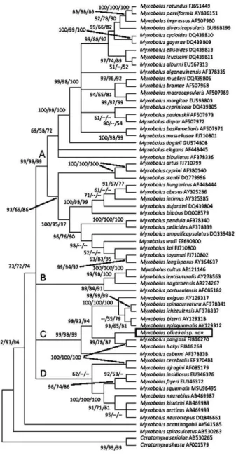
In the phylogenetic analyses, the Myxobolus species clustered into three distinct lineages. Myxobolus spirosul-catus represented the basal group and Myxobolus acan-thogobii clustered as the sister group of the remaining species of Myxobolus (Fig. 6). These remaining species clustered in a monophyletic group composed of numer-ous species divided into four smaller clades (A-D) (Fig. 6). M. oliveirai sp. nov. formed a monophyletic unit with eight other Myxobolus species in Clade C: five species parasites of hosts from the family Mugilidae; two para-sites of Pangasiidae hosts and one parasite of Centrarchi-dae. Mean genetic divergence within Clade C (Table) was 20.4%. In this clade, the smallest distance in reference to M. oliveirai sp. nov. was with Myxobolus hakyi (16.6%) and the largest was with Myxobolus osburni (28.6%).
Type host - B. hilarii (Valenciennes, 1850) (Characi-dae, Bryconinae).
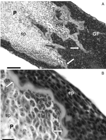
Etymology - The specific epithet name is in homage to Ricardo Afonso Torres de Oliveira, research assistant of the laboratory of Saúde e Bem Estar dos Peixes of the Fig. 1: light photomicrographs of Myxobolus oliveirai sp. nov.,
para-site of Brycon hilarii.A: formalin-fixed gill filaments showing plas-modia in the distal extremity (arrows). Bar = 3 mm; B: mature fresh spores. Bar = 10 µm; C: histological section showing plasmodia (P) in the distal extremity. Note the expansion of the gill filaments with compression and thinning of the tissue of the extremity distal (black arrows) as well as invaginations in the filament tissue in the proximal extremity (white arrows). Bar = 20 µm. Sirius red staining.
Centro Nacional de Pesquisa e Conservação de Peixes Continentais of Instituto Chico Mendes de Conservação da Biodiversidade, who, in recent years, has provided great support to the fieldstrip of our research group.
Type dataand depository - Pantanal Mato-Grossense (Aquidauana, Cuiabá, Miranda and Paraguay Rivers) in the Paraguay basin, Brazil.
Specimens deposited - Slides with stained spores (syntype) have been deposited in the collection of the Museum of Natural History, Institute of Biology, State University of Campinas, SP, Brazil (accession ZUEC 28). The 18S rDNA sequence was deposited in GenBank under the accession HM754633.
Prevalence - Fourty-eight of 216 B. hilarii examined (22.2%) had plasmodia of an unknown parasite from the genus Myxobolus - 48/126 wild specimens (38.1%) and 0/90 (0%) cultivated specimens.
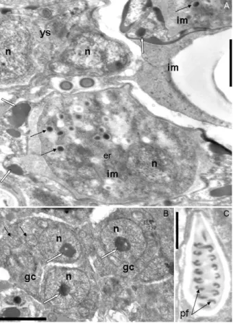
Site of infection - Distal extremity of the gill filaments. Fig. 3: electron micrographs of Myxobolus oliveirai sp. nov. parasite of gill filaments in Brycon hilarii.A-C: host-parasite interface; A: showing the capsule of connective tissue (ct) surrounding the plas-modium (P). Note the ectoplasm area (ec) showing pinocytotic ca-nals (arrow) and mitochondria (m), sporogenic stages (sst) and an in-ner layer of young spores (ys). Bar = 1 µm; B: smaller magnification showing the host-parasite interface (arrow), sst, ys and transversal sections of mature spores (msp). Bar = 5 µm; C: detail of P showing pinocytotic canals terminating in pinocytotic vesicles (arrows) and m. Bar = 0.5 µm; H: host.
Fig. 4: electron micrographs of Myxobolus oliveirai sp. nov. para-site of gill filaments in Brycon hilarii. A: plasmodium showing transversal section of young spore (ys) with two nuclei (n) and immature spores (im) showing sporoplasmossomes (thin arrows), sporoplasm nucleus and valve-forming material (white arrows). Bar = 1 µm; B: generative cells (gc) with n containing conspicuous nucleolus (white arrows) and mitochondria (thin arrows). Bar = 2 µm; C: longitudinal section of a polar capsule with its polar fila-ments (pf ) sections. Bar = 1 µm.
DISCUSSION
oliveirai sp. nov. Although the description of Myxobolus sp. parasite of Serrasalmus sp. (Walliker 1969) is poor, a superficial comparison reveals that the anterior end of the spores of Myxobolus sp. are more pointed than in M. oliveirai sp. nov. Furthermore, the infection site (kidney) and host (Serrasalmus sp.) of the parasite described by Walliker (1969) differ from those of M. oliveirai sp. nov., which infects gill filaments in B. hilarii. Regarding M. cordeiroi, the differences were in the number of polar filament turns, infection sites (connective tissue of sever-al organs) and host (a pimelodid fish). The spore dimen-sions of the different species do not fit the values found in the present material. Therefore, our specimens cannot be identified with any of the aforementioned species.
When comparing M. oliveirai sp. nov. with species from other continents (Eiras et al. 2005), we selected those species that most resembled our material in their sets of characteristics. Thus, we included Myxobolus bel-lus infecting the integument of Carpioides carpio in the USA (Kudo 1934), Myxobolus bramaeformis described from the kidneys and gut of Hypophthalmichthys moli-trix in the Amur Basin (Akhmerov 1960), Myxobolus fahmii infecting the gills of Barbus bynni in Egypt (Ali et al. 2002) and Myxoboluspseudosquamae parasitizing the gills and kidney of Sinocyclochilus grahami tingi in China (Ma & Zhao 1993). Despite some similarities, the features of our specimens do not match the character-istics of any of the aforementioned species. Moreover, their hosts are phylogenetically very different and their geographic locations are also quite different. In particu-lar, only a very few number of species have as small a spore thickness (4.6 ± 0.6 µm) as that described for M. oliveirai sp. nov. Considering the 744 nominal species reported by Eiras et al. (2005), only 21 have a similar spore thickness. In these cases, however, the other spore features are very different. Furthermore, this is the first report of a myxosporean species infecting fish from the genus Brycon and the BLAST search using the partial 18S rDNA sequence of M. oliveirai sp. nov. did not match any of the Myxozoa available in GenBank.
Fig. 5: schematic representation of mature spores of Myxobolus olivei-rai sp. nov. A: frontal view; B: lateral view. Bar = 3 µm.
Fig. 6: condensed phylogenetic tree (bootstrap > 50) of consensus among neighbor-joining, maximum parsimony and maximum likeli-hood showing relationship between Myxobolus oliveirai sp. nov. and other Myxobolus spp based on partial 18S rDNA. Genbank accessions are given after species name. Numbers above nodes indicate boot-strap confidence levels.
(A-D), with unsolved groups, despite the high bootstrap values supporting the relationships.
M. oliveirai sp. nov. clustered in Clade C together with eight other species, forming a monophyletic unit, where it appears as sister group of the clade formed by M. pangasii and M. hakyi, which are parasites of Pan-gasianodon hypophthalmus (Pangasiidae: Siluriformes) and, together, these three species compose the sister group of the clade formed by five species (Myxobolus exiguus, Myxobolus spinacurvatura, Myxobolus ichkeu-lensis, Myxobolus bizerti and Myxobolus episquamalis), parasites of mugilid hosts. M. osburni, a parasite of Le-pomis gibbosus, which is a Perciformes from the family Centrarchidae, appears as the basal unit of Clade C.
The mean genetic divergence estimated in Clade C was 20.4%. With regard to the new species, the small-est divergence was with M. hakyi (16.6%), a sister taxa of M. oliveirai sp. nov., whereas the largest divergence was with M. osburni (28.6%) (Table) which is the basal species for Clade C.
The phylogenetic tree obtained here is in accordance with results described by Ferguson et al. (2008) and Liu et al. (2010), who report M. acanthogobii as a species with the greatest divergence among Myxobolus spp as well as in relation to the clustering of M. exiguus, M. spinacurvatura, M. ichkeulensis, M. bizerti, M. episqua-malis, M. pangasii and M. hakyi in a monophyletic unit. The results of the present study show the phyloge-netic relation of the new species with other Myxobolus species available in GenBank. This is the first phyloge-netic study on a Myxobolus species parasite of a South American characid host considering Myxozoa parasites of fresh water fish. From this region, only the 18S rDNA gene sequence of M. cordeiroi (Adriano et al. 2009a), with around 500 bp, has been deposited in GenBank. Thus, additional molecular and phylogenetic studies of Myxobolus spp parasites of this fish family are needed to identify the true position of the Myxobolus parasites of characid hosts in relation to the Myxobolus species parasites of other families of fish.
There was a much lower prevalence of M. oliveirai sp. nov. in the southern region of the Pantanal than that found in the central and northern areas, demonstrating
that environmental characteristics may influence the distribution of this myxosporean species in the Brazil-ian Pantanal. Environmental parameters (water tem-perature and water flow) have been reported to influ-ence the prevalinflu-ence of Myxobolus cerebralis (Baldwin et al. 2000, Hallett & Bartholomew 2008). The Brazilian Pantanal is the largest floodplain area in the world, with approximately 150,000 km2 (Silva 1986, Ceccarelli et al. 2007), and this very large ecosystem has areas with heterogeneous features, which may have contributed to-ward the lower prevalence of M. oliveirai sp. nov. in the southern region. However, the nature of the parameters that influence the prevalence of infection is unknown.
Regarding the seasons studied, the variation in prev-alence was non-significant, revealing that season had no influence over infection by M. oliveirai sp. nov. in the present study. This finding is in contrast to results described by Gbankoto et al. (2001), who report the oc-currence of significant seasonal variations in Myxobo-lus sp. and Myxobolus zillii parasites of Sarotherodon melanotheron melanotheron and Tilapia zillii, respective-ly, and Gbankoto et al. (2003), who report seasonal differ-ences in the infection of S. melanotheron melanotheron by Myxobolus heterospora. However, the results of the present study are in agreement with those described by Gbankoto et al. (2003), who report no seasonal variation in the infection of T. zillii by M. heterospora.
The comparison of infection between female and male hosts revealed no influence of sex on the preva-lence of M. oliveirai sp. nov. This finding is in agree-ment with accounts describing some Myxozoa species (Gbankoto et al. 2001, Viozzi & Flores 2003), but con-trasts with others (Muzzall 1995, Gbankoto et al. 2003) that reported a significant difference between females and males regarding the prevalence of infection.
The histological analysis of piraputanga gills in-fected with M. oliveirai sp. nov. revealed numerous large cysts primarily in the distal area of the gill filaments, but no pronounced inflammatory response was found in the infection site, which is similar to findings described for other South American myxoporean species (Barassa et al. 2003, Eiras et al. 2008, 2009, Adriano et al. 2009a, b). However, the development of the plasmodia caused
ex-Myxobolus exiguus 0.206 0.155 0.305 0.171 0.226 0.222 0.171
pansion and compression of the gill filaments, producing a thinning of the tissue at the distal extremity, whereas the growth of the plasmodia in proximal extremity oc-curred through invaginations, pervading and occupying the filament tissue. These structural alterations in the gills are similar to structural changes reported for other myxosporean species (Feist & Longshaw 2006).
The ultrastructural analysis revealed that sporogen-esis in M. oliveirai sp. nov. followed the general pattern of other Myxobolus species (Current et al. 1979, Casal et al. 1996, 2002, Adriano et al. 2006) and the plasmo-dium wall consisted of a single membrane, which was continuous with pinocytic canals extending to the ecto-plasm, as seen in some Myxobolus species (Current et al. 1979), but differed from others delimited by a double membrane (Casal et al. 2002, 2006).
Gill infections by Myxoporea in farmed fish can cause significant tissue damage (Martins et al. 1997, 1999, Adriano et al. 2005a, b, 2006, Feist & Longshaw 2006) and occasionally death (Martins et al. 1999, Feist & Longshaw 2006). Although the histopathological changes produced by M. oliveirai sp. nov. impaired the function of the gill surface, the infection apparently did not significantly affect the general health of the fish. However, the histopathological analysis only addressed the wild host specimens (farmed fish were not infect-ed) and we do not know the impact that infection by M. oliveirai sp. nov. may have on farmed specimens, espe-cially in intensive farming. Thus, the presence and dis-persion of M. oliveirai sp. nov. needs to be monitored closely by commercial fish farmers.
ACKNOWLEDGEMENTS
To Ricardo Afonso Torres de Oliveira (CEPTA/ICMBio), for help in dissecting the fish, and to Dr Laerte Batista de Oliveira Alves, manager of the National Center for Research and Conservation of Continental Fishes (CEPTA/ICMBio), and Dr José Augusto Ferraz de Lima, the manager of Pantanal National Park, for support during the fieldwork.
REFERENCES
Adriano EA, Arana S, Alves AL, Silva MR, Ceccarelli PS, Henrique-Silva F, Maia AA 2009a. Myxobolus cordeiroi n. sp., a parasite of Zungaro jahu (Siluriformes: Pimelodiade) from Brazilian Pan-tanal: morphology, phylogeny and histopathology. Vet Parasitol 162: 221-229.
Adriano EA, Arana S, Carriero MM, Naldoni J, Ceccarelli PS, Maia AA 2009b. Light, electron microscopy and histopathology of
Myxobolus salminus n. sp., a parasite of Salminus brasiliensis
from the Brazilian Pantanal. Vet Parasitol165: 25-29.
Adriano EA, Arana S, Cordeiro NS 2005a.Histology, ultrastructure and prevalence of Henneguya piaractus (Myxosporea) infecting the gills of Piaractus mesopotamicus (Characidae) cultivated in Brazil. Dis Aquat Organ64: 229-235.
Adriano EA, Arana S, Cordeiro NS 2005b. Histophatology and ultra-Histophatology and ultra-structure of Henneguya caudalongula sp. n. infecting Prochilo-dus lineatus (Pisces: Prochilodontidae) cultivated in the state of São Paulo, Brazil. Mem Inst Oswaldo Cruz100: 177-181.
Adriano EA, Arana S, Cordeiro NS 2006. Myxobolus cuneus n. sp. (Myxosporea) infecting the connective tissue of Piaractus meso-potamicus (Pisces: Characidae) in Brazil: histopathology and ul-trastructure. Parasite13: 137-142.
Akhmerov AK 1960. Myxosporidia of fishes of the Amur River Basin. Rybnoe Khozyaistvo Vnutrikh Vodoemov Latviiskoi SSR5: 239-308.
Ali MA, Al-Rasheid KA, Sakran T, Abdel-Baki AA, Abdel-Ghaf-far FA 2002. Some species of the genus Myxobolus (Myxozoa: Myxosporea) infecting freshwater fish of the River Nile, Egypt, and the impact on their hosts. Parasitol Res88: 9-15.
Altschul SF, Madden TL, Schäffer AA, Zhang J, Zhang Z, Miller W, Lipman DJ 1997. Gapped BLAST and PSI-BLAST: a new generation of protein database search programs. Nucleic Acids Res25: 3389-3402.
Andree KB, Székely C, Molnár K, Gresoviac SJ, Hedrick RP 1999. Relationships among members of the genus Myxobolus (Myxo-zoa: Bivalvidae) based on small subunit ribosomal DNA sequenc-es. J Parasitol85: 68-74.
Azevedo C, Casal G, Matos P, Ferreira I, Matos E 2009. Light and elec-Light and elec-tron microscopy of the spore of Myxobolus heckelii n. sp. (Myxo-zoa), parasite from the Brazilian fish Centromochlus heckelii (Te-leostei, Auchenipteridae). J Eukaryot Microbiol56: 589-593.
Azevedo C, Casal G, Mendonça I, Carvalho E, Matos P, Matos E 2010. Light and electron microscopy of Myxobolus sciades n. sp. (Myxozoa), a parasite of the gills of the Brazilian fish Sciades herzbergii (Block, 1794) (Teleostei: Ariidae). Mem Inst Oswaldo Cruz105: 203-207.
Baldwin TJ, Vincent ER, Silflow RM, Stanek D 2000. Myxobolus cerebralis infection in rainbow trout (Oncorhynchus mykiss) and brown trout (Salmo trutta) exposed under natural stream condi-tions. J Vet Diagn Invest12: 312-321.
Barassa B, Adriano EA, Arana S, Cordeiro NS 2003. Henneguya cur-vata n. sp. (Myxosporea: Myxobolidae) parasitizing the gills of
Serrasalmus spilopleura (Characidae: Serrasalminae), a South American freshwater fish. Folia Parasitol50: 151-153.
Casal G, Matos E, Azevedo C 1996. Ultrastructural data on the life cycle stages of Myxobolus braziliensis n. sp., parasite of an Ama-zonian fish. Europ J Protistol 32: 123-127.
Casal G, Matos E, Azevedo C 2002. Ultrastructural data on the spore of Myxobolus maculatus n. sp. (phylum Myxozoa), parasite from the Amazonian fish Metynnis maculatus (Teleostei). Dis Aquat Organ51: 107-112.
Casal G, Matos E, Azevedo C 2006. A new myxozoan parasite from the Amazonian fish Metynnis argenteus (Teleostei, Characidae): light and electron microscope observations. J Parasitol 92: 817-821.
Ceccarelli PS, Adriano EA, Santos SMC, Rego RF, Silva LOL 2007. Levantamento quali-quantitativo da fauna parasitológica de pei-xes do Pantanal Mato-Grossense. In Centro Nacional de Pesquisa e Gestão de Recursos Pesqueiros Continentais-Cepta (ed.), Pes-quisas patológicas e genéticas em recursos pesqueiros da Bacia do Alto Paraguai, IBAMA, Pirassununga, p. 16-116.
Current WL, Janovy Jr. J, Knight SA 1979. Myxosoma funduli Kudo (Myxosporida) in Fundulus kansae: ultrastructure of the plasmo-dium wall and of sporogenesis. J Protozool 26: 574-583.
Eiras JC, Molnár K, Lu YS 2005. Synopsis of the species of Myxobo-lus Butschli, 1882 (Myxozoa: Myxosporea: Myxobolidae). Syst Parasitol 61: 1-46.
Eiras JC, Monteiro CM, Brasil-Sato MC 2010. Myxobolus francis-coi sp. nov. (Myxozoa: Myxosporea: Myxobolidae), a parasite of
Prochilodus argenteus (Actinopterygii: Prochilodontidae) from the upper São Francisco River, Brazil, with a revision of the
Myxobolus spp from South America. Zoologia 27: 131-137.
Eiras JC, Takemoto RM, Pavanelli GC 2008. Henneguya caudicula
Ferguson JA, Atkinson SD, Whipps CM, Kent ML 2008. Molecu-lar and morphological analysis of Myxobolus spp of salmonid fishes with the description of a new Myxobolus species. J Para-sitol94: 1322-1334.
Gbankoto A, Pampoulie C, Marques A, Sakiti GN 2001. Occurrence of myxosporean parasites in the gills of two tilapia species from Lake Nokoué (Bénin, west Africa): effect of host size and sex, and seasonal patterns of infection. Dis Aquat Organ 44: 217-222.
Gbankoto A, Pampoulie C, Marques A, Sakiti GN, Dramane KL 2003. Infection patterns of Myxobolus heterospora in two tilapia species (Teleostei: Cichlidae) and its potential effects. Dis Aquat Organ 55: 125-131.
Hallett SL, Bartholomew JL 2008. Effects of water flow on the infection dynamics of Myxobolus cerebralis.Parasitology 135: 371-384.
IBAMA - Instituto Brasileiro do Meio Ambiente e dos Recursos Na-turais Renováveis 2008. Estatística da pesca 2006 Brasil: gran-des regiões e unidagran-des da federação, IBAMA, Brasília, 181 pp.
Kent ML, Andree KB, Bartholomew JL, El-Matbouli M, Desser SS, Devlin RH, Feist SW, Hedrick RP, Hoffmann RW, Khattra J, Hallett SL, Lester RJ, Longshaw M, Palenzeula O, Siddall ME, xiao C 2001. Recent advances in our knowledge of the Myxozoa.
J Eukaryot Microbiol48: 395-413.
Kudo RR 1934. Studies on some protozoan parasites of fishes of Il-linois. Ill Biol Monogr 13: 7-44.
Liu Y, Whipps CM, Gu ZM, Zeng LB 2010. Myxobolus turpisrotun-dus (Myxosporea: Bivalvulida) spores with caudal appendages: investigating the validity of the genus Henneguya with
morpho-1997. Pathology and behavioral effects associated with Henneguya
sp. (Myxozoa: Myxobolidae) infections of captive pacuPiaractus mesopotamicus in Brazil. J World Aquac Soc 28: 297-300.
Muzzall PM 1995. Distribution of Myxobolus scleroperca (Myxoboli-dae: Myxosporea) in yellow perch (Perca flavescens) in the Great Lakes. J Parasitol 81: 498-499.
Resende EK 2003. Mygratory fishes of the Paraguay-Paraná Basin excluding the upper Paraná Basin. In J Carolsfeld, B Harvey, C Ross, A Baer (eds.), Migratory fishes of South America: biology, fisheries and conservation status, IDRC, Ottawa, 372 pp.
Schmahl G, Mehlhorn H, Tarachewski H 1989. Treatment of fish par-asites. 7. Effects of sym. triazinone (toltrazuril) on development stages of Myxobolus sp. Bütschli, 1882 (Myxosporea, Myxozoa): a light and electron microscopic study. Eur J Protistol 25: 26-32.
Silva MV 1986. Mitos e verdades sobre a pesca no Pantanal Sul-Mato-Grossense, Fiplan, Campo Grande, 146 pp.
Tamura K, Dudley J, Nei M, Kumar S 2007. MEGA4: Molecular Evo-MEGA4: Molecular Evo-lutionary Genetics Analysis (MEGA) software version 4.0. Mol Biol Evol24: 1596-1599.
Viozzi GP, Flores VR 2003. Myxidium biliare sp. n. (Myxozoa) from gall bladder of Galaxias maculatus (Osmeriformes: Galaxiidae) in Patagonia (Argentina). Folia Parasitol (Praha) 50: 190-194.
Walliker D 1969. Myxosporidea of some Brazilian freshwater fishes.
J Parasitol 55: 942-948.