1 – Doctorate student on the Postgraduate course in Biotechnology, Universidade Federal de São Carlos - São Carlos, SP, Brazil. 2 – Master’s degree student of the Postgraduate course in Health Sciences, Universidade Federal de São Paulo - Santos, SP, Brazil. 3 – Physiotherapist, Universidade Federal de São Paulo - Santos, SP, Brazil.
4 – Doctorate student on the Postgraduate course in Physiological Sciences, Universidade Federal de São Carlos - São Carlos, SP, Brazil. 5 – Full Professor of the Universidade Federal de São Carlos - São Carlos, SP, Brazil.
6 – Tenured Professor of the Universidade Federal de São Paulo - Santos, SP, Brazil. Work carried out at the Universidade Federal de São Carlos – SP.
Correspondência: Ana Claudia Muniz Renno - Av. Ana Costa, 95, Vila Mathias – 11050- 240 – Santos, SP. E-mails: a.renno@unifesp.br / polifisio@hotmail.com Received for publication: 03/23/2011, accepted for publication: 07/13/2011.
COMPARATIVE STUDy OF THE EFFECTS OF LOW-LEVEL LASER AND
LOW-INTENSITy ULTRASOUND ASSOCIATED WITH
bIOSILICATE
®ON
THE PROCESS OF bONE REPAIR IN THE RAT TIbIA
Poliani de Oliveira1, Kelly Rosseti Fernandes2, Evandro Fornias Sperandio3, Fabio Alexandre Casarin Pastor4, Keico Okino Nonaka5,
Nivaldo Antonio Parizotto5, Ana Claudia Muniz Renno6
The authors declare that there was no conflict of interest in conducting this work
This article is available online in Portuguese and English at the websites: www.rbo.org.br and www.scielo.br/rbort
ABSTRACT
Objective: Verify the effects of the association between Biosilicate® and ultrasound and, Biosilicate® and laser in bone consolidation process of rats, through the biome-chanical and histological analysis. Methods: Forthy male rats wereused. The animals were randomized into four groups (n=10): control group fracture no treated (CGF); group treated with Biosilicate® (BG); group treated with Biosilicate®andlaser (BLG); group treated with Biosili-cate® and ultrasound (BUG). Results: The biomechani-cal analysis showed no significant difference among any groups after 14 days post-surgery. In the morphometric analysis, the control group showed moderate presence of new formed bone tissue inside the defects areas and the
Biosilicate®group showed similar results. Despite those facts, the biomaterial osteogenic potential was demon-strated by the great amount of cells and bone tissue around the particles. Curiously, the Biosilicate®plus laser or ul-trasound groups showed lower amounts of bone tissue deposition when compared with control fracture and Bio-silicate®groups. Conclusion: The data from this study can conclude that Biosilicate® was able to accelerate and optimized the bone consolidation, through the modulation of the inflammatory process and the stimulation of new bone formation. However, when resources were associated, there are no positive results.
Keywords - Bone Tissue; ultrasound; Laser; Biocompat-ible Materials; Rats
INTRODUCTION
Bone repair is a highly complex regenerative process that includes interactions between a series of biological events, such as active gene synthesis and the action of a large number of cells and proteins, which will give rise to restoration of the integrity of the bone tissue(1).
How-ever, over the course of this process, changes culminat-ing in regenerative deficiencies and consequent delayed consolidation and even bone nonunion may occur. It has been estimated that in the United States, out of the 6.2 million fractures that occur every year, around 10% evolve to non-consolidation and pseudarthrosis(1).
Within this context, various biophysical and
bio-chemical advances have been studied in an attempt to minimize the bone consolidation time and diminish the chances of possible complications stemming from the abnormal regeneration process(2). Among the topics
stu-died have been the effects of treatments such as applica-tion of morphogenetic bone proteins, bioactive materials, use of intensity ultrasound (US) and use of low-level laser therapy (LLLT)(3).
respon-103 ASSOCIATED WITH BIOSILICATE® ON THE PROCESS OF BONE REPAIR IN THE RAT TIBIA
ses(4,5). In the 1990s, our research group developed a
highly bioactive glass ceramic with high-performance mechanical properties, named Biosilicate®(6). Some
studies have been demonstrating the osteogenic po-tential of Biosilicate®, both in vitro and in vivo(5,7).
In addition, LLLT and US have also been high-lighted through their osteogenic potential. The effects from this therapy have been reported by several au-thors. Renno et al(8) and Stein et al(9) showed that there
was a significant increase in osteoblast proliferation after irradiation with laser at 830 nm and 20 J/cm2.
Moreover, the laser seemed to accelerate the fracture repair process and caused increases in callus volume and bone mineral density(10). Pinheiro et al(11) showed
that laser (830 nm, 40 mW, 4.8 J/cm2) was capable of
increasing the quantity of mineralized bone tissue in fractures induced in the femurs of rats. US was found to have the effects of increasing cell proliferation and accelerating bone consolidation after a fracture, and increasing the mechanical strength of the bone callus in rats and rabbits(12,13). Takikawa et al(12) observed
in an experimental study that, after six weeks of US treatment, it had significantly increased the consolida-tion rate in fractures with bone nonunion, compared with the control group. The same results were found by Sun et al(14) and Lirani et al(15).
As stated above, biomaterials, US and LLLT have been emerging as promising alternatives for treating bone fractures. These resources not only present great osteogenic potential but also constitute noninvasive treatment methods and present relatively low cost. Studies investigating the effects from associating these resources in the consolidation process are scarce in the literature. Within this context, the present study had the aim of ascertaining the effects from associ-ating Biosilicate® with US and LLLT in the bone consolidation process in rats, from biomechanical and histological analyses.
METHODOLOGY
Forty male Wistar rats weighing between 280 and 320 g were used in this study. The animals were kept under controlled environmental conditions (dark/light cycle of 12 hours each, cleaned environment, tem-perature of 24 ± 2 oC and adequate ventilation), where
they received ordinary feed and water ad libitum. This study was approved by the Ethics Committee for Ani-mal Experimentation of the Federal University of São
Carlos (Opinion Report 023/2006).
The animals were randomly distributed into four groups (n = 10): control group with fracture (FG): the rats in this group were subjected to a fracture but did not receive any treatment; Biosilicate® group (BG): the rats were subjected to a fracture and were treated with Biosilicate®; Biosilicate® + laser group (BLG): the rats were subjected to a fracture and were treated with an association of Biosilicate® + laser; Biosilicate® + US (BUSG): the rats were subjected to a fracture and were treated with an association of Biosilicate® + US.
To produce the bone defects, the rats were anes-thetized in accordance with their body weight, using a mixture of ketamine and xylazine (80/10 mg/kg). After anesthesia, shaving and asepsis, an incision was made in both tibias. With the aid of a mini-drill fitted with a milling disc of 2.0 mm in diameter, irrigated with physiological serum, bone defects were made in the middle third of the tibia (10 mm below the knee joint). The procedure was completed by suturing the muscle and skin using monofilament 4.0 nylon thread, with a distance of 1 cm between the stitches, and cleaning the site. The animals continued to be provided with free access to water and food until the time of sacrifice.
TREATMENTS
Biomaterial
The bioactive material used was Biosilicate®. This
is a highly bioactive glass ceramic composed of the chemical elements silicon, oxygen, sodium, calcium and phosphorus (Si, O, Na, Ca and P), which are released into solution in the form of Si(OH)4, Na+,
Ca2+ and PO
42-. Biosilicate® was used in this study
in granulated form, with grain size of approximately
180-212 μm. This was introduced into the defect by
means of an appropriate spatula, just after the defect was made, and the circular fracture was completely filled. The details of the composition of Biosilicate® and the thermal treatment are described in the patent WO 2004/074199.
low-level laser
output power of 100 mW, power density of 3.57 W/ cm2, beam area of 0.028 cm² and divergence of
1.5°. The application was made at just one point, with a fluence of 120 J/cm², for 34 seconds, thereby supplying 3.4 J of energy.
low-intensity ultrasound
A portable ultrasound device was used (Exogen, United States), in pulsed mode with a burst of 1.5 MHz, pulse width of 200 us, pulse repetition fre-quency of 1 kHz and intensity of 30 mW/cm2, for
20 minutes.
The treatments started immediately after the opera-tive procedure, with subsequent applications on the 2nd, 4th, 6th, 8th, 10th and 12th days after the operation,
thus totaling seven applications in 14 days.
On the 14th day after the experimental procedure,
the animals were sacrificed by means of a lethal dose of anesthesia, injected intraperitoneally. The tibias were then extracted and the soft tissue was removed for subsequent analyses.
Biomechanical analysis
The biomechanical analysis was done by means of the three-point flexion test. The test was done on the animals’ right tibia in all the groups. The biomechani-cal tests were performed on the Instron universal test-ing machine (model 4444), at room temperature. To carry out the tests, a load cell of maximum capacity 1 kN and a preload of 5 N were used. A metal support of 3.8 cm in length was used, thus exposing only 1.8 cm of the test body. The tibias were positioned with the defect region on the underside, such that this re-gion would be subjected to traction. From this test, the following variables were obtained: maximum load (kN), resilience (J) and tenacity (J).
Histological analysis
The tibias were subjected to decalcification in MORSE decalcifying solution (20% sodium citrate and 50% formic acid in equal parts) and were then processed in paraffin. The paraffin blocks were cut longitudinally into slices of standardized thickness
5.0 μm and histological slides were produced. The
sections were then stained with hematoxylin and eo-sin (HE) for qualitative analysis, and with Masson’s trichrome (MT) for morphometry. Each animal was represented by two histological slides, each contain-ing a series of at least three consecutive slices.
The histopathological analysis was carried out using an optical microscope (Olympus, Optical
Co. Ltd, Tokyo, Japan). Modiications such as the
presence of neoformed bone, medullary tissue,
pres-ence of inlammatory iniltrate and granulation tissue
were taken into consideration.
The morphometry of the neoformed bone area in the region of each animal’s defect was measured us-ing the Motican 5.0 image analysis system. The slides were observed in two areas of the cortical region of the defect. The two areas were selected and named C1 and C2, corresponding to the upper and lower cortical areas of the defect. The neoformed bone tissue present in these regions was measured and the area seen in the 10X objective lens was registered. After this, the areas were summed, thus resulting in the total area of neoformed tissue in the defect.
STATISTICAL ANALYSIS
The data were analyzed statistically using descrip-tive techniques such as tables and graphs, in the form of means and standard deviations. The Shapiro-Wilk W test was used on all the variables to ascertain whether they presented normal distribution. If the dis-tribution was normal, comparisons were made using one-way ANOVA with the post-hoc Tukey-HSD test. If the distribution was not normal, the Kruskal-Wallis test was used. The analyses were performed using the Statistica software, version 7.0. The significance level
was taken to be p ≤ 0.05.
RESULTS
Biomechanical analysis
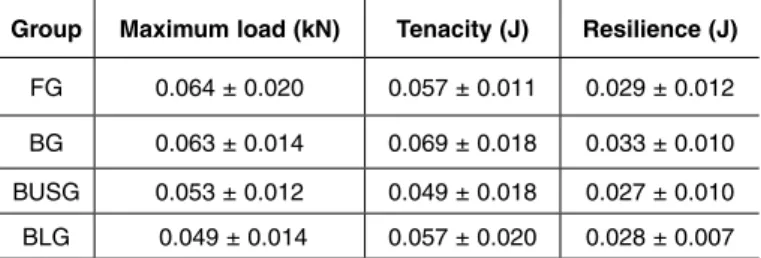
Table 1 presents the means and standard deviations of the mechanical properties of all the experimental groups. The statistical analysis did not demonstrate any significant differences between any of the groups after the experimental period of 14 days.
Table 1 – Biomechanical properties of the tibias of the experi-mental groups (mean ± SD).
Group Maximum load (kN) Tenacity (J) Resilience (J)
FG 0.064 ± 0.020 0.057 ± 0.011 0.029 ± 0.012
BG 0.063 ± 0.014 0.069 ± 0.018 0.033 ± 0.010 BUSG 0.053 ± 0.012 0.049 ± 0.018 0.027 ± 0.010
BLG 0.049 ± 0.014 0.057 ± 0.020 0.028 ± 0.007
105
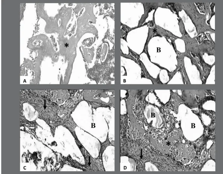
Figure 1 – Photomicrograph of the morphological findings from the different experimental groups. In the region of the defect, granu-lation tissue (arrows) and neoformed tissue (*) can be observed, and in the respective groups, the presence of the biomaterial (B). (A) FG: fracture control; (B) BG: Biosilicate®; (C) BUSG: Biosilicate® + ultrasound; (D) BLG: Biosilicate® + laser. Staining: H.E. ASSOCIATED WITH BIOSILICATE® ON THE PROCESS OF BONE REPAIR IN THE RAT TIBIA
Qualitative histological analysis
It can be seen from Figure 1A that the control group had moderate presence of neoformed bone tis-sue inside the defects and moderate presence of in-flammatory infiltrate. The animals treated only with Biosilicate® presented histopathological findings similar to those of the animals in the control group, with a moderate quantity of neoformed bone tissue (Figure 1B). Nonetheless, the osteogenic potential of the biomaterial was seen, given that there was great presence of cells and bone tissue around the particles. Interestingly, the groups with Biosilicate® in associa-tion with therapeutic laser and with US demonstrated some presence of cellular infiltrate, but with smaller amounts of neoformed bone tissue deposition than
in the control group and the group with Biosilicate® alone (Figures 1C and 1D).
morphometric analysis
It can be seen in Figure 2 that the groups treated with the associations of Biosilicate® + laser and Bio-silicate® + US presented significantly smaller quanti-ties of neoformed bone tissue area than shown by the untreated control group and the group treated only with Biosilicate®. However, no difference was found between these groups.
DISCUSSION
Figure 2 - Means and standard deviations of the area neoformed bone of the different experimental groups. a p < 0.05 vs FG; * p < 0.05 vs BG. FG: fracture control; BG: Biosilicate®; BLG: Bio-silicate® + laser; BUSG: BioBio-silicate® + ultrasound.
showed the osteogenic potential of Biosilicate®,
given that it was possible to see the deposition of neoformed bone tissue around the biomaterial par-ticles. However, the quantity of bone tissue was no greater than in the control group, possibly because of the short experimental period used in this study. Moreover, no modifications to the biomechanical properties were observed.
In the present study, we used a highly bioactive glass ceramic material called Biosilicate®. It has been reported in the literature that when a bioactive mate-rial comes into contact with body fluids, a layer of hy-droxycarbonate apatite is formed on the surface, thus promoting osteogenesis(17). Previous studies (both in
vivo and in vitro) have demonstrated that Biosilicate® has high osteogenic potential. Moura et al(5) showed
that Biosilicate® can induce bone tissue formation in a culturing system for osteoblastic cells. In an in vivo study, Granito et al(7) observed that there was
greater bone formation in defects filled with Bio-silicate® than in those filled with bioglass 45S5®,
which is currently considered to be the gold standard for bioactive materials.
Following the positive results from Biosilicate® in the bone repair process, a hypothesis that the effect from this material could be boosted through laser ir-radiation and/or ultrasound energy was raised. How-ever, the results from the present study demonstrated that the animals whose bone defects were filled with Biosilicate® also received LLLT and US therapy pre-sented a significant decrease in the quantity of neo-formed bone tissue, compared with the control group.
The mechanism through which this occurs is un-clear. It is known that Biosilicate® is a highly bioac-tive glass ceramic(5) and that both LLLT and US have
high osteogenic potential(11,18). One hypothesis may be
that associating these types of treatment might have caused excessive stimulation to the lesion site. In ad-dition, parameters like power, wavelength and flu-ence may have influflu-enced this process and may have inhibited cell migration and growth on the surface of the glass ceramic.
Similar results were found by Rennó et al(19), who
conducted an in vitro evaluation on the effects of laser therapy at 830 nm and 10 J/cm2 on the proliferation
of osteoblastic cells on Biosilicate® scaffolds. It was demonstrated that MC3T3 cells grew successfully on scaffolds composed of Biosilicato®, with osteoblast cells presenting normal morphology and easily adhe-ring and proliferating by means of the disc surfaces. On the other hand, laser irradiation produced a 13% decrease in osteoblast proliferation (MC3T3) in the Biosilicate® discs.
Some resent studies have reported that there is an increase in osseointegration between bone tissue and implants subsequent to laser and US. In a study asses-sing the effects of LLLT on hydroxyapatite implants, satisfactory results were found. LLLT using GaAsAl at 780 nm was found to promote a greater degree of osseointegration at the bone-hydroxyapatite interface, thus suggesting that LLLT may be considered to be a good tool for boosting the bone-implant interface in orthopedic surgery(20). AboElsaad et al(21) evaluated
the effects of LLLT using 830 nm and bioactive glass implants in periodontal defects. The results showed that LLLT in association with biomaterial had a posi-tive effect with regard to accelerating the periodontal repair. In a study evaluating the action of US on bone growth in hydroxyapatite implants, Iwai et al(22) found
positive results from US associated with the bioma-terial. US was capable of increasing the number of osteoblasts and the quantity of bone in hydroxyapatite implants.
CONCLUSION
Based on the results from this study, we can con-clude that Biosilicate® was capable of accelerating and boosting bone recovery, through modulating the inflammatory process and stimulating new bone tissue formation. However, when LLLT or US were used in association, no positive results were found.
FG BG BLG BUSG
107 ASSOCIATED WITH BIOSILICATE® ON THE PROCESS OF BONE REPAIR IN THE RAT TIBIA
REFERENCES
1. Sena K, Leven RM, Mazhar K, Sumner DR, Virdi AS. Early gene response to low-intensity pulsed ultrasound in rat osteoblastic cells. Ultrasound Med Biol. 2005;31(5):703-8.
2. Hadjiargyrou M, McLeod K, Ryaby JP, Rubin C. Enhancement of fracture healing by low intensity ultrasound. Clin Orthop Relat Res. 1998;(355 Suppl):S216-29. 3. Brighton CT, McCluskey WP. Response of cultured bone cells to a capacitively
coupled electric field: inhibition of cAMP response to parathyroid hormone. J Orthop Res. 1988;6(4):567-71.
4. Hench LL, Polak JM. Third-generation biomedical materials. Science. 2002;295(5557):1014-7.
5. Moura J, Teixeira LN, Ravagnani C, Peitl O, Zanotto ED, Beloti MM, et al. In vitro osteogenesis on a highly bioactive glass-ceramic (Biosilicate). J Biomed Mater Res A. 2007;82(3):545-57.
6. FUNDAÇÃO UNIVERSIDADE FEDERAL DE SÃO CARLOS; UNIVERSIDADE DE SÃO PAULO. Process and compositions for preparing particulate, bioac-tive or resorbable biosilicates for use in the treatment of oral ailments. Int. C. C03C10/00, 20 Feb. 2004, WO2004/074199.
7. Granito RN, Ribeiro DA, Rennó AC, Ravagnani C, Bossini PS, Peitl-Filho O, et al. Effects of biosilicate and bioglass 45S5 on tibial bone consolidation on rats: a biomechanical and a histological study. J Mater Sci Mater Med. 2009;20(12):2521-6.
8. Renno AC, McDonnell PA, Parizotto NA, Laakso EL. The effects of laser ir-radiation on osteoblast and osteosarcoma cell proliferation and differentiation in vitro. Photomed Laser Surg. 2007;25(4):275-80.
9. Stein A, Benayahu D, Maltz L, Oron U. Low-level laser irradiation promotes proliferation and differentiation of human osteoblasts in vitro. Photomed Laser Surg. 2005;23(2):161-6.
10. Liu X, Lyon R, Meier HT, Thometz J, Haworth ST. Effect of lower-level laser therapy on rabbit tibial fracture. Photomed Laser Surg. 2007;25(6):487-94. 11. Pinheiro AL, Oliveira MG, Martins PP, Ramalho LM, Oliveira MA, Novaes Júnior
A, et al. Biomodulatory effectos of LLLT on bone regeneration. Laser therapy. 2001; 13(1):73-79.
12. Takikawa S, Matsui N, Kokubu T, Tsunoda M, Fujioka H, Mizuno K, Azuma Y. Low-intensity pulsed ultrasound initiates bone healing in rat nonunion fracture model. J Ultrasound Med. 2001;20(3):197-205.
13. Li JK, Chang WH, Lin JC, Ruaan RC, Liu HC, Sun JS. Cytokine release from osteoblasts in response to ultrasound stimulation. Biomaterials. 2003;24(13):2379-85.
14. Sun JS, Tsuang YH, Lin FH, Liu HC, Tsai CZ, Chang WH. Bone defect healing enhanced by ultrasound stimulation: an in vitro tissue culture model. J Biomed Mater Res. 1999;46(2):253-61.
15. Lirani-Galvão AP, Jorgetti V, da Silva OL. Comparative study of how low-level laser therapy and low-intensity pulsed ultrasound affect bone repair in rats. Photomed Laser Surg. 2006;24(6):735-40.
16. Ratner BD. Biomaterials science: an introduction to materials in medicine. 2nd. ed. Amsterdam: Elsevier Academic Press; 2004.
17. Hench LL. Bioceramics: from concept to clinic. J Am Ceram Soc. 1991;74(7):1487-510.
18. Luger EJ, Rochkind S, Wollman Y, Kogan G, Dekel S. Effect of low-power laser irradiation on the mechanical properties of bone fracture healing in rats. Lasers Surg Med. 1998;22(2):97-102.
19. Renno AC, McDonnell PA, Crovace MC, Zanotto ED, Laakso L. Effect of 830 nm laser phototherapy on osteoblasts grown in vitro on Biosilicate scaffolds. Photomed Laser Surg. 2010;28(1):131-3.
20. Guzzardella GA, Torricelli P, Nicoli Aldini N, Giardino R. Laser technology in orthopedics: preliminary study on low power laser therapy to improve the bone-biomaterial interface. Int J Artif Organs. 2001;24(12):898-902.
21. AboElsaad NS, Soory M, Gadalla LM, Ragab LI, Dunne S, Zalata KR, et al. Effect of soft laser and bioactive glass on bone regeneration in the treatment of infra-bony defects (a clinical study). Lasers Med Sci. 2009;24(3):387-95. 22. Iwai T, Harada Y, Imura K, Iwabuchi S, Murai J, Hiramatsu K, et al. Low-intensity