Supplementation with the histone deacetylase inhibitor trichostatin
A during
in vitro
culture of bovine embryos
Clara Slade Oliveira
1, Naiara Zoccal Saraiva
2, Marcela Maria de Souza
2, Tatiane de Almeida
Drummond Tetzner
2, Marina Ragagnin de Lima
2and Joaquim Mansano Garcia
2Department of Preventive Veterinary Medicine and Animal Reproduction, Faculty of Agricultural and Veterinary Sciences, Sao Paulo State University, Access Road Professor Paulo Donato Castellane, Jaboticabal, Brazil
Date submitted: 15.07.2010. Date accepted: 13.05.2011
Summary
Trichostatin A (TSA) is a histone deacetylase inhibitor that induces histone hyperacetylation and increases gene expression levels. The aim of the present study was to establish a suitable condition
for the use of TSA inin vitrocultures of bovine embryos, and to determine whether TSA would increase
blastocyst rates by improvement of chromatin remodelling during embryonic genome activation and by increasing the expression of crucial genes during early development. To test this hypothesis, 8-cell embryos were exposed to four concentrations of TSA for different periods of time to establish adequate protocols. In a second experiment, three experimental groups were selected for the evaluation of embryo quality based on the following parameters: apoptosis, total cell number and blastocyst hatching. TSA promoted embryonic arrest and degeneration at concentrations of 15, 25 and 50 nM. All treated groups presented lower blastocyst rates. Exposure of embryos to 5 nM for 144 h and to 15 nM for 48 h decreased blastocyst hatching. However, the terminal deoxynucleotidyl transferase-mediated dUTP nick-end labeling assay (TUNEL) assay revealed similar apoptosis rates and total cell numbers in all groups studied. Although, in the present study, TSA treatment did not improve the parameters studied,
the results provided background information on TSA supplementation duringin vitroculture of bovine
embryos and showed that embryo quality was apparently not affected, despite a decrease in blastocyst rate after exposure to TSA.
Keywords: Cattle, Embryonic development, Histone deacetylase inhibition, Histone hyperacetylation, Trichostatin A
Introduction
Although the in vitro production of bovine embryos
has been established since the 1980s (Brackett et al.,
1982), the percentage of embryos that reach blastocyst
stage is still much lower than that obtained in
vivo (Lequarre et al., 2003). The high rates of
1All correspondence to: Clara Slade Oliveira. UNESP– Via de Acesso Professor Paulo Donato Castellane, CEP 14884–900, Jaboticabal, São Paulo, Brazil. Tel: +55 16 32092633/+55 16 97242262. Fax: +55 21 25258804. E-mail: [email protected]
2Department of Preventive Veterinary Medicine and Animal Reproduction, Faculty of Agricultural and Veterinary Sciences, Sao Paulo State University, Access Road Professor Paulo Donato Castellane, zip code 14884–900, Jaboticabal, Brazil.
embryo mortality might be due to developmental block, a phenomenon described in many species that occurs concomitantly with major genome activation
(Meirelles et al., 2004). Embryonic arrest during
major genome activation occurs in cattle during the transition from the 4th to the 5th cell cycle due to the inability to modify the repressive chromatin structure and to activate the transcription of genes that are important for development (Betts & King, 2001).
Chromatin remodelling is regulated in part
by epigenetic modifications, which include post-translational histone modification. The acetylation of histones is related to an increase in gene expression by permitting the access of transcription factors to DNA
(Schubeleret al., 2004). After fertilization, the embryo
genome is methylated and genes are expressed at
reaches its peak at the time of embryonic genome activation, which coincides with an overall increase of
gene expression levels (Maaloufet al., 2008). Histone
acetylation is regulated by two main enzymes: histone acetyltransferase, which adds acetyl groups to the histone tail, neutralizing its positive charges and weakening its binding to nucleosomes; and histone deacetylase (HDAC), which removes acetyl groups and causes chromatin compaction and silencing of the DNA segment at that site (Johnstone, 2002). Experiments that use RNAi-mediated reduction of HDAC confirmed the role of this enzyme in histone acetylation and gene expression in blastomeres (Ma & Schultz, 2008).
Studying bovine embryos produced in vitro, Ikeda
et al. (2009) found no effect of the HDAC inhibitor
trichostatin A (TSA) administered during in vitro
fertilization (IVF) on blastocyst rate, although the drug increased the ratio of inner cell mass cells. However, TSA has been applied successfully to the production of bovine clones in order to increase the efficiency of epigenetic recombination of adult cells in nucleus
donor cells (Enrightet al., 2003; Weeet al., 2007; Ding
et al., 2008), and in embryos immediately after embryo
fusion (Dinget al., 2008; Iager et al., 2008). Thus, TSA
affects chromatin remodelling by increasing histone acetylation.
The objectives of the present study were to determine an adequate concentration and exposure time of TSA during embryonic genome activation and
to evaluate the effects of TSA on thein vitroproduction
of bovine embryos. We tested the hypothesis that TSA would improve blastocyst rates by overcoming chromatin repression.
Material and methods
Supplements
Reagents and culture media were purchased from Sigma Chemical Co. unless otherwise stated.
Preparation and selection of oocytes
Bovine ovaries were collected at a local slaughterhouse and processed within 2 h after slaughter. The ovaries
were washed in saline (37◦C) and follicles that
measured 3 to 8 mm in diameter were aspirated with an 18-gauge needle coupled to a 20-ml syringe. Cumulus–oocyte complexes (COCs) presenting at least three layers of cumulus cells and homogenous cytoplasm were selected under a stereomicroscope. The COCs were washed in HEPES-buffered TCM-199 (Gibco BRL) supplemented with 10% fetal bovine
serum (FBS; Cripion, Andradina, Brazil), 16 g/ml
sodium pyruvate, and 83.4g/ml amikacin (Instituto
Biochimico).
In vitromaturation (IVM)
Groups of 15 COCs were transferred to 100-l droplets
of medium that contained sodium bicarbonate-buffered TCM-199 supplemented with 10% FBS,
1.0 g/ml follicle-stimulating hormone (FSH)
(FollitropinTM, Bioniche Animal Health), 50 g/ml
human chorionic gonadotrophin (hCG; ProfasiTM),
1.0g/ml estradiol, 16g/ml sodium pyruvate, and
83.4 g/ml amikacin. The COCs were covered with
sterile mineral oil (Dow Corning Co.) and incubated
for 24 h at 38.5◦C in an atmosphere of 5% CO
2 in air
under saturated humidity.
In vitrofertilization
After IVM, groups of 25 oocytes were washed twice
and transferred to 30-l droplets of TALP-IVF medium
supplemented with 0.6% bovine serum albumin (BSA),
10 g/ml heparin, 18 M penicillamine, 10 M
hypotaurine and 1.8 M epinephrine, and covered
with sterile mineral oil. Frozen straws of semen from Nellore bulls were centrifuged on a discontinuous
45/90 Percoll gradient for 5 min at 3600 g. The
pellet was resuspended in 2 ml TALP stock medium
and again centrifuged for 5 min at 3600 g. After
centrifugation, 80 l of the medium containing the
pellet were collected from the bottom of the tube and homogenized in a conical tube. The final suspension was adjusted to a final concentration of approximately
104 live spermatozoa per oocyte. The plates were
incubated at 38.5◦C for 20 h in an atmosphere of 5%
CO2in air under saturated humidity.
In vitroculture (IVC)
After IVF, presumptive zygotes were denuded of cumulus cells by vigorous pipetting and cultured in synthetic oviductal fluid (SOF) medium supplemented
with 2.5% FBS and 5 mg/ml BSA at 38.5◦C in
an atmosphere of 5% CO2 in air under saturated
humidity. Groups of 15 to 20 presumptive zygotes
were cultured in 100-l droplets until the time of
treatment with TSA. The cleavage rate, blastocyst development, and blastocyst hatching were evaluated 48 h, 7 days and 9 days after IVF, respectively.
Treatment with trichostatin A
The embryos were washed and transferred to 100-l
medium. One half of the medium was changed every 48–72 h.
Assessment of embryo quality by an apoptosis assay (TUNEL) and determination of total blastocyst cell number
Blastocysts were evaluated by the terminal deoxynuc-leotidyl transferase-mediated dUTP nick-end labelling
(TUNEL) assay using theIn SituCell Death Detection
Kit (Fluorescein, Roche Applied Science) as described by Paula-Lopes & Hansen (2002). Blastocysts were in-cubated with 0.5% Triton X-100 in phosphate-buffered saline (PBS) for 30 min at room temperature. Next, the embryos were transferred to the final solution
containing 10 l of the enzyme solution (terminal
deoxynucleotidyl transferase) and 90l of the labelling
solution (fluorescein-dUTP) and incubated for 1 h at
38.5◦C, followed by incubation with 50
g/ml RNase
A for 1 h at room temperature. The nuclei were
stained with 10l/ml Hoechst 33342 for 10 min and
the blastocysts were washed three times in PBS. The positive control was incubated with 50 IU/ml DNase for 1 h before incubation with the final enzyme/dUTP solution. The negative control was incubated without the enzyme. The number of apoptotic cells and total number of nuclei was determined under a fluorescence microscope (Olympus IX-70) at wavelengths of 330– 385 nm (Hoechst 33342) and 420–490 nm (fluorescein).
Statistical analysis
Differences in blastocyst, apoptosis and hatching rates between groups were analysed by the
chi-squared (2) test using the MINITAB software, Release
14.1 (Minitab Inc., State College, PA). The total cell number was analysed by one-way analysis of variance (ANOVA) and means were compared by the Tukey test using GraphPad Prism 4.0 software (GraphPad Prism, Inc.).
Results
Effect of TSA concentration and time of exposure on blastocyst development
Three replicates corresponding to 1136 oocytes (n =
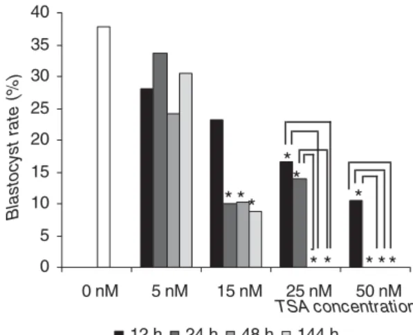
58–110 per experimental group) were analysed. First, the adequate concentration and exposure time of TSA during IVC of bovine embryos were determined (Fig. 1).
No difference was observed in the blastocyst rate between 8-cell embryos exposed to 5 nM TSA for 12,
24, 48 or 144 h and the control group (p > 0.05). For
embryos treated with 15 nM TSA, exposure for 12 h
did not affect blastocyst rate (p > 0.05). However,
0 5 10 15 20 25 30 35 40
0 nM 5 nM 15 nM 25 nM 50 nM
12 h 24 h 48 h 144 h
Blastocyst rate (%) *
TSA concentration
* * *
*
* * *
* **
Figure 1 Effects of different concentrations and times of exposure to trichostatin A (TSA) on the development of bovine embryos.In vitroculture medium was supplemented 70 h after in vitro fertilization (IVF) with 5, 15, 25 or 50 nM TSA for 12, 24, 48 or 144 h. Asterisk (∗) indicates significant differences compared with the control group and connectors indicate significant differences within the same concentration (p<0.05, chi-squared test).
blastocyst development was lower than in the control group after 24, 48 and 144 h of treatment with 15 nM
TSA (p < 0.05). Treatment with 25 nM TSA for 12
or 24 h reduced the blastocyst rate compared with
the control group (p <0.05). Embryos did not reach
the blastocyst stage when exposed to 25 nM TSA
for longer periods of time (48 or 144 h) (p < 0.05).
The administration of 50 nM TSA resulted in lower blastocyst development compared with the control
group when the embryos were exposed for 12 h (p<
0.05). Embryos did not reach the blastocyst stage when 50 nM TSA was administered for longer periods of
time (24, 48 and 144 h) (p<0.05).
In a second experiment, we carefully investigated the effects of adequate TSA treatment on embryonic development. For this purpose, 1732 cleaved embryos
obtained in nine replicates (n = 371–555 cleaved
embryos per experimental group) were exposed to 5 nM for 48 h, 15 nM for 48 h, and 5 nM for 144 h. All of the TSA concentrations tested promoted a decrease in blastocyst rate (Table 1).
Effects of TSA on blastocyst hatching
Five replicates corresponding to 418 blastocysts (n =
68–159 per experimental group) were analyzed. Treatment with 5 nM TSA for 48 h had no effect
on the percentage of blastocyst hatching (p < 0.05)
(Table 2). However, TSA promoted a decrease in blastocyst hatching at concentrations of 15 nM applied
Table 1Effects of supplementation ofin vitroculture medium with trichostatin A on the development of bovine embryos (Experiment 2)
Cleaved embryos Blastocysts
TSA n(%) n(%)
0 (control) 433 (85.91) 193 (44.57)a
5 nM 48 h 371 (81.89) 135 (36.38)b
15 nM 48 h 555 (83.33) 119 (21.44)c
5 nM 144 h 373 (82.15) 92 (24.66)c
Trichostatin A (TSA) was added to thein vitroculture medium 70 h post-fertilization. The percentage of blastocysts refers to the total number of cleaved embryos.
a,b,cMeans followed by the same letter did not differ
significantly (p<0.05, chi-square test). Data from nine
replicates.
n=number of structures.
Table 2Effects of supplementation ofin vitroculture medium with trichostatin A on blastocyst hatching of bovine embryos
Hatched Total Hatching
TSA blastocysts (n) blastocysts (n) rate (%)
Control 96 159 60.37a
5 nM 48 h 55 100 55.00a
15 nM 48 h 35 91 38.46b
5 nM 144 h 15 68 22.05c
Trichostatin A (TSA) was added to thein vitroculture medium 70 h post-fertilization.
a,b,cMeans followed by the same letter did not differ
significantly (p<0.05, chi-squared test). Data from five
replicates.
n=number of structures.
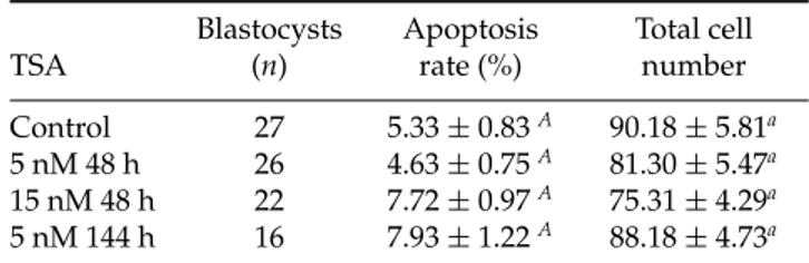
Effects of TSA on total cell number and apoptosis rates of bovine blastocysts
In this experiment, four replicates corresponding to
91 blastocysts (n = 16–27 per experimental group)
were analysed. No difference was observed in apoptosis rates or total cell number between the four
experimental groups (p > 0.05) (Table 3). Therefore,
harmful effects on cell cycle or apoptosis were not observed in the experimental groups studied.
Discussion
The present study provides data regarding the effect of TSA supplementation on bovine preimplantation development. The results showed that even at low concentrations TSA decreased the blastocyst rate, although no harmful effects on embryo quality were observed.
The first cycle of DNA replication after embryonic genome activation is critical for the maximum
Table 3Effects of supplementation ofin vitroculture medium with trichostatin A on apoptosis rate and total cell number of bovine blastocysts
Blastocysts Apoptosis Total cell
TSA (n) rate (%) number
Control 27 5.33±0.83A 90.18±5.81a
5 nM 48 h 26 4.63±0.75A 81.30±5.47a
15 nM 48 h 22 7.72±0.97A 75.31±4.29a
5 nM 144 h 16 7.93±1.22A 88.18±4.73a
Trichostatin A (TSA) was added to thein vitroculture medium 70 h post-fertilization. Apoptosis rate and total cell number are reported as the mean±standard error.
A,aMeans followed by the same letter did not differ
significantly (p<0.05, one-way analysis of variance
(ANOVA) and Tukey post test). Data from four replicates. n=number of structures.
expression of endogenous genes (Aoki et al., 1997).
Thus, the time of TSA supplementation chosen in the present experiment was 70 h post-IVF, when most embryos were in the 8-cell stage. At this time, HDAC inhibition and the consequent increase in gene expression may contribute to overcome chromatin repression.
The results of the first experiment showed that higher concentrations of TSA can be applied for 12 h. However, our aim was to use the drug for longer periods of time when most embryos are in the fourth and fifth cell cycle and to include the remaining cycles of preimplantation development. For this purpose, administration of 5 nM TSA for 48 h and 144 h and 15 nM for 48 h was chosen for Experiment 2.
In a previous study, we demonstrated that male and female embryos respond in a different manner to TSA
treatment (Oliveira et al., 2010). Therefore, TSA was
administered to in vitro produced embryos fertilized
with non-sexed sperm, which produces both male and female embryos in the same drop. The objective was to determine a concentration that would not negatively affect blastocyst development, blastocyst hatching, total cell number or apoptosis.
In conventional systems, IVC increases the propor-tion of male embryos, which develop faster (Avery
et al., 1992) and reach the blastocyst stage more
frequently (Xu et al., 1992). It is possible that the
negative effect on blastocyst rate observed was due to an altered sex proportion of embryos that developed in the presence of TSA, as male embryos seem to be more
sensitive to the negative effects of TSA (Oliveiraet al.,
2010).
by trapoxin promotes histone hyperacetylation and increases gene transcription in mouse embryos (Aoki
et al., 1997). In addition, other HDAC inhibitors have
been shown to act as reprogramming agents in cloned
bovine embryos (Enrightet al., 2003; Weeet al., 2007;
Dinget al., 2008; Iageret al., 2008).
Unfortunately, HDAC inhibition does not target specific mechanisms that would result in the increased expression of specific genes. Thus, both beneficial and harmful genes are stimulated and their in-creased expression activates different pathways that improve or worsen embryonic development. The ideal concentration of TSA in IVC should increase gene expression in such a way that the balance between harmful and beneficial genes is shifted in favour of the latter. This shift was not achieved with the TSA concentrations tested in the present study (5–50 nM). Higher concentrations blocked blastocyst development completely when applied for 24 h (50 nM) or 48 h (25 nM).
In conclusion, the most suitable concentration of TSA and period of administration are 5 nM and 48 h, although there was no beneficial effect on the parameters studied. Further studies are needed to investigate embryo implantation and birth, as well as the gene expression profile in TSA treated embryos, as they present histone hyperacetylation and are as viable as control embryos.
Acknowledgements
The authors thank Roberta Vantini for technical assistance in the IVF laboratory. This study was supported by the State of Sao Paulo Research Foundation (FAPESP), Brazil.
References
Aoki, F., Worrad, D.M. & Schultz, R.M. (1997). Regulation of transcriptional activity during the first and second cell cycles in the preimplantation mouse embryo.Dev. Biol.181, 296–307.
Avery, B., Jorgensen, C.B., Madison, V. & Greve, T. (1992). Morphological development and sex of bovine in vitro-fertilized embryos.Mol. Reprod. Dev.32, 265–70.
Betts, D.H. & King, W.A. (2001). Genetic regulation of embryo death and senescence.Theriogenology55, 171–91. Brackett, B.G., Bousquet, D., Boice, M.L., Donawick, W.J.,
Evans, J.F. & Dressel, M.A. (1982). Normal development followingin vitrofertilization in the cow.Biol. Reprod.27, 147–58.
Dean, W., Santos, F., Stojkovic, M., Zakhartchenko, V., Walter, J., Wolf, E. & Reik, W. (2001). Conservation of methylation reprogramming in mammalian development: aberrant reprogramming in cloned embryos.Proc. Natl. Acad. Sci. USA98, 13734–38.
Ding, X., Wang, Y., Zhang, D., Wang, Y., Guo, Z. & Zhang, Y. (2008). Increased pre-implantation development of cloned bovine embryos treated with 5-aza-2’-deoxycytidine and trichostatin A.Theriogenology70, 622–30.
Enright, B.P., Kubota, C., Yang, X. & Tian, X.C. (2003). Epigenetic characteristics and development of embryos cloned from donor cells treated by trichostatin A or 5-aza-2′-deoxycytidine.Biol .Reprod.69, 896–901.
Iager, A.E., Ragina, N.P., Ross, P.J., Beyhan, Z., Cunniff, K., Rodriguez, R.M. & Cibelli, J.B. (2008). Trichostatin A improves histone acetylation in bovine somatic cell nuclear transfer early embryos.Cloning Stem Cells10, 371– 79.
Ikeda, S., Tatemizo, A., Iwamoto, D., Taniguchi, S. & Hoshino, Y. (2009). Enhancement of histone acetylation by trichostatin A duringin vitrofertilization of bovine oocytes affects cell number of the inner cell mass of the resulting blastocysts.Zygote17, 209–15.
Johnstone, R.W. (2002). Histone-deacetylase inhibitors: novel drugs for the treatment of cancer.Nat. Rev. Drug. Discov.1, 287–99.
Lequarre, A.S., Marchandise, J., Moreau, B., Massip, A. & Donnay, I. (2003). Cell cycle duration at the time of maternal zygotic transition for in vitro produced bovine embryos: effect of oxygen tension and transcription inhibition.Biol. Reprod.69, 1707–13.
Maalouf, W.E., Alberio, R. & Campbell, K.H.S. (2008). Differ-ential acetylation of histone H4 lysine during development of in vitro fertilized, cloned and parthenogenetically activated bovine embryos.Epigenetics3, 199–209.
Ma, P. & Schultz, R.M. (2008). Histone deacetylase 1 (HDAC1) regulates histone acetylation, development, and gene expression in preimplantation mouse embryos.Dev. Biol.319, 110–20.
Meirelles, F.V., Caetano, A.R., Watanabe, Y.F., Ripamonte, P., Carambula, S.F., Merighe, G.K. & Garcia, S.M. (2004). Genome activation and developmental block in bovine embryos.Ann. Reprod. Sci.82–83, 13–20.
Oliveira, C.S., Saraiva, N.Z., Souza, M.M., Tetzner, T.A.D., Lima, M.R. & Garcia, J.M. (2010). Effects of histone hyperacetylation on the preimplantation development of male and female bovine embryos.Reprod. Fertil. Dev.22, 1041–8.
Paula-Lopes, F.F. & Hansen, P.J. (2002). Apoptosis is an adaptive response in bovine preimplantation embryos that facilitates survival after heat shock.Biochem. Biophys. Res. Commun.295, 37–42.
Schubeler, D., Macalpine, D.M., Scalzo, D., Wirbelauer, C., Kooperberg, C., Van Leeuwen, F., Gottschling, D.E., O’Neil, L.P., Turner, B.M., Delrow, J., Bell, S.P. & Groudine, M. (2004). The histone modification pattern of genes revealed through genome-wide chromatin analysis of a higher eukaryote.Genes Dev.18, 1263–71.
Wee, G., Shim, J.J., Koo, D.B., Chae, J.I. & Lee, K.K. (2007). Epigenetic alteration of the donor cells does not recapitulate the reprogramming of DNA methylation in cloned embryos.Reproduction134, 781–87.