Streptococcus agalactiae
in the Pathogenesis of Infective
Endocarditis
Ho Seong Seo1, Yan Q. Xiong2,3, Paul M. Sullam1*
1Division of Infectious Diseases, Veterans Affairs Medical Center and the University of California San Francisco, San Francisco, California, United States of America,
2Department of Medicine, Los Angeles Biomedical Research Institute at Harbor-UCLA Medical Center, Torrance, California, United States of America,3Geffen School of Medicine at UCLA, Los Angeles, California, United States of America
Abstract
The binding of bacteria to fibrinogen and platelets are important events in the pathogenesis of infective endocarditis. Srr1 is a serine-rich repeat glycoprotein ofStreptococcus agalactiaethat binds directly to the Aachain of human fibrinogen. To assess the impact of Srr1 on the pathogenesis of endocarditis due toS. agalactiae, we first examined the binding of this organism to immobilized human platelets. Strains expressing Srr1 had significantly higher levels of binding to human plateletsin vitro, as compared with isogenicDsrr1mutants. In addition, platelet binding was inhibited by pretreatment with anti-fibrinogen IgG or purified Srr1 binding region. To assess the contribution of Srr1 to pathogenicity, we compared the relative virulence ofS. agalactiaeNCTC 10/84 strain and itsDsrr1mutant in a rat model of endocarditis, where animals were co-infected with the WT and the mutant strains at a 1:1 ratio. At 72 h post-infection, bacterial densities (CFU/g) of the WT strain within vegetations, kidneys, and spleens were significantly higher, as compared with theDsrr1mutant. These results indicate that Srr1 contributes to the pathogenesis of endocarditis due toS. agalactiae, at least in part through its role in fibrinogen-mediated platelet binding.
Citation:Seo HS, Xiong YQ, Sullam PM (2013) Role of the Serine-Rich Surface Glycoprotein Srr1 ofStreptococcus agalactiaein the Pathogenesis of Infective Endocarditis. PLoS ONE 8(5): e64204. doi:10.1371/journal.pone.0064204
Editor:Robert A. Burne, University of Florida, United States of America
ReceivedFebruary 26, 2013;AcceptedApril 12, 2013;PublishedMay 23, 2013
This is an open-access article, free of all copyright, and may be freely reproduced, distributed, transmitted, modified, built upon, or otherwise used by anyone for any lawful purpose. The work is made available under the Creative Commons CC0 public domain dedication.
Funding:This study was supported by the Department of Veterans Affairs and the VA Merit Review program, the Northern California Institute for Research and Education, NIH grants R01-AI41513 (P.M.S.), R01-AI057433 (P.M.S.), and a Fellowship Award from the American Heart Association, Western Affiliate (H.S.S). The funders had no role in study design, data collection and analysis, decision to publish, or preparation of the manuscript.
Competing Interests:The authors have declared that no competing interests exist.
* E-mail: paul.sullam@ucsf.edu
Introduction
Streptococcus agalactiae (Group B streptococcus [GBS]) is a frequent cause of neonatal meningitis and sepsis. In recent years, however, GBS infections in nonpregnant adults are being increasingly reported. Individuals at greater risk for this disease include the elderly, immunosuppressed patients, and diabetics [1– 3]. Although GBS is a relatively uncommon cause of endocarditis (accounting for 1–2% of culture-positive cases), endovascular infection due to this organism is associated with a high mortality rate (34–50%), especially in the setting of prosthetic valve infection [4–7]. Complications such as sepsis, valvular destruction, cardiac failure, and embolic phenomena are also frequent in this disease [8].
The pathogenesis of endocarditis is a complex process, involving multiple host-pathogen interactions. A central aspect of virulence in this disease is the ability of organisms to bind host components, such as fibrinogen, fibronectin, and platelets [9–13]. These binding events appear to be important both for the initial attachment of bacteria to the endovascular surface, and for the subsequent progression of infection. For several Gram-positive bacteria, binding to human platelets is mediated in part by an adhesin belonging to the serine-rich repeat (SRR) glycoprotein [14–16]. For example, strains of Streptococcus gordonii can bind platelets directly via the interaction of the SRR adhesins GspB or
Hsa with a receptor (GPIb) on the platelet membrane [15]. The binding ofStaphylococcus aureusto platelets is mediated in part by the SRR protein SraP, though the receptor for this adhesin remains unidentified [17]. In addition,S. aureuscan attach to platelets via fibrinogen and fibrin, which act as a molecular bridge between the bacteria and platelet surface [18–20].
Two SRR glycoproteins (Srr1 and Srr2) have been identified thus far inS. agalactiae[21]. Expression of Srr1 has been shown to contribute to colonization and virulence in models of GBS bacteremia and meningitis infection [22–25]. In addition, we have recently demonstrated that Srr1 binds to human fibrinogen via its interaction with the Aa chain of the protein, and that loss of fibrinogen binding is associated with decreased attachment to brain microvascular endothelial cellsin vitro, as well as attenuated virulence, in an experimental model of meningitis [26]. In view of the importance of fibrinogen binding for endovascular infection, we examined the impact of Srr1 on platelet bindingin vitro, and its role in the pathogenesis of infective endocarditis.
Materials and Methods
Reagents
Rabbit anti-fibrinogen IgG was acquired from Innovative Research.
Strains, plasmids, and growth conditions
The bacteria and plasmids used in this study are listed in Tables 1. S. agalactiae strains were grown in Todd-Hewitt broth (Difco) supplemented with 0.5% yeast extract (THY broth). All mutant strains grew at a comparable ratein vitroas compared with respective parental strains (data not shown).Escherichia colistrains DH5a, BL21 and BL21(DE3) were grown at 37uC under aeration
in Luria Bertani broth (LB; Difco). Antibiotics were added to the media as required. All isolates were stored at280uC until thawed just prior to use.
Cloning and expression of the Srr1 binding region (Srr1-BR)
Genomic DNA was isolated from GBS NCTC 10/84 using Wizard Genomic DNA purification kits (Promega), according to the manufacturer’s instructions. PCR products were cloned into either pET28FLAGor pET22(+) to express FLAG-tagged or
His6-tagged versions of Srr1-BR (amino acids [AA] 303–641 of the SRR1). Proteins were purified by either Ni-NTA (Promega) or anti-FLAG M2 agarose affinity chromatography (Sigma-Aldrich).
Binding ofS. agalactiaeto immobilized human platelets and rat fibrinogen
Overnight cultures ofS. agalactiae were harvested by centrifu-gation, washed in PBS, and adjusted to a concentration of 106CFU/ml. Purified rat fibrinogen or washed, fixed human platelets were immobilized in 96-well microtiter plates as described previously (46). The plates were then treated with casein-based blocking solution (Roche) at 37uC for 1 h and washed three times with PBS. Purified recombinant Srr1-BR (200mg/ml), anti-Srr1 IgG (100mg/ml), or anti-fibrinogen IgG (100mg/ml) were added
to the wells for 30 min, followed by washing and the addition of 100ml of the bacterial suspension. The plates were incubated at room temperature for 1 h, and the wells were washed three times
with PBS to remove nonadherent organisms. The wells were treated with 100ml of trypsin (2.5 mg/ml) for 10 min at 37uC to
release the attached bacteria, and the number of bound bacteria was determined by plating serial dilutions of the recovered bacteria onto blood agar plates as previously described [27].
Binding of recombinant Srr1-BR to human platelets
Fixed human platelets were immobilized in 96 well cell culture plates as described previously [28]. After treatment with a casein-based blocking reagent, the wells were incubated withFLAG
Srr1-BR (0–4mM) in PBS for 1 h at room temperature, followed by
washing. Bound protein was detected by ELISA with anti-FLAG monoclonal antibody. For some studies, the wells were preincu-bated with His6-tagged Srr1-BR (0–50mM) or anti-fibrinogen IgG
(0–100mg/ml) for 0.5 h at RT followed by washing, prior to
addingFLAGSrr1-BR (1mM).
Rat model of infective endocarditis
The relative virulence of S. agalactiae NCTC 10/84 parental strain and its isogenic variant (NCTC 10/84Dsrr1) was assessed in
a competition model of IE in rats, as described previously [29,30]. In brief, Sprague-Dawley female rats (250 to 300 g, Harlan Laboratory, Inc.) were first anesthetized with ketamine (35 mg/kg) and xylazine (10 mg/kg). A sterile polyethylene catheter was surgically placed across the aortic valve of each animal, such that the tip was positioned in the left ventricle, to induce the formation of sterile valvular vegetations (nonbacterial thrombotic endocar-ditis) [27,29]. The catheters were left in place throughout the study. Three days post-catheterization, the animals were infected intravenously with an inoculum of approximately 56105CFU containing a 1:1 mixture ofS. agalactiaeNCTC 10/84 and itsDsrr1
isogenic variant. At 72 hr post-infection, the rats were euthanized with thiopental (100 mg IP). Animals were included in the final analysis only if the catheters were correctly positioned across the aortic valve at the time of sacrifice, and if macroscopic vegetations were visible. At sacrifice, all cardiac vegetations, as well as kidneys and spleens, were harvested, weighed, homogenized in saline,
Table 1.Strains and plasmids.
Strains Genotype or descriptiona Source
Escherichia coli
DH5a F2r2m+
Ø80dlacZDM15 Gibco BRL
BL21 (DE3) expression host, inducible T7 RNA polymerase Novagen
Streptococcus agalactiae
COH31 serotype III, clinical isolate [51]
PS954 COH31Dsrr1, CmR this study
NCTC 10/84 serotype V, clinical isolate [52]
PS2645 NCTC 10/84Dsrr1, CmR [23]
Plasmids Descriptiona Source
pDE123 streptococcal shuttle vector, ErmR [26]
pDE123-srr1 vector for expression of Srr1, ErmR [26]
pET22b(+) expression vector, AmpR Novagen
pET28FLAG expression vector with FLAG-tag, KanR [27]
pET22-Srr1-BR vector for expression of Srr1-BR, AmpR [26]
pET28FLAGSrr1-BR vector for expression of FLAG-tagged Srr1303–641 this study
aCmR, chloramphenicol resistance; ErmR, erythromycin resistance; AmpR, ampicillin resistance; KanR, kanamycin resistance.
serially diluted, and plated onto 8% sleep blood Todd Hewitt agar (with or without 2.5mg/ml of chloramphenicol) for quantitative culture. The plates were incubated for 48 h at 37uC, and bacterial densities were expressed as the log10CFU per gram of tissue.
Differences in means +/2 SD were compared for statistical significance by the paired t-test. The data were also analyzed by calculating a ‘‘competition index,’’ which was defined as the ratio of the paired strains within tissues for each animal, normalized by the ratio of organisms in the inoculum [27,29]. The mean of the log10 normalized ratios was tested against the hypothesized ‘no
effect’ mean value of 0, using a paired t-test, withP#0.05 as the threshold for statistical significance.
Animals were maintained in accordance with the American Association for Accreditation of Laboratory Animal Care (AAALAC) criteria. All animal studies were approved by the Animal Research Committee (IACUC) of the Los Angeles Biomedical Research Institute at Harbor-UCLA Medical Center.
Statistical methods
Data expressed as means6standard deviations were compared for statistical significance by the paired or unpaired t test, as indicated.
Results
Binding of GBS to human platelets is mediated by fibrinogen
To assess whether GBS binding to human platelets is mediated by Srr1, we compared two GBS strains (COH31 and NCTC 10/ 84) and their respective srr1 deletion variants for adherence to these cells in vitro (Fig. 1A). Both strains bound platelets significantly above background levels, with 28.062.6% and 12.062.6% (mean 6 SD) of the inoculum bound for COH31 and NCTC 10/84, respectively. Levels of binding by both srr1
mutant strains were significantly lower than those of the parent strains, with a 79.263.4% reduction in platelet binding for COH31Dsrr1and a 71.464.2% reduction for NCTC 10/84Dsrr1.
Figure 1. GBS binding to immobilized human platelets is mediated by glycoprotein Srr1.(A) Platelet binding by GBS strains COH31 and NCTC 10/84, theirDsrr1isogenic variants, and the mutant strains complementedin transwithsrr1(pSrr1). (B) GBS binding to human platelets was inhibited by pretreating the monolayers with 100mg/ml of anti-fibrinogen IgG (Anti-Fg). Normal IgG (IgG) served as a control. (C) Inhibition of binding by recombinant Srr1 binding region (Srr1-BR). Levels of binding were calculated as relative to the WT strains (mean6SD). Values shown represent the means (6S.D.) of triplicate measurements. * = P,0.01.
doi:10.1371/journal.pone.0064204.g001
Figure 2. Recombinant Srr1-BR interacts with human platelets.(A) Binding ofFLAGSrr1-BR protein to immobilized platelets. (B) Inhibition of FLAGSrr1-BR binding to platelets by His6 tagged Srr1-BR. Platelets were pretreated with the indicated concentrations of His6 tagged Srr1-BR. (C)
Binding ofFLAGSrr1-BR to immobilized platelets pretreated with anti-fibrinogen IgG or preimmune rabbit IgG. Values represent relative binding of FLAGSrr1-BR binding as compared with untreated platelets. Bars indicate the means (6S.D.). * = P,0.01.
Complementation of thesrr1mutationin transrestored binding by both mutant strains, thereby demonstrating that the loss of binding observed withsrr1disruption was not due to polar or pleiotropic effects. In addition, GBS binding to human platelets was inhibited by rabbit anti-fibrinogen IgG, but not by normal rabbit IgG (Fig. 1B), with WT GBS binding levels reduced to those seen with
the srr1 deletion strains. We then examined the impact of preincubating the platelet monolayers with the recombinant binding domain of Srr1 (Srr1-BR). As shown in Fig. 1C, pretreating the immobilized platelets with recombinant Srr1-BR inhibited subsequent binding by both GBS strains. Since previous studies have shown that human platelets express membrane-associated fibrinogen [11,31,32], our results indicate that GBS binding to human platelets is mediated by the interaction of Srr1 with fibrinogen on the surface of these cells.
Binding of Srr1-BR to immobilized platelets
To further assess the role of Srr1, we evaluated the binding of FLAG-tagged Srr1-BR (FLAGSrr1-BR) to immobilized human
platelets. We found thatFLAGSrr1-BR interacted with platelets in a
concentration-dependent manner, when tested over a range of 0– 4mM (Fig. 2A). In addition, binding was significantly inhibited by preincubating the platelets with His6-tagged Srr1-BR, (Fig. 2B) or anti-fibrinogen IgG (Fig. 2C). These results demonstrate that Srr1-BR can bind platelets via its interaction with fibrinogen, and that this interaction is specific.
Effect of Srr1 expression on streptococcal endocarditis
Some fibrinogen binding proteins, such as ClfA ofS. aureus, bind fibrinogen from only certain animal species [33]. With a view towardsin vivostudies, we next sought to assess whether Srr1 had a similar impact on the interaction of GBS with rat fibrinogen. PSI-BLAST analysis indicated that the predicted binding region in rat fibrinogen is located at AA294–334 of the Aa chain, which has 49% identity with the Srr1 binding site on human fibrinogen (Fig. 3A). When testedin vitro, binding of the isogenic mutants to rat fibrinogen was found to be significantly lower than that of wild type strains COH31 and NCTC 10/84 (Fig. 3B). In addition,
FLAGSrr1-BR was bound to immobilized rat fibrinogen in a
concentration-dependent manner, as was seen previously with human fibrinogen (Fig. 3C) [26].
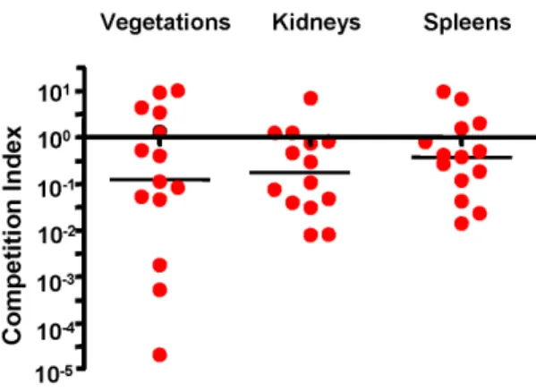
To examine the impact of Srr1 expression on the pathogenesis of endocarditis, we compared the relative virulence of GBS NCTC 10/84 with its isogenic mutant (Dsrr1), as measured by a rat
co-infection model of this disease. Animals (n = 14) had significantly lower densities of the mutant strain (mean log10 CFU/g 6
SD = 7.4661.63) within vegetations as compared with the parent strain (8.6261.25). Levels of the mutant strain were also significantly reduced within kidneys and spleens (Table 2). We then re-analyzed these data by comparing the ratio of the isogenic strains within tissues, with the CFU of each strain normalized to the number of CFU within the inoculum (competition index) (Fig. 4). When assessed by this approach, the levels of the srr1
mutant (Dsrr1) remained significantly reduced in all tissues, as compared with WT. Thus, Srr1 appears to be a significant virulence determinant for the pathogenesis of endocarditis due to GBS.
Figure 3. GBS binding to rat fibrinogen.(A) Alignment of Srr1 binding domain in the human fibrinogen Aa chain, with the homologous region of the rat protein; (B) Rat fibrinogen binding by wild type GBS and their isogenic variants (Dsrr1). (C) Rat fibrinogen binding byFLAGSrr1-BR protein over a range of concentrations. Casein
served as a negative control. doi:10.1371/journal.pone.0064204.g003
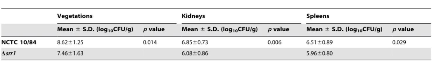
Table 2.Impact of Srr1 on virulence in an animal model of endocarditis.
Vegetations Kidneys Spleens
Mean±S.D. (log10CFU/g) pvalue Mean±S.D. (log10CFU/g) pvalue Mean±S.D. (log10CFU/g) pvalue
NCTC 10/84 8.6261.25 0.014 6.8560.73 0.006 6.5160.89 0.029
Dsrr1 7.4661.63 6.0860.86 5.9660.80
Infective endocarditis was induced in rats, using an inoculum of 56105CFU containing GBS NCTC10/84 and its isogenicDsrr1mutant, at a 1:1 ratio. Animals were
sacrified 72 h post-infection, and log10CFU/g of tissue for each strain was determined by plating onto selective media.
Discussion
A number of bacterial surface structures have been shown to mediate binding to fibrinogen, such as ClfA, ClfB, FnbA and Efb ofS. aureus, and the Fss proteins ofEnterococcus faecalis[33–38]. We recently identified Srr1 of GBS as a fibrinogen-binding protein that was important for bacterial attachment to microvascular endothelial cells and CNS invasion [26]. Although the binding region of Srr1 has limited homology to other adhesins, analysis of its predicted secondary structure indicated that the conformation of this domain would resemble the binding region of ClfB. As has been shown for several other adhesins, the binding pocket of ClfB is formed by two Ig folds that engage the Aachain of fibrinogen via a ‘‘dock, lock, and latch’’ mechanism [35,39]. Srr1 appears to interact with fibrinogen Aaby a similar mechanism, since deletion
of the predicted latch region abrogates fibrinogen binding by the protein, and markedly reduces virulence in an animal model of meningitis [26].
Our results indicate that Srr1-mediated binding to fibrinogen also contributes to the pathogenesis of infective endocarditis. Reduced fibrinogen bindingin vitrowas associated with decreased virulence, as measured by our co-infection (competition) model of endovascular infection. In particular, densities (CFU/g) of an Srr1 deletion mutant were significantly lower, as compared with its parent strain, both within vegetations and in kidneys and spleens. Of note, the mutant strain was not entirely avirulent, as it still produced disease in the infected animals. This indicates that GBS expresses other factors that contribute to its virulence, and is consistent with other studies on the role of microbial binding in endocarditis, where mutation or deletion of a single adhesin
produces only a partial reduction in pathogenicity [17,29,30]. GBS are known to express other fibrinogen binding proteins (FbsA and FbsB), which may have contributed to the residual virulence of ourDsrr-1mutant strain [37,38]. Moreover it is likely that GBS
express additional surface components that can mediate binding to cardiac valves, or enhance virulence by other mechanisms.
Binding to fibrinogen may be important for a number of events in the pathogenesis of endovascular infections [10,40,41]. First, bacterial attachment to the endocardium generally requires prior alteration of the valve surface, such that it is covered with a matrix of platelets and host proteins, including fibrinogen [20,42–45]. Studies withS. aureushave shown that fibrinogen immobilized on the valve surface is likely to contribute to the attachment of circulating bacteria, thereby initiating infection [46–50]. Our current results indicate that fibrinogen may have a similar role for GBS. In addition, fibrinogen in plasma could also serve to crosslink GBS to platelets that have aggregated at sites of valve injury. The subsequent progression of endovascular infection may also be enhanced by GBS binding to fibrinogen. Bacteria proliferating on the valve surface bacteria are thought to induce the further deposition of fibrinogen onto the infected valve, which in turn, is likely to trigger platelet attachment and aggregation. These processes, in combination with bacterial growth, result in the production of vegetations [9,10]. In view of ourin vitrostudies, where Srr-1 enhanced the binding of bacteria to both fibrinogen and platelets, is possible that Srr1-fibrinogen binding may be one mechanism for the continued recruitment of plateletsin vivoto the infected endocardium, thereby stimulating disease progression.
A longstanding therapeutic goal has been to develop agents that block bacterial binding to host tissues, thereby preventing or attenuating subsequent infection. Fibrinogen binding is an appealing target for disruption, in view of the importance of this interaction for the pathogenesis of infective endocarditis. Although inhibitory agents could target specific adhesins individually, such as Srr1, an alternative strategy might be to develop drugs that interfere with a larger number of ‘‘dock, lock, and latch’’ adhesins. Although the binding clefts of these adhesins vary in terms of primary amino acid sequence, it may still be possible to generate agents that block binding, either by preventing docking, or by inhibiting the latching process. If successful, this approach would yield an inhibitor that could be used for a variety of pathogens.
Acknowledgments
We thank Dr. Barbara Bensing and Dr. Arnold S. Bayer for their helpful scientific and editorial advice, and Dr. Kelly Doran for providing the GBS isolates used in these studies.
Author Contributions
Conceived and designed the experiments: HSS YQX PMS. Performed the experiments: HSS YQX. Analyzed the data: HSS YQX PMS. Contributed reagents/materials/analysis tools: HSS YQX PMS. Wrote the paper: HSS YQX PMS.
References
1. Sunkara B, Bheemreddy S, Lorber B, Lephart PR, Hayakawa K, et al. (2012) Group B Streptococcus infections in non-pregnant adults: the role of immunosuppression. Int J Infect Dis 16: e182–186.
2. Farley MM (2001) Group B streptococcal disease in nonpregnant adults. Clin Infect Dis 33: 556–561.
3. Munoz P, Llancaqueo A, Rodriguez-Creixems M, Pelaez T, Martin L, et al. (1997) Group B streptococcus bacteremia in nonpregnant adults. Arch Intern Med 157: 213–216.
4. Siciliano RF, Cais DP, Navarro RC, Strabelli TM (2010) Acute Streptococcus agalactiaeendocarditis: outcomes of early surgical treatment. Heart Lung 39: 331–334.
5. Ivanova Georgieva R, Garcia Lopez MV, Ruiz-Morales J, Martinez-Marcos FJ, Lomas JM, et al. (2010)Streptococcus agalactiaeleft-sided infective endocarditis. Analysis of 27 cases from a multicentric cohort. J Infect 61: 54–59. 6. Rollan MJ, San Roman JA, Vilacosta I, Sarria C, Lopez J, et al. (2003) Clinical
profile ofStreptococcus agalactiaenative valve endocarditis. Am Heart J 146: 1095– 1098.
7. Sambola A, Miro JM, Tornos MP, Almirante B, Moreno-Torrico A, et al. (2002) Streptococcus agalactiaeinfective endocarditis: analysis of 30 cases and review of the literature, 1962–1998. Clin Infect Dis 34: 1576–1584.
8. Kannan R, Komaranchath AM, Mathew T, Ramprakash B, Sundararaman T, et al. (2001) Streptococcus agalactiae endocarditis. J Assoc Physicians India 49: 1125–1126.
Figure 4. Competitive index (CI) analysis of WT and Dsrr1
mutant obtained in the rat model of endocarditis.Competition index (CI) was calculated as the ratio of the WT to theDsrr1mutant in each tissue, normalized for the ratio of strains within the inoculum. Circles represent data from individual animals. A CI above 100(dashed
line) indicates a competitive disadvantage ofDsrr1compared with WT. Horizontal black bars indicates means of CIs.
9. Fitzgerald JR, Loughman A, Keane F, Brennan M, Knobel M, et al. (2006) Fibronectin-binding proteins ofStaphylococcus aureusmediate activation of human platelets via fibrinogen and fibronectin bridges to integrin GPIIb/IIIa and IgG binding to the FcgammaRIIa receptor. Mol Microbiol 59: 212–230. 10. Loughman A, Fitzgerald JR, Brennan MP, Higgins J, Downer R, et al. (2005)
Roles for fibrinogen, immunoglobulin and complement in platelet activation promoted byStaphylococcus aureusclumping factor A. Mol Microbiol 57: 804–818. 11. Fitzgerald JR, Foster TJ, Cox D (2006) The interaction of bacterial pathogens
with platelets. Nat Rev Microbiol 4: 445–457.
12. Moreillon P, Que YA, Bayer AS (2002) Pathogenesis of streptococcal and staphylococcal endocarditis. Infect Dis Clin North Am 16: 297–318. 13. Veloso TR, Chaouch A, Roger T, Giddey M, Vouillamoz J, et al. (2012) Use of
a Human-like Low-grade Bacteremia Model of Experimental Endocarditis to Study the Role ofStaphylococcus aureusAdhesins and Platelet Aggregation in Early Endocarditis. Infect Immun 81(3): 697–703.
14. Zhou M, Wu H (2009) Glycosylation and biogenesis of a family of serine-rich bacterial adhesins. Microbiology 155: 317–327.
15. Bensing BA, Lopez JA, Sullam PM (2004) The Streptococcus gordoniisurface proteins GspB and Hsa mediate binding to sialylated carbohydrate epitopes on the platelet membrane glycoprotein Ibalpha. Infect Immun 72: 6528–6537. 16. Lizcano A, Sanchez CJ, Orihuela CJ (2012) A role for glycosylated serine-rich
repeat proteins in gram-positive bacterial pathogenesis. Mol Oral Microbiol 27: 257–269.
17. Siboo IR, Chambers HF, Sullam PM (2005) Role of SraP, a Serine-Rich Surface Protein ofStaphylococcus aureus, in binding to human platelets. Infect Immun 73: 2273–2280.
18. Heilmann C, Niemann S, Sinha B, Herrmann M, Kehrel BE, et al. (2004) Staphylococcus aureusfibronectin-binding protein (FnBP)-mediated adherence to platelets, and aggregation of platelets induced by FnBPA but not by FnBPB. J Infect Dis 190: 321–329.
19. Miajlovic H, Loughman A, Brennan M, Cox D, Foster TJ (2007) Both complement- and fibrinogen-dependent mechanisms contribute to platelet aggregation mediated byStaphylococcus aureusclumping factor B. Infect Immun 75: 3335–3343.
20. Pietrocola G, Schubert A, Visai L, Torti M, Fitzgerald JR, et al. (2005) FbsA, a fibrinogen-binding protein fromStreptococcus agalactiae, mediates platelet aggre-gation. Blood 105: 1052–1059.
21. Tazi A, Bellais S, Tardieux I, Dramsi S, Trieu-Cuot P, et al. (2012) Group B Streptococcus surface proteins as major determinants for meningeal tropism. Curr Opin Microbiol 15: 44–49.
22. Mistou MY, Dramsi S, Brega S, Poyart C, Trieu-Cuot P (2009) Molecular dissection of the secA2 locus of group B Streptococcus reveals that glycosylation of the Srr1 LPXTG protein is required for full virulence. J Bacteriol 191: 4195– 4206.
23. van Sorge NM, Quach D, Gurney MA, Sullam PM, Nizet V, et al. (2009) The group B streptococcal serine-rich repeat 1 glycoprotein mediates penetration of the blood-brain barrier. J Infect Dis 199: 1479–1487.
24. Seifert KN, Adderson EE, Whiting AA, Bohnsack JF, Crowley PJ, et al. (2006) A unique serine-rich repeat protein (Srr-2) and novel surface antigen (epsilon) associated with a virulent lineage of serotype III Streptococcus agalactiae. Microbiology 152: 1029–1040.
25. Sheen TR, Jimenez A, Wang NY, Banerjee A, van Sorge NM, et al. (2011) Serine-rich repeat proteins and pili promoteStreptococcus agalactiaecolonization of the vaginal tract. J Bacteriol 193: 6834–6842.
26. Seo HS, Mu R, Kim BJ, Doran KS, Sullam PM (2012) Binding of glycoprotein Srr1 of Streptococcus agalactiae to fibrinogen promotes attachment to brain endothelium and the eevelopment of meningitis. PLoS Pathog 8: e1002947. 27. Seo HS, Xiong YQ, Mitchell J, Seepersaud R, Bayer AS, et al. (2010)
Bacteriophage lysin mediates the binding ofStreptococcus mitisto human platelets through interaction with fibrinogen. PLoS Pathog 6: e1001047.
28. Seo HS, Sullam PM (2011) Characterization of the fibrinogen binding domain of bacteriophage lysin fromStreptococcus mitis. Infect Immun 79: 3518–3526. 29. Xiong YQ, Bensing BA, Bayer AS, Chambers HF, Sullam PM (2008) Role of
the serine-rich surface glycoprotein GspB of Streptococcus gordonii in the pathogenesis of infective endocarditis. Microb Pathog 45: 297–301.
30. Mitchell J, Siboo IR, Takamatsu D, Chambers HF, Sullam PM (2007) Mechanism of cell surface expression of theStreptococcus mitisplatelet binding proteins PblA and PblB. Mol Microbiol 64: 844–857.
31. Mitchell J, Sullam PM (2009)Streptococcus mitisphage-encoded adhesins mediate attachment toa2-8-linked sialic acid residues on platelet membrane gangliosides. Infect Immun 77: 3485–3490.
32. Que YA, Moreillon P (2011) Infective endocarditis. Nat Rev Cardiol 8: 322– 336.
33. Geoghegan JA, Ganesh VK, Smeds E, Liang X, Hook M, et al. (2009) Molecular characterization of the interaction of staphylococcal microbial surface components recognizing adhesive matrix molecules (MSCRAMM) ClfA and Fbl with fibrinogen. J Biol Chem 285: 6208–6216.
34. Shannon O, Flock JI (2004) Extracellular fibrinogen binding protein, Efb, from Staphylococcus aureusbinds to platelets and inhibits platelet aggregation. Thromb Haemost 91: 779–789.
35. Ganesh VK, Barbu EM, Deivanayagam CC, Le B, Anderson AS, et al. (2011) Structural and biochemical characterization of Staphylococcus aureusclumping factor B/ligand interactions. J Biol Chem 286: 25963–25972.
36. Sillanpaa J, Nallapareddy SR, Houston J, Ganesh VK, Bourgogne A, et al. (2009) A family of fibrinogen-binding MSCRAMMs fromEnterococcus faecalis. Microbiology 155: 2390–2400.
37. Devi AS, Ponnuraj K (2010) Cloning, expression, purification and ligand binding studies of novel fibrinogen-binding protein FbsB ofStreptococcus agalactiae. Protein Expr Purif 74: 148–155.
38. Pietrocola G, Visai L, Valtulina V, Vignati E, Rindi S, et al. (2006) Multiple interactions of FbsA, a surface protein from Streptococcus agalactiae, with fibrinogen: affinity, stoichiometry, and structural characterization. Biochemistry
45: 12840–12852.
39. Xiang H, Feng Y, Wang J, Liu B, Chen Y, et al. (2012) Crystal structures reveal the multi-ligand binding mechanism ofStaphylococcus aureusClfB. PLoS Pathog 8: e1002751.
40. O’Brien L, Kerrigan SW, Kaw G, Hogan M, Penades J, et al. (2002) Multiple mechanisms for the activation of human platelet aggregation byStaphylococcus aureus: roles for the clumping factors ClfA and ClfB, the serine-aspartate repeat protein SdrE and protein A. Mol Microbiol 44: 1033–1044.
41. Sullam PM, Valone FH, Mills J (1987) Mechanisms of platelet aggregation by viridans group streptococci. Infect Immun 55: 1743–1750.
42. Widmer E, Que YA, Entenza JM, Moreillon P (2006) New concepts in the pathophysiology of infective endocarditis. Curr Infect Dis Rep 8: 271–279. 43. Moreillon P, Que YA (2004) Infective endocarditis. Lancet 363: 139–149. 44. Nobbs AH, Lamont RJ, Jenkinson HF (2009) Streptococcus adherence and
colonization. Microbiol Mol Biol Rev 73: 407–450.
45. Santoro J, Levison ME (1978) Rat model of experimental endocarditis. Infect Immun 19: 915–918.
46. Sullam PM, Bayer AS, Foss WM, Cheung AL (1996) Diminished platelet binding in vitro byStaphylococcus aureusis associated with reduced virulence in a rabbit model of infective endocarditis. Infect Immun 64: 4915–4921. 47. Xiong YQ, Fowler VG, Yeaman MR, Perdreau-Remington F, Kreiswirth BN,
et al. (2009) Phenotypic and genotypic characteristics of persistent methicillin-resistant Staphylococcus aureus bacteremia in vitro and in an experimental endocarditis model. J Infect Dis 199: 201–208.
48. Piroth L, Que YA, Widmer E, Panchaud A, Piu S, et al. (2008) The fibrinogen-and fibronectin-binding domains of Staphylococcus aureus fibronectin-binding protein A synergistically promote endothelial invasion and experimental endocarditis. Infect Immun 76: 3824–3831.
49. Que YA, Haefliger JA, Piroth L, Francois P, Widmer E, et al. (2005) Fibrinogen and fibronectin binding cooperate for valve infection and invasion in Staphylococcus aureusexperimental endocarditis. J Exp Med 201: 1627–1635. 50. Moreillon P, Entenza JM, Francioli P, McDevitt D, Foster TJ, et al. (1995) Role
of Staphylococcus aureus coagulase and clumping factor in pathogenesis of experimental endocarditis. Infect Immun 63: 4738–4743.
51. Wessels MR, Haft RF, Heggen LM, Rubens CE (1992) Identification of a genetic locus essential for capsule sialylation in type III group B streptococci. Infect Immun 60: 392–400.