R E S E A R C H
Open Access
Tissue apoptosis in mice infected with
Leptospira
interrogans
serovar Icterohaemorrhagiae
Márcia Marinho
1*, Cilene Vidovix Táparo
1†, Itamar S. Oliveira-Júnior
2†, Silvia Helena Venturoli Perri
1†and Tereza Cristina Cardoso
1†Abstract
Background:This investigation aimed to evaluate the occurrence of some apoptotic features induced by Leptospira interrogansserovar Icterohaemorrhagiae infection in young BALB/c mice during 2, 4, 7, 10, 14 and 21 days post-infection (dpi).
Methods:The animals were euthanized and lung, liver and kidneys were harvested to histopathology analysis and immunohistochemistry to caspase-3 antigen detection was performed.
Results:Chromatin condensation in kidney and liver tissues, but not in lung tissue, was observed. Caspase-3 reactive cells, mainly characterized as renal epithelial cells, were detected in the days 14 and 21 at high levels when compared to days 2, 4 and 7 (p = 0.025; p < 0.05). Lung sections revealed caspase-3 labeled alveolar cells in 10 and 14 days post-infection was higher than observed at 7 days (p = 0.0497; p < 0.05). Liver sections demonstrated reactive cells at a highest level at 14 and 21 days post-infection when comparison to 2, 4, 7 and 10 days (p = 0.0069; p < 0.05).
Conclusions:Our results suggest that infection ofL. interrogansinduce in kidney, liver and lung an activation of apoptosis mediated by caspase-3 dependent pathway in later phases of infectious process.
Keywords:Leptospirosis, Caspases, Programmed cell death, Mice, Inflammatory response
Background
Leptospirosis is an important zooanthroponotic disease spread worldwide which infection is recognized as a re-emergent disease [1]. The spirochetes of the genus
Leptospiraare responsible for human and animal lepto-spirosis characterized as mild febrile illness to severe multiorgan failure, especially pulmonary hemorrhage and renal failure [2]. Pathogenic leptospires are highly motile and invasive spirochetes that have the capacity to survive and grow in tissue by escaping natural defense mechanisms [3]. The disease is transmitted to humans by direct or indirect exposure to contaminated urine from mammalian reservoir hosts such as rodents but also farm, wild, and domestic mammals [4].
Asymptomatic form of leptospirosis with fever, head-ache, and myalgia that can spontaneously resolve is one
of clinical presentations. However, more severe cases, with sepsis and multiple organ failure, including hepatic and renal dysfunctions associated to pulmonary he-morrhage, are also reported [2].
Studies in vivohave been performed to reproduce the clinical disease in order to understand Leptospira inva-sion and colonization during pathogenesis due to the variability of clinical presentation of leptospirosis in humans [2].
In spite of hamsters being considered the most suscep-tible animal for in vivo studies, different select mice strains have also been used, since they are considered re-sistant to disease development [5–9]. It is known that selected mice strains, high and low antibody producers against complex natural antigens, show different host cellular and antibody response after Leptospira interro-gansinfection when compared with BALB/c mice at five weeks of age, probably due to immunological system competence [5, 6]. However, it is also known that mice aged one week or less are susceptible to infection when
* Correspondence:mmarinho@fmva.unesp.br
†
Equal contributors
1Department of Support, Production and Animal Health, Veterinary Medicine School, São Paulo State University (UNESP–Univ Estadual Paulista),
Araçatuba, São Paulo, Brazil
Full list of author information is available at the end of the article
their immune system is still under development [5–7]. The importance of outbreed BALB/c mice on leptospir-osis pathogenesis studies is due to their heterogeneous phenotype and also to susceptibility to be infected at one week of age as demonstrated before [5].
Cellular apoptosis, or programmed cell death, is an es-sential mechanism for embryonic development and host response against many infectious and non-infectious dis-ease [10]. Apoptosis events followed by tissue injury are well documented, including in many renal diseases [11]. Caspases are a family of cysteine proteases that mediate apoptosis induced by a variety of stimuli [12]. Based on their structures and order in cell death pathways, caspases can be divided into initiators (caspase-2, −8, −9, −10
and−12) and effectors (caspase-3,−6 and−7). Two
path-ways, intrinsic and extrinsic death pathpath-ways, have been identified in most cases of caspase-dependent apoptosis [10]. Moreover, the effector caspase-3 can be activated by both death pathways, directly by intrinsic pathway and in-directly by extrinsic death pathway [12].
L. interrogans serovar Icterohaemorrhagiae has been described to invade Vero cells and induce macrophages apoptosis [13]. Besides,in vivo apoptosis of hepatocytes of guinea pigs infected by the same serovar has been de-scribed [14]. It has been demonstrated that L. interro-gans induces apoptosis in J774A1 cells by activation of caspases-3 through activation of caspase-8 [15]. The role of apoptosis in the pathogenesis of leptospirosis remains unclear. Similarly, the mechanisms by which leptos-pirosis induces the activation of cell death needs to be elucidate [4, 16, 17].
The aim of the present study was to investigate whether apoptosis is close related toL. interrogansserovar Ictero-haemorrhagiae by infection of young BALB/c mice at 2, 4, 7, 10, 14 and 21 days post-infection (dpi). Leptospira-induced lung, kidney and liver dysfunctions were assessed by histological parameters and anti-caspase-3 antigens were detected by immunohistochemistry assay.
Methods
Mice and inoculum
The animal experiments were conducted with approval from the Research Ethics Committee of São Paulo State University (UNESP). Thirty young BALB/c mice at five days of age, male and female, were divided into six groups of five mice each. For each experimental group, the same number of animals was considered as control group.
Animals were intraperitoneally injected with 1 × 10⁸
L. interrogansserovar Icterohaemorrhagie (strain 11437), which was previously cultured in Fletcher medium for seven days. Negative control for each group studied was injected with 1 mL of Fletcher medium alone. The
Leptospira sample was provided by the Laboratory of
Oswaldo Cruz Foundation (FIOCRUZ), maintained in semi-solid Fletcher medium and quantified according to the Faine’s technique. Mice were bled from the retro-orbital plexus at 2, 4, 7, 10, 14, and 21 dpi. During nec-ropsy, lung, liver and kidneys were removed and fixed in 10 % neutral buffered formalin and paraffin-embedded for histological and immunohistochemistry. Tissue sam-ples were inoculated into Fletcher medium and incu-bated at 28 °C to assess bacterial recovery and confirm infection byLeptospiraspp.
Histological
The 3μm-thin sections from the lung, liver and kidneys
collected at 2, 4, 7, 10, 14 and 21 dpi were analyzed after stained according to hematoxylin and eosin (HE) stan-dard procedure described before [6]. The main micro-scopic alterations related to apoptosis were documented: adherence to the extracellular matrix and neighboring cells, chromatin condensation, intranuclear DNA frag-mentation and formation of apoptotic bodies [18]. All analyses were “blind”, i.e., the researchers did not know which samples belonged to which experimental group (infected and uninfected). The images were collected under an AxioImager® A.1 light and a microscope con-nected to AxioCam®MRc (Carl Zeiss, Germany).
Immunohistochemistry
For immunohistochemistry, endogenous peroxidase ac-tivity was blocked after dewaxing by incubating the sec-tions in 2 % (v/v) hydrogen peroxidase (30 vol.) diluted in 50 % (v/v) methanol for 30 min. Pre-treatment for antigen retrieval were performed according to the speci-fications for primary antibody anti-caspase-3 at dilution of 1:200 in phosphate buffered solution (PBS), pH 7.2, added with 0.5 % Tween (Sigma-Aldrich, USA). Non-specific binding was blocked with 3 % (w/v) nonfat dry milk in PBS pH 7.2 for 30 min. The sections were incu-bated with the primary antibody for 18–22 h at 4 °C in a humidified chamber. The slides were washed in PBS and incubated with biotinylated secondary antibody and streptavidin-HRP complex (LSAB+ Kit, K0690, Dako) according to the manufacturer’s instructions.
in a histological field. All analyses were“blind”, i.e., the re-searchers did not know which samples belonged to which experimental group (infected and uninfected).
For caspase-3 positive cells, significant differences be-tween groups, and respective control animals, were deter-mined by one-way ANOVA followed by Tukey’s multiple comparison test. A value of p < 0.05 was considered statis-tically significant. Data are expressed as mean ± standard deviation (SD). All statistical analyses were performed using Prism software (GraphPad®, USA).
Ethics committee approval
The present study was approved by the Research Ethics Committee of São Paulo State University (FMVA- UNESP).
Results and discussion
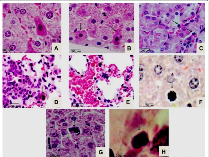
Hepatocytes with eosinophilic cytoplasm and nuclei of chromatin fragmentation were observed at 21 dpi (Fig. 1a).
At the same time, hepatocytes with cell retraction and dif-fuse necrosis restricted to the cortical renal tubules (Fig. 1b) and tubular necrosis near to cortical area were observed (Fig. 1c). Lung sections presenting capillary congestion and mononuclear cells infiltrates followed by polymorphonuclear cells were observed during disease progression (Fig. 1d). By 21 dpi, inflammatory cell infil-trate in pulmonary parenchyma followed by hemorrhagic areas were observed in lung sections (Fig. 1e). From 14 to 21 dpi chromatin fragmentation and apoptotic bodies were frequently observed in liver and kidney slides (Fig. 1f, g and h). The histological features from control group re-vealed no microscopic lesions.
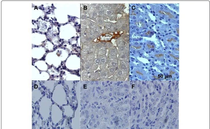
Caspase-3 reactive cells in the lung sections were ob-served in alveolar epithelium from 7 to 14 dpi (Fig. 2a). Centrilobular vein in liver sections were considered the most reactive to caspase-3 antigens from 10 to 14 dpi in liver slides (Fig. 2b). In kidney sections, the cortical
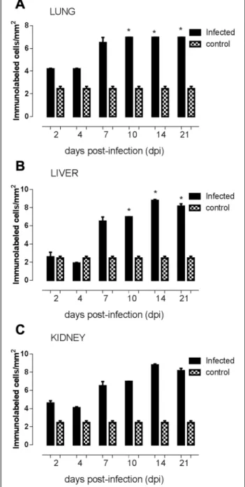
region of renal tubules revealed intense reactive cells to caspase-3 antigen at all dpi (Fig. 2c). The control group is demonstrated by negative results in the lung, liver and kidney slides (Fig. 3a, b and c; black bars). At 4 dpi, the number of caspase-3 positive cells was considered sig-nificantly lower (p = 0.0497) in comparison to 7, 10 and 14 dpi, and also related to control group in lung slides (Fig. 3a). Regarding to the number of caspase-3 positive labeled cells in liver sections, a significant difference (p = 0.0069) was detected at 14 and 21 dpi when com-pared to control group (Fig. 3b). Analysis of kidney sec-tions showed a higher number of caspase-3 positive labeled cells (p = 0.025) at 14 and 21 dpi, in comparison to early stages of infection and to control group (Fig. 3c).
Leptospirosis is responsible for a severe infectious dis-ease in humans. In spite of great importance of leptos-pirosis for public health, the mechanism involved in the pathogenesis remains unclear [1]. Several animal models are available to studyin vivo Leptospirainfection [5–7, 9]. The hamster model is considered susceptible to develop severe fatal leptospirosis similar to what is observed in human cases [5–7]. Moreover, at early age, BALB/c mice are susceptible to the virulent strain ofLeptospira interrogans serovar Icterohaemorrhagie, different from
animals at 5 to 7 weeks of age when they can be con-sidered resistant [5–7].
Pathological changes observed in this study demon-strated the BALB/c susceptibility toLeptospira interrogans
serovar Icterohaemorrhagie infection, especially after seven days after infection. Herein, pulmonary lesions, charac-terized as edema, capillary hemorrhage and hemoptysis are manifestations of severe leptospirosis in humans, confir-ming the severe disease developed by young BALB/c mice model [7, 19, 20]. Same histological lesions have been reported in hamsters consistent with our results and contrary to what have been demonstrated in resistant ani-mals [7, 9, 20, 21].
The mechanism by which leptospires cause host tissue damage or, conversely, the pathway used by infected hosts to avoid organ failure is still unclear. Few studies have focused in the liver histological features of animal models of leptospirosis. Inflammation of the portal tract in the liver of hamsters with numerous inflammatory elements, ending in a massive necrosis, has been de-scribed [7, 9]. In this study, liver histological changes were observed with an increase at late periods of obser-vation. These achievements are in agreement with pre-vious studies in which leptospirosis is well characterized
Fig. 2Immunodetection of caspase-3 reactive cells in BALB/c lung, liver and kidney tissue infected withL. interrogansserovar Icterohaemorrhage.
as a biphasic illness [4]. The acute phase is represented by a great number of circulating bacterial forms followed by clearance/initial apoptosis spread in target organs such as lung, liver and kidney [16]. The clearance phase of Leptospirainfection in vivo has been associated with antibody increase and high levels of inflammatory
cytokines, such as TNF-αand IL-6 [5–7, 9, 22, 23]. Both
kidney and liver histological analysis show an increase of chromatin fragmentation and apoptotic bodies at late periods of observation afterL. interrogansinfection.
Acute renal failure in a hamster model ofLeptospira in-fection has been previously observed, which exhibited the same histological damages observed in kidney tissue in the present work [5–7]. In contrast, resistant BALB/c mice infected with a virulent strain of L. interrogans serovar Canicola revealed less histological lesions when compared toL. interrogansserovar Icterohaemorrhagie in this study [5, 8]. Previous studies have demonstrated that an early regulation of chemokines and proinflammatory cytokines in mouse kidneys might be associated with limited patho-logical lesions; however, no study measured these factors in BALB/c at early age [5–7, 9, 22, 23].
Apoptosis of host cells plays an important role in modulating the pathogenesis of many infectious diseases. It has been reported that L. interrogans induces apop-tosis in macrophages in vitro and hepatocytes in vivo
[13, 14, 24, 25]. This study represents the first descrip-tion of apoptotic features in lung, liver and kidney ob-tained from BALB/c mice infected with L. interrogans
serovar Icterohaemorrhagie. Caspase-3, serving as an ex-ecutor caspase, plays a pivotal role in the apoptosis in-duced by various pathogens. In addition,in vitro studies have demonstrated that activation of caspase-3 is not the only mechanism leading to L. interrogans-induced apoptosis [24]. However, once caspase-3 is activated, it will be very difficult to reverse the programmed cell death [10].
The present findings demonstrated an increase of re-active caspase-3 cells distributed in lung, liver and kid-ney sections at after seven days post-infection, contrary to studies performed in vitro, whereas reactive cells were detected at early stages after L. interrogans infec-tion [12, 17, 24, 26]. It is important to emphasize that
in vivo infections reproduce the clinical signs according to the bacterial spread into the body, contrary toin vitro
models, whereas intrinsic pathogen-cell signaling is spe-cifically characterized. Both models have a great of im-portance; however, their differences must be taken into account when comparisons are made.
The results presented here revealed an increase of re-active caspase-3 cells in the lung, liver and kidney at late periods after infection withL. interrogans. These results corroborate previously described histological changes related to apoptosis; however, they contradict other
in vitro studies [13–15, 18, 19, 24]. Further studies should be conducted to determine the factors involved in activation or inhibition of apoptosis pathways in
L. interrogans infection. Blocking these keys responsible for self-destructive reactions would minimize tissue in-jury and inflammatory response.
Conclusions
Our results suggest that infection withL. interrogans in-duce an activation of apoptosis mediated by caspase-3 dependent pathways in later phases of the infectious process in the kidney, liver and lung tissues.
Competing interests
The authors declare that there are no competing interests.
Authors’contributions
MM, CVT, ISO Jr and TCC performed the experiments, drafted the manuscript and carried out the histological and immunohistochemical assays. SHVP participated in the statistical analysis. All authors read and approved the final manuscript.
Acknowledgments
The authors would like to thank the State of São Paulo Research Foundation (FAPESP–process n. 011/10423-0) for the financial support. T. C. Cardoso is a CNPq fellowship recipient.
Author details
1Department of Support, Production and Animal Health, Veterinary Medicine School, São Paulo State University (UNESP–Univ Estadual Paulista),
Araçatuba, São Paulo, Brazil.2Department of Inflammatory Mediators, Anesthesiology, Pain and Intensive Care, UNIFESP, São Paulo, São Paulo, Brazil.
Received: 15 December 2014 Accepted: 30 June 2015
References
1. Adler B, de la Peña Moctezuma A.Leptospiraand leptospirosis. Vet Microbiol. 2010;140(3–4):287–96.
2. Bharti AR, Nally JE, Ricaldi JN, Matthias MA, Diaz MM, Lovett MA, et al. Leptospirosis: a zoonotic disease of global importance. Lancet Infect Dis. 2003;3(12):757–71.
3. McBried AJ, Athanazio DA, Reis MG, Ko AI. Leptospirosis. Curr Opin Infect Dis. 2005;18(5):376–86.
4. Levett PN. Leptospirosis. Clin Microbiol Rev. 2001;14(2):296–326. 5. Marinho M, Silva C, Lima VMF, Peiró JR, Perri SHV. Cytokine and antibody
production during murine leptospirosis. J Venom Anim Toxins incl Trop Dis. 2006;12(4):595–603 [http://www.scielo.br/pdf/jvatitd/v12n4/06.pdf] 6. Marinho M, Monteiro CMR, Peiró JR, Machado GF, Oliveira-Júnior IS. TNF-α
and IL-6 immunohistochemistry in rat renal tissue experimentally infected withLeptospira interrogansserovar Canicola. J Venom Anim Toxins incl Trop Dis. 2008;14(3):533–40 [http://www.scielo.br/scielo.php?script=sci_arttext&-pid=S1678-91992008000300012]
7. Marinho M, Oliveira-Júnior IS, Monteiro CMR, Perri SHV, Salomão R. Pulmonary disease in hamsters infected withLeptopira interrogans: histopathologic findings and cytokine mRNA expressions. Am J Trop Med Hyg. 2009;80(5):832–6.
8. Silva EF, Santos CS, Athanazio DA, Seyffert N, Seixas FK, Cerqueira GM, et al. Characterization of virulence ofLeptospiraisolates in a hamster model. Vaccine. 2008;26(31):3892–6.
9. Matsui M, Rouleau V, Bruyère-Ostells L, Goarant C. Gene expression profiles of immune mediators and histopathological findings in animal models of leptospirosis: comparison between susceptible hamsters and resistant mice. Infect Immun. 2011;79(11):4480–92.
10. Boatright KM, Salvesen GS. Mechanisms of caspaseactivation. Curr Opin Cell Biol. 2003;15(6):725–31.
11. Jo SK, Yun SY, Chang KH, Cha DR, Cho WY, Kim HK, et al.α-MSH decreases apoptosis in ischaemic acute renal failure in rats: possible mechanism of this beneficial effect. Nephrol Dial Transplant. 2001;16(8):1583–91.
12. Niquet J, Allen SG, Baldwin RA, Wasterlain CG. Evidence of caspase-3 activation in hyposmotic stress-induced necrosis. Neurosci Lett. 2004;356(3):225–7.
13. Merien F, Baranton G, Perolat P. Invasion of vero cells and induction of apoptosis in macrophages by pathogenicLeptospira interrogansare correlated with virulence. Infect Immun. 1997;65(2):729–38.
14. Merien F, Truccolo J, Rougier Y.In vivoapoptosis of hepatocytes in guinea pigs infected withLeptopira interrogansserovar Icterohaemorrhagiae. FEMS Microbiol Lett. 1998;169(1):95–102.
15. Li SJ, Hu Y, Yan J, Mao YF, Li LW. Upregulation of FasL/Fas expression and FasL/Fas-associated apoptosis in J774.1 cells induced byLeptospira interrogans. Zhejiang Da Xue Xue Bao Yi Xue Ban. 2008;37(6):551–7 [Article in Chinese].
16. Ko AI, Coarant C, Picardeau M. Leptospira: the dawn of the molecular genetics era for an emerging zoonotic pathogen. Nat Rev Microbiol. 2009;7(10):736–47.
17. Ortiz A, Justo P, Catalán MP, Sanz AB, Lorz C, Egido J. Apoptotic cell death in renal injury: the rationale for intervention. Curr Drug Targets Immune Endocr Metabol Disord. 2002;2(2):181–92.
18. Carvalho SM, Macedo NA, Silva SMMS, Quessada AM, Mineiro ALBB, Costa FAL. Apoptose na nefropatia da leptospirose em ovinos. Rev Port Cienc Vet. 2008;103(565–566):47–54.
19. Kuwano K, Yoshimi M, Maeyama T, Hamada N, Yamada M, Nakanishi Y. Apoptosis signaling pathways in lung diseases. Med Chem. 2005;1(1):49–56. 20. Kuwano K. Epithelial cell apoptosis and lung remodeling. Cell Mol Immunol.
2007;4(6):419–29.
21. Martinez-Lopez DG, Fahey M, Coburn J. Responses of human endothelial cells to pathogenic and non-pathogenicLeptospiraspecies. PLoS Negl Trop Dis. 2010;4(12):e918.
22. Marangoni AR, Aldini V, Sambri L, Giacani L, Di Leo K, Cevenini R. Production of tumor necrosis factor alpha byTreponema pallidum, Borrelia burgdorferis.l., andLeptospira interrogansin isolated rat Kupffer cells. FEMS Immunol Med Microbiol. 2004;40(3):187–91.
23. Uchiyama T, Otani H, Okada T, Ninomya H, Kido M, Imamura H, et al. Nitric oxide induces caspase-dependent apoptosis and necrosis in neonatal rat cardiomyocytes. J Mol Cell Cardiol. 2002;34(8):1049–61.
24. Jin D, Ojcius DM, Sun D, Dong H, Luo Y, Mao Y, et al.Leptospira interrogans induces apoptosis in macrophages via caspase-8- and caspase-3-dependent pathways. Infect Immun. 2009;77(2):799–809.
25. Xue F, Zhao X, Yang Y, Zhao J, Yang Y, Cao Y, et al. Responses of murine and human macrophages to leptospiral infection: a study using comparative array analysis. PLoS Negl Trop Dis. 2013;7(10):e2477. 26. Wong VY, Keller PM, Nuttall ME, Kikly K, DeWolf Jr WE, Lee D, et al. Role of
caspases in human renal proximal tubular epithelial cell apoptosis. Eur J Pharmacol. 2001;433(2–3):135–40.
Submit your next manuscript to BioMed Central and take full advantage of:
• Convenient online submission
• Thorough peer review
• No space constraints or color figure charges
• Immediate publication on acceptance
• Inclusion in PubMed, CAS, Scopus and Google Scholar
• Research which is freely available for redistribution