RESEARCH ARTICLE
Proteomic Analysis of the Relationship
between Metabolism and Nonhost
Resistance in Soybean Exposed to
Bipolaris
maydis
Yumei Dong1, Yuan Su2, Ping Yu3, Min Yang1, Shusheng Zhu1, Xinyue Mei1, Xiahong He1, Manhua Pan1, Youyong Zhu1, Chengyun Li1*
1Key Laboratory of Agro-Biodiversity and Pest Management of Education Ministry of China, Yunnan Agricultural University, Kunming, 650201, China,2The Life Science and Technology Department of Kunming University, Kunming, 650214, China,3Institute of Biotechnology and Germplasm Resources, Yunnan Academy of Agricultural Sciences, Kunming 650223, China
Abstract
Nonhost resistance (NHR) pertains to the most common form of plant resistance against pathogenic microorganisms of other species.Bipolaris maydisis a non-adapted pathogen affecting soybeans, particularly of maize/soybean intercropping systems. However, no experimental evidence has described the immune response of soybeans againstB.maydis. To elucidate the molecular mechanism underlying NHR in soybeans, proteomics analysis based on two-dimensional polyacrylamide gel electrophoresis (2-DE) was performed to identify proteins involved in the soybean response toB.maydis. The spread ofB.maydis
spores across soybean leaves induced NHR throughout the plant, which mobilized almost all organelles and various metabolic processes in response toB.maydis. Some enzymes, including ribulose-1,5-bisphosphate carboxylase/oxygenase (RuBisCO), mitochondrial pro-cessing peptidase (MPP), oxygen evolving enhancer (OEE), and nucleoside diphosphate kinase (NDKs), were found to be related to NHR in soybeans. These enzymes have been identified in previous studies, and STRING analysis showed that most of the protein func-tions related to major metabolic processes were induced as a response toB.maydis, which suggested an array of complex interactions between soybeans andB.maydis. These find-ings suggest a systematic NHR against non-adapted pathogens in soybeans. This response was characterized by an overlap between metabolic processes and response to stimulus. Several metabolic processes provide the soybean with innate immunity to the non-adapted pathogen,B.maydis. This research investigation on NHR in soybeans may foster a better understanding of plant innate immunity, as well as the interactions between plant and non-adapted pathogens in intercropping systems.
PLOS ONE | DOI:10.1371/journal.pone.0141264 October 29, 2015 1 / 21
a11111
OPEN ACCESS
Citation:Dong Y, Su Y, Yu P, Yang M, Zhu S, Mei X, et al. (2015) Proteomic Analysis of the Relationship between Metabolism and Nonhost Resistance in Soybean Exposed toBipolaris maydis. PLoS ONE 10(10): e0141264. doi:10.1371/journal.pone.0141264
Editor:Wei Wang, Henan Agricultural Univerisity, CHINA
Received:May 27, 2015
Accepted:October 5, 2015
Published:October 29, 2015
Copyright:© 2015 Dong et al. This is an open access article distributed under the terms of the
Creative Commons Attribution License, which permits unrestricted use, distribution, and reproduction in any medium, provided the original author and source are credited.
Data Availability Statement:All relevant data are within the paper and its Supporting Information files.
Funding:This study was financially supported by the 973 Program (2011CB100400) of the Ministry of Science and Technology of China.
Introduction
Nonhost resistance (NHR) is a plant immune response against major microorganisms that are pathogenic to other plant species [1,2]. UnlikeR-gene-mediated resistance, which is governed by either a single or a few genes, NHR is mainly controlled by multiple genes expressed in mul-tiple patterns and durable defense responses [2]. Due to its highly robust and exceptional resis-tance properties, NHR is of scientific and economic imporresis-tance. Information regarding the mechanism underlying NHR may serve as a foundation for the development of novel strategies for crop disease management and crop distribution [3].
Soybeans are an important crop throughout the world because of their high plant protein and oil content [4]. Growing a combination of different crops and species in one field can boost yield and suppress crop diseases [5,6]. Maize/soybean intercropping is a common combination used in some countries to address issues of limited arable land and increasing food demand [6]. In the maize/soybean intercropping system, maize is susceptible to infec-tion byBipolaris maydis, whereas soybeans present no detectable symptoms of disease [7].
B.maydisdoes not have a soybean pathogen phenotype, nor is it related to any known soy-bean pathogens. In addition, it does not infect soysoy-beans via touch interactions. A previous microscopy analysis of infected soybeans showed that successful callose deposition and hydrogen peroxide (H2O2) production (S1 Fig) may be the first line of defense againstB. maydisgrowth [7]. Based on these findings, we hypothesized that soybean resistance toB.
maydisis established via NHR, which is the most robust and durable forms of plant resis-tance in nature. It allows soybean plants to protect themselves against a wide variety of para-sitic microorganisms [8–9].
In the past few decades, several studies have confirmed the existence of NHR in plants [7,
9–12]. Unlike the well-studied host resistance conferred by plant resistance (R) genes, the molecular basis of NHR remains elusive. In this present study, the foundation of soybean NHR againstB.maydis, a pathogen affecting a remotely related plant species, was characterized at the proteomic level. The availability of complete soybean genome sequences and recent devel-opments in various sequencing technologies have allowed us to investigate the complex mecha-nisms underlying NHR in soybeans at the protein level [13–15].
Plants respond to stresses via differential expression of sets of genes, which results in changes in protein levels. The proteome represents the global protein expression profile of a species at a given time and under a set of conditions [16,17]. Proteomic analysis involves identifying proteins involved in stress responses, determining their functions, and identifying possible regulatory networks that can be used to interpret stress-responsive processes that occur in plants [16,17].
In a previous study, although there were no visible symptoms in soybean leaves inoculated withB.maydis, microscopic observations showed that successful callose deposition and H2O2
production (S1 Fig) may be the immediate response of a plant to restrict the growth ofB. may-dis[7]. Information on the molecular mechanism underlying this kind of robust and durable defense in whole soybean plants is currently limited. To better understand the mechanism of NHR in soybean, it is important to identify proteins that might be involved in the soybean response to exposure toB.maydis, determine the types of metabolic proteins involved in the NHR in soybeans, compare the findings of the present study and those of previous NHR inves-tigations in plants, and determine whether any organelle was involved in the response toB.
maydis. This present study addresses these issues and summarizes the metabolic processes involved in NHR in soybeans. The results may facilitate further our understanding of NHR in soybean plants.
Proteomic Analysis of the Relationship between Metabolism and NHR in Soybeans
Materials and Methods
Plant growth and inoculation
Seeds of the soybean cultivar Hua Chun 6 were sown in floriculture substrate in pots [15 cm (D) × 15 cm (H)] and grown in a greenhouse (70% relative humidity, 25°C/20°C day/night temperature, 14 h/10 h photoperiod) at a density of two plants per pot. These pots contained a mixture of sand and floriculture substrate at a ratio of 1:1 [18]. Samples ofB.maydisrace O (Y664) were collected from Wenshan Prefecture (east longitude 104°350and northern latitude
23°180) in China
’s Yunnan Province and preserved at the Yunnan Agricultural University Col-lege of Plant Protection. TheB.maydisisolate was grown for 8 days at 27°C in a culture medium containing potato dextrose agar (PDA) and in the dark. The PDA medium was pre-pared as described elsewhere [19]. Conidia were collected with sterilized water (with 0.1% Tween-20) and the concentration was adjusted to 1 × 10⁵conidia per mL. This mixture served as the inoculating solution. Then, 200 mL of sterilized water (with 0.1% Tween-20) was used for mock inoculation of the controls, and 200 mL of the conidia solution was used to inoculate the treatment groups. At 72 h post-inoculation, soybean leaves, stems, and roots were collected from bothB.maydis-inoculated and mock-inoculated (control) soybean seedlings, and the stems and roots were cut into 1-cm sections for easier grounding. Three replicates were pre-pared for each experiment.
Protein extraction from soybean leaves
One gram of leaf tissue (1.5 g of stem and root tissue) was ground to a fine powder using a mor-tar and pestle and in liquid nitrogen. The powder was transferred to a 50-mL centrifuge tube and supplemented with 20 mL of a pre-cooled mixture of 10% trichloroacetic acid and 2 mM phenylmethanesulfonyl fluoride (PMSF) and 50 mM DL-dithiothreitol (DTT), vortex mixed with acetone, and incubated overnight (or for 16 h–18 h) at -20°C. After incubation, the sus-pension was centrifuged at 20,000 rpm for 20 min at 4°C, and the protein pellet was washed three times with 2 mM PMSF and 50 mM DTT in 80% acetone. The pellet was dried with a cir-culating water vacuum pump (SHZ-IIID, Shanghai, China). A 20-mg fraction of the protein powder was then resuspended in 450μL of lysis buffer (7 M urea, 2 M thiourea, 5% (3-[(3-cho-lamidopropyl)dimethylammonio]propanesulfonate (CHAPS), 2 mM DTT, and 0.3% Biolyte) by flicking the tube wall with fingers, and then incubated for 1 h at 25°C (or room tempera-ture). The suspension was later centrifuged at 22,000 rpm for 20 min at 25°C, and then the resulting supernatant was collected. The protein content of the samples was quantitated using the Bradford method with bovine serum albumin as protein standard [20]. For each treatment, three independent proteins were prepared, and triplicate 2-DE gels were used for each sample.
2-DE used for protein separation
Six samples were individually run in triplicate to control gel variation. Approximately 600μg of the purified protein extract was adjusted to a total volume of 500μL with a rehydration buffer containing 7 M urea, 5% (w/v) CHAPS, 0.5% (v/v) immobilized pH gradient (IPG) buffer (pH 4–7 NL) (GE Healthcare, Munich, Germany), and freshly prepared 20 mM DTT for every 24-cm IPG strip (GE Healthcare, Munich, Germany). The adjusted protein solution was vortexed and centrifuged for 5 min at 22,000 rpm at 4°C and then was directly loaded into a focusing tray. IPG strips (4–7 NL, 24 cm; GE Healthcare, Munich, Germany) were passively rehydrated for 14 h at 20°C. Then, the proteins (three replicates per treatment) were separated in the first dimension by isoelectric focusing (IEF), and then in the second dimension via sodium dodecyl sulfate-polyacrylamide gel electrophoresis (SDS-PAGE).
Proteomic Analysis of the Relationship between Metabolism and NHR in Soybeans
IEF was conducted using the IPGphor system (GE Healthcare, Munich, Germany) under the following conditions [21–22]: 250 V for 30 min with a linear ramp, 1,000 V for a minimum of 1 h with a rapid ramp, 9,000 V for 4.5 h with a linear ramp, and finally, 9,000 V at 65,000 V/ h with a rapid ramp; the entire system was kept at 20°C. After IEF, the strips were equilibrated with 6 M urea, 2% SDS, 1.5 M Tris-HCl (pH 8.8), 30% glycerol, 0.02% bromophenol blue, and 130 mM DTT for 20 min. The second equilibration step was performed in a solution consisting of 6 M urea, 2% SDS, 1.5 M Tris–HCl (pH 8.8), 30% glycerol, 0.02% bromophenol blue, and 135 mM iodoacetamide (IAA) for 20 min. The equilibrated strips were placed on 12.5% SDS-polyacrylamide gels and sealed with 1% low-melting agarose. SDS-PAGE was performed at 20 mA/gel first and then adjusted to a constant current of 30 mA/gel after 45 min. The 2-DE gels were fixed with the mixture solution (40% ethanol, 10% acetic acid) for 3 h at 16°C, and then stained overnight with Coomassie brilliant blue (CBB) G-250. The protein spots were detected by destaining with a mixture solution (5% w/v ammonium sulfate and 0.5% methanol).
SDS-PAGE analysis
Gels were then carefully washed with double-distilled water, scanned at an optical resolution of 300 dpi (or 600 dpi), and saved as.tiff files. For spot detection and volume quantification, 300 dpi.tiff files were analyzed using the PDQuest software version 7.4. To correct for variability in CBB staining, the individual spot volumes were normalized by dividing each spot’s optical den-sity (OD) value by the sum total OD of all the spots in the respective gels. Automated and man-ual spot matching were also performed [23].
The relative volumes were used to identify individual spots that were significantly differen-tially expressed between the two groups of gels (at least two-fold higher or lower and statisti-cally significant as calculated by the student’st-test, atP<0.05) (|ratio|2,P<0.05). Only
those with significant and reproducible changes were considered differentially expressed pro-teins [21].
Protein spot in-gel digestion, identification, and prediction of protein
function
Soybean leaf, stem, and root proteins with differential expression patterns (|ratio|2,P<0.05)
on gels were manually excised, washed three times with Millipore1pure water, and subjected to in-gel digestion and MALDI-TOF/TOF MS analysis, as described elsewhere [21,22]. The resulting peptide sequence data were submitted to the online MASCOT search engine (Matrix Science, London, U.K.) (http://www.phytozome.net/soybean) and queried against NCBInr databases [22].
The minimum requirements of our protein analysis were as follows: (i) at least three peptide sequences with identities higher than the threshold matched; and (ii)≧9% coverage of the total
protein sequence by matching peptide-positive matches were BLASTP searched against the NCBI protein database (http://www.ncbi.nlm.nih.gov) for updated annotation and identifica-tion of homologous proteins [22,23]. Some of the predicted cellular locations were determined using the Plant-PLoc version 2.0 (http://www.csbio.sjtu.edu.cn/bioinf/plant-multi/). STRING (http://www.string-db.org/) was used for predicting protein-protein interactions
Results
2-DE was used to screen for differentially expressed proteins that were associated withB.maydis
infection. A total of 68 proteins were differentially expressed in the roots, stems, and leaves of soybeans at 72 h after inoculation. Some of these 68 proteins were similar to those of NHR com-ponents reported in previous studies, whereas others differed from those earlier described [9].
Proteomic Analysis of the Relationship between Metabolism and NHR in Soybeans
Sixteen proteins in soybean leaves were induce after exposure to
B
.
maydis
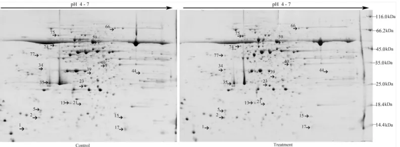
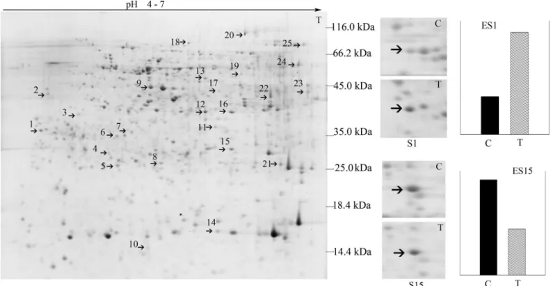
Leaf proteomic analysis of soybean NHR was performed to determine the mechanism underly-ing the response of soybean plants toB.maydis. Approximately 1,300 ± 50 protein spots were separated on 2-DE gels (Fig 1).
The 18 protein spots showed significant changes (|ratio|>2,P<0.05). Among these, 16
proteins were further classified by using MALDI-TOF-MS as provided by AmiGO into five groups, including proteins related to metabolic processes, catalytic activity, developmental pro-cesses, cellular components (such as structural proteins), and defense response (Fig 1,Table 1). Eight of the nine metabolic proteins were located in chloroplasts. Of these, 6 proteins showed increased staining intensity and 2 proteins showed decreased staining intensity.
Nucleoside diphosphate kinase (here considered indicative of catalytic activity) was located in the nucleus, and its concentration increased by 9.2-fold. The spot representing the stress-response protein oxygen-evolving enhancer protein 1 increased in staining intensity [24]. The spots representing 2 defense proteins, including acidic chitinase and trypsin inhibitor, also increased in staining intensity, mainly in the vacuole. The intensity of spot 74, which repre-sented the V-type proton ATPase subunit B 1-like isoform 2, showed a decrease in staining intensity in the cytoplasm, whereas that of its subunit, B 2-like isoform 2, increased (Table 1). Some of these differentially expressed proteins are involved in metabolic processes, develop-mental processes, organization of cellular components, and catalytic activity. Expression of these particular proteins in soybean leaves was induced in response toB.maydis, together with the production of acidic chitinase and trypsin inhibitor.
Twenty-five proteins were expressed in the stems of soybean plants in
response to
B
.
maydis
Several studies have documented that leaf and root proteomics can be used to interpret the responses of soybeans to biotic and abiotic stress [24]. However, the stem protein response to
Fig 1. Representative 2-DE gel pattern of soybean leaves at 72 h post-inoculation withB.maydis.For isoelectric focusing, a total of 500μg/600μL proteins were loaded onto each 24-cm IPG strip (pH 4–7). This was followed by SDS-PAGE on a 12.5% gel and Coomassie staining. 2-DE maps of proteins from untreated control leaves or leaves treated withB.maydisare shown. Proteins differentially regulated in response toB.maydiswere numbered in pairs of control and treatment maps. The MW (kDa) and pI of each protein was determined using a 2-DE marker.
doi:10.1371/journal.pone.0141264.g001
Proteomic Analysis of the Relationship between Metabolism and NHR in Soybeans
Table 1. Predicted functions and subcellular locations of soybean leaf proteins in response toB.maydis.
Spot No. (a) Up
(+) /down (-) Ratio (b) Protein score SC (%) (c) PM (d) Mr(e) theoretical/ observed
PI (f) theoretical/
observed
Function (g) Species
(h)
GenBank Acc. No. (i)
Predicted cellular location (j)
Metabolic process
23 + 2.8 180 32.93 6 26.77/31.1 6.21/5.14 Chlorophyll a-b binding protein 6A Chloroplastic-like Glycine max gi| 356559472 Chloroplast
2 + 13.3 105 35.97 3 11.44/14.36 4.36/4.52 Unknown Glycine
max gi| 255626375
Chloroplast
15 + 4.4 487 76.97 12 20.23/23.2 8.87/6 Uncharacterized
protein Glycine max gi| 351725817 Chloroplast
39 + 2.0 289 38.87 9 32.5/36.84 5.93/5.32 Unknown Glycine
max gi| 255641907
Chloroplast
59 + 2.1 307 61.97 12 30.24/56.14 5.43/5.92 Unnamed protein product Glycine max gi| 257670630 Chloroplast Mitochondrion
66 - 5.7 189 35.94 11 49.87/56.14 5.4/5.92 BAHD
acyltransferase DCR-like Glycine max gi| 356530840 Cytoplasm
77 - 5.6 178 43.94 16 52.39/66.01 5.26/5.38 4-hydroxy-3-methylbut-2-enyl diphosphate reductase-like Glycine max gi| 356538819 Chloroplast
5 - 2.6 118 21.8 4 26.33/18.19 6.93/4.65
Ribulose-1,5-bisphosphate Carboxylase/ oxygenase large subunit Glycine max gi| 290586534 Chloroplast
44 + 4.1 583 56.42 23 53.03/50.09 6/5.58
Ribulose-1,5-bisphosphate carboxylase/ oxygenase large subunit Glycine max gi| 91214125 Chloroplast Developmental process
34 + 2.5 422 55.26 10 35.27/23.52 6.66/5.37 Oxygen-evolving enhancer protein 1 Chloroplastic-like Glycine max gi| 356559442 Chloroplast Response to stimulus
13 + 7.7 1020 148.8 13 17.47/20.58 5.41/5.9 Unknown Glycine
max gi| 255626437
Cytoplasm
35 + 2.3 92 21.15 5 32.19/34.2 5.01/5.3 Acidic chitinase Glycine
max gi|4835584 Vacuole Cellular component organization or biogenesis
74 - 2.7 741 91.39 26 54.27/68.09 4.92/6.51 V-type proton ATPase subunit B 1-like isoform 2
Glycine max
gi| 356536394
Cytoplasm
75 + 2.6 352 62.97 16 54.29/70.09 4.95/5.13 V-type proton ATPase subunit B 2-like isoform 2
Glycine max gi| 356568551 Cytoplasm (Continued) Proteomic Analysis of the Relationship between Metabolism and NHR in Soybeans
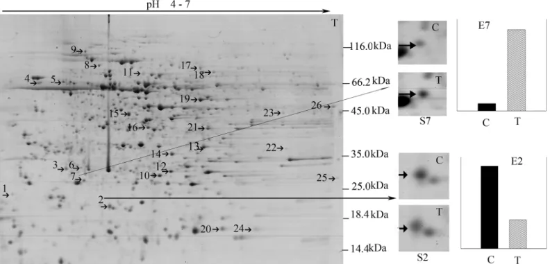
pathogens is remains elusive. Here, 72 hpi stem proteomic analysis was performed to produce a more systemic understanding of how the three major organs (roots, stems, and leaves) of soy-bean plants are involved in the responses to the non-adapted pathogenB.maydis. Approxi-mately 1,000 ± 50 protein spots were separated on 2-DE gels (Fig 2).
Among these 26, 25 differentially expressed (|ratio|>2,P<0.05) proteins were successfully
identified and sorted into five functional groups, including proteins associated with metabolic processes, cellular components, defense response, developmental processes, and molecular function (Table 2). Here, 48% of the stem proteins were determined to be involved in metabolic processes. The spots associated with half of these showed an increase in staining intensity with
B.maydisinfection, whereas half showed a decrease. Around 16% of the evaluated stem mito-chondrial proteins showed a change in expression after infection, suggesting that NHR in soy-beans is involved in significant energy consumption. Two of the mitochondrial proteins were related to heat shock 70, which is the protein chaperone that is responsible for the refolding and misfolding of proteins. A putative cysteine protease was grouped into the metabolic pro-cess group, although a previous study reported that it is involved in both developmental and pathogen-related PCD [25]. Protein spots 4, 8, and 19 had functional annotations similar to those of Hsp70, although these were classified in different functional groups and were pre-dicted to localize to different locations (mitochondrion and chloroplast), which was consistent with the diverse locations and functions of Hsps. The staining intensity of spot 19 (Hsp 70) showed a 12-fold increase relative to that of the controls.
Table 1. (Continued)
Spot No. (a) Up
(+) /down
(-)
Ratio (b)
Protein score
SC (%) (c)
PM (d)
Mr(e) theoretical/
observed
PI (f) theoretical/
observed
Function (g) Species
(h)
GenBank Acc. No. (i)
Predicted cellular location (j)
Defense response
21 + 2.1 179 64.29 6 18.25/22.11 6.12/5.08 Trypsin inhibitor Glycine max
gi|9367042 Vacuole
Catalytic activity (molecular function)
17 + 9.2 320 52.35 6 16.40/15.69 6.91/5.19 Nucleoside
diphosphate kinase
Glycine max
gi| 26245395
Mitochondrion Nucleus
(a) Protein spots increased (+) or decreased (-) in staining intensity in response toB.maydis (b) Ratio was equal to the relative expression of (Treatment / Control)
(c) SC, sequence coverage by PMF (d) PM, number of peptides matched
(e) Theoretical molecular mass was from report of identification and NCBI database, and experimental molecular mass was observed from real 2-DE gel image combined with analysis of PDQuest software
(f) Theoretical isoelectric point was from reports and from the NCBI database; experimental isoelectric points were observed from real 2-DE gel images combined with analysis of PDQuest software
(g) Predicted function of the protein obtained via the MASCOT software from the NCBI database (h) Species of the proteins obtained via the MASCOT software from the NCBI database (i) GenBank Acc. No., accession number in NCBI database
(j) Plant-mPLoc: Predicting subcellular localization of plant proteins including those with multiple sites (predicted at the website:http://www.csbio.sjtu.edu. cn/bioinf/plant-multi/)
doi:10.1371/journal.pone.0141264.t001
Proteomic Analysis of the Relationship between Metabolism and NHR in Soybeans
This phenomenon indicates that NHR in soybean is related to protein functional diversity. Protein spot 10 (isopentenyl-diphosphate delta-isomerase), protein spot 17 (26S protease regu-latory subunit 6B homolog), and protein spot 25 (peptidyl-prolylcis-transisomerase CYP20-2, chloroplastic-like isoform 2) were predicted to have more than one subcellular location. Pro-tein spot 7 (oxygen-evolving enhancer proPro-tein 1, chloroplastic-like) was found to have been caused by the same protein as leaf protein spot 34, which showed a 2.5-fold increase in expres-sion in leaves and a 11-fold increase expresexpres-sion in stems. This finding suggests that protein spot 34 might play an important role in soybean response toB.maydis. The staining intensity of protein spot 14 increased by 10-fold and that of protein spot 24 decreased by 6-fold in the nuclei of soybean stems. All 25 stem proteins were predicted in various important subcellular locations, including the nucleus, cytoplasm, endoplasmic reticulum, Golgi apparatus, vacuole, chloroplast, and mitochondrion, which indicated that the response proteins might include pro-teins related to metabolism.
Root proteins activate metabolic processes in response to
B
.
maydis
challenge on leaves
We investigated the patterns of root protein expression in response toB.maydischallenge on leaves (Fig 3).
Almost 1,000 proteins were detected in the CSS gel using PDQuest software 7.4, and all rep-licate gels showed highly similar distribution patterns in the 2-DE images. Around 25 proteins were differentially expressed (|ratio|2,P<0.05). Overall analysis revealed that out of all the
protein spots that matched in the treatment and control groups, 39% showed increased
Fig 2. Two DE patterns of stem proteins responding toB.maydis.S7 and S2 show enlarged images of protein spots 7 and 2, which were differentially expressed underB.maydisstress conditions;“C”refers to the protein pattern dissolved on the control gel, and“T”to the protein pattern dissolved on the treatment gel. The differential abundance of proteins was read using the PDQuest software and is plotted as the relative intensity enumerated in E7 and E2. E7 and E2 represent the relative expression of protein spots 7 and 2 in the control and treatment groups.“C”also refers to the control, and“T”to treatment. S7/E7 and S2/E2 are representative images of the differentially expressed proteins in the stems of soybean plants.
doi:10.1371/journal.pone.0141264.g002
Proteomic Analysis of the Relationship between Metabolism and NHR in Soybeans
Table 2. Predicted functions and subcellular locations of soybean stem proteins responsive toB.maydis.
Number of spots (a) Up (+) /down (-) Ratio (b) Protein score SC (%) (c) PM (d) Mr(e) theoretical/ observed PI (f) theoretical/ observed
Function (g) Species (h)
Acc. no. (i) Predicted cellular location (j)
Metabolic process
1 - 2 116 35.43 4 14.27/25.56 4.13/4.11 C2 domain-containing
protein At1g63220-like Glycine max gi| 356522642 Cytoplasm
3 - 3 99 20.28 5 39.70/31.44 7.01/4.62 Putative cysteine protease Glycine
max gi| 149393486
Vacuole
8 - 2 182 27.88 14 73.38/72.28 5.37/4.73 Heat shock cognate 70
kDa protein 2-like
Glycine max
gi| 356502432
Mitochondrion
9 - 6 551 41.03 27 93.29/84.63 4.86/4.9 Endoplasmin homolog Glycine
max gi| 356564371
Endoplasmic reticulum
10 + 3 336 64.75 14 33.79/31.41 5.84/5.18 Isopentenyl-diphosphate
Delta-isomerase Glycine max gi| 356531894 ChloroplastNucleus
16 + 2 543 47.52 18 54.53/51.75 5.89/5.54 Mitochondrial-processing
peptidase subunit alpha-like Glycine max gi| 356522822 Mitochondrion
18 + 2 1030 95.05 25 47.91/48.5 5.49/5.76 Enolase-like Glycine
max gi| 356505318
Cytoplasm
19 + 12 484 42.84 24 72.68/69.06 5.82/5.53 Heat shock 70 kDa protein Glycine
max gi| 356524786
Mitochondrion
21 + 3 84 11.08 3 38.34/40.41 5.3/5.83 Adenosine kinase 2-like Glycine
max gi| 356505238
Mitochondrion
22 - 2 192 25.26 5 33.13/33.89 6.14/6.52 Uncharacterized protein
LOC100781644 Glycine max gi| 356563399 Cell wallCytoplasm
25 - 3 436 18.45 7 22.92/23.69 9.1/6.89 Peptidyl-prolyl cis-trans
isomeraseCYP20-2, chloroplastic-like isoform 2
Glycine max gi| 356576933 ChloroplastEndoplasmic reticulum
13 + 2 233 62.95 12 30.81/31.24 5.9/5.58 Carbonic anhydrase,
chloroplastic-like Glycine max gi| 356501896 Chloroplast Cellular component
2 - 3 98 25.7 4 23.77/24.03 5.55/4.62 Uncharacterized protein
LOC100305490 Glycine max gi| 351727673 Chloroplast
11 - 3 112 20.00 7 55.78/57.38 5.15/5.14 ATP synthase CF1 alpha
subunit Glycine max gi| 91214148 Chloroplast
14 + 10 259 59.75 14 35.30/40.1 5.47/5.54 Probable carboxylesterase
2-like Glycine max gi| 356521488 Nucleus
17 + 2 366 46.25 15 46.56/51.98 5.61/5.7 26S protease regulatory
subunit 6B homolog
Glycine max
gi| 356539717
CytoplasmNucleus
24 - 6 396 51.01 6 16.49/14.48 5.93/6.85 Nucleoside diphosphate
kinase Glycine max gi| 351720837 Nucleus Defense response
4 - 4 346 37.56 17 71.20/72.59 5.1/4.59 Heat shock cognate 70
kDa protein-like Glycine max gi| 356569000 Chloroplast
(Continued)
Table 2. (Continued)
Number of spots (a) Up (+) /down (-) Ratio (b) Protein score SC (%) (c) PM (d) Mr(e) theoretical/ observed PI (f) theoretical/ observed
Function (g) Species (h)
Acc. no. (i) Predicted cellular location (j)
6 - 2 743 56.86 11 29.00/29.76 6.34/4.8 Unknown Glycine
max gi| 255644428
Chloroplast
12 + 4 472 96.95 10 17.20/23.8 5.61/5.66 Unknown Glycine
max gi| 255628729
Chloroplast
20 + 3 847 141.35 13 17.50/17.73 5.64/5.97 Unnamed protein product Glycine
max gi| 227247706
Cytoplasm
23 - 3 522 55.43 11 36.88/39.24 8.18/6.49 Malate dehydrogenase 1
mitochondrial-like Glycine max gi| 356543225 Mitochondrion
26 - 2 459 36.52 10 36.20/39.53 8.22/6.86 Malate dehydrogenase
mitochondrial-like Glycine max gi| 356517066 Mitochondrion Developmental process
7 + 11 834 79.46 16 35.27/28.73 6.66/4.87 Oxygen-evolving enhancer
protein 1 chloroplastic-like
Glycine max gi| 356559442 Chloroplast Molecular_function
15 + 3 476 60.87 18 42.25/42.22 5.52/5.66 Alpha-1,4-glucan-protein
synthase Glycine max gi| 356495127 Golgi apparatus
(a) Protein spots increased (+) or decreased (-) in staining intensity in response toB.maydis (b) Ratio was equal to the relative expression of (Treatment / Control)
(c) SC, sequence coverage by PMF (d) PM, number of peptides matched
(e) Theoretical molecular mass was from report of identification and NCBI database, and experimental molecular mass was observed from real 2-DE gel image combined with analysis of PDQuest software
(f) Theoretical isoelectric point was from reports and from the NCBI database; experimental isoelectric points were observed from real 2-DE gel images combined with analysis of PDQuest software
(g) Predicted function of the protein obtained via the MASCOT software from the NCBI database (h) Species of the proteins obtained via the MASCOT software from the NCBI database (i) GenBank Acc. No., accession number in NCBI database
(j) Plant-mPLoc: Predicting subcellular localization of plant proteins including those with multiple sites (predicted at the website:http://www.csbio.sjtu.edu.cn/bioinf/plant-multi/)
intensity and 61% presented a decrease in intensity. Twenty-three of these differentially expressed proteins were successfully identified in six functional groups, including proteins related to metabolic processes, cellular components, regulation of cellular processes, trans-membrane transport, defense response, and molecular function (Table 3). These proteins were detected in the nucleus, cytoplasm, cell wall, cell membrane, mitochondrion, and Golgi appara-tus of root cells in response to aB.maydischallenge on leaves, which might contribute to the multiple NHR responses to non-adapted pathogens.
Protein spots 1 and 19 were identified as elongation factor 1-delta-like (2.7-fold increase) and elongation factor 1-gamma-like (16.7-fold increase) proteins, which were predicted to local-ize to the mitochondrion and cytoplasm, respectively. This indicated that the metabolic pro-cesses related to protein synthesis in soybean roots might be different in response toB.maydis.
Protein spot 25, which is a copper amino oxidase precursor, showed a significant increase in intensity (33.1-fold) in soybean root organs. Chalcone isomerase 4-like, proteasome subunit beta type-6-like, and alcohol dehydrogenase 1-like proteins are related to flavone and alcohol metabolic processes, showed an increase in spot intensity in response toB.maydis, which indi-cated that several proteins in the roots might be bifunctional under stress conditions.
The spot intensities of proliferating cell nuclear antigen-like and auxin-induced protein decreased, suggesting that the soybean root cells underwent a reduction in growth to increase its defense response to the infection. On the other hand, the intensity of protein spot 8, which is related to a protease, increased. This finding indicated that the protein degradation process also increased in response toB.maydis(Table 3).
Fig 3. Two-DE map of root proteins responding toB.maydis.S1 and S15 were zoomed in the protein spot 1 and spot 15 differentially expressed under theB.maydisstress;“C”refers to the protein spot excised from the control gel, and“T”refers to the protein spot excised from the treatment gel. The differential abundance of proteins was determined using the PDQuest software and was plotted as the relative intensity enumerated as ES1 and ES15. ES1 and ES15 represent the relative expression between control and treatment of protein spot 1 and spot 15,“C”means control, and“T”indicates treatment. S1/ ES1 and S15/ES15 are representative differentially expressed root proteins.
doi:10.1371/journal.pone.0141264.g003
Proteomic Analysis of the Relationship between Metabolism and NHR in Soybeans
Table 3. Proteins in soybean roots in response to the challenge ofB.maydison leaves.
No. of spots Up
(+)/down (-) Ratio Protein score SC (%)
PM Species Acc. no. Protein name Predicted cellular
location
Metabolic process
1 + 2.7 923 69.6 12 Glycine
max
gi| 356516563
Elongation factor 1-delta-like Mitochondrion
4 + 2.2 262 32.3 7 Glycine
max
gi| 255646685
Unknown Chloroplast
5 + 2.4 502 57.9 8 Glycine
max
gi| 351723871
Chalcone isomerase 4-like Chloroplast
8 + 2.1 639 37.8 8 Glycine
max
gi| 356507848
Proteasome subunit beta type-6-like Nucleus
17 - 2.5 322 34.3 9 Glycine
max
gi| 356516166
Caffeic acid 3-O-methyltransferase-like isoform 1
Chloroplast
19 + 16.7 527 47.6 15 Glycine
max
gi| 356543373
elongation factor 1-gamma-like Cytoplasm
22 + 2.4 721 50.0 16 Glycine
max
gi| 356509324
Alcohol dehydrogenase 1-like Cytoplasm
24 - 2.4 519 68.2 14 Glycine
max gi| 356505332 Fructose-bisphosphate Aldolase, cytoplasmic isozyme Cytoplasm
25 + 33.1 500 21.3 9 Glycine
max
gi| 351721496
Copper amino oxidase precursor Cell wall
Cellular component
9 - 2.1 694 67.9 16 Glycine
max
gi|1498340 Actin Cytoplasm
10 - 3.7 432 45.1 12 Glycine
max
gi| 255641658
Unknown Cytoplasm
11 - 3.6 364 42.3 11 Glycine
max
gi| 356509393
L-ascorbate peroxidase T, chloroplastic-like isoform 1
Chloroplast Mitochondrion
18 - 2.9 666 39.7 15 Glycine
max gi| 356532109 2,3-bisphosphoglycerate-independent phosphoglycerate mutase-like Cytoplasm
Regulation of cellular process
2 - 6 112 57.4 3 Glycine
max
gi| 356560765
Cnkyrin repeat domain-containing protein 2-like
Chloroplast
3 - 2.1 276 30.8 7 Glycine
max
gi| 356566211
Proliferating cell nuclear antigen-like Nucleus
21 - 2.1 498 64.6 11 Glycine
max
gi| 356537146
Ubiquitin-conjugating enzyme E2 variant 1D-like
Nucleus
Transport
6 + 2.2 207 38.0 7 Glycine
max
gi| 356556130
Plasma membrane-associated cation-binding protein 1-like
Cell membrane
Response to stimulus (defense response)
12 - 2.9 219 9.29 2 Glycine
max gi| 359807323 Uncharacterized protein LOC100814078 Chloroplast. Mitochondrion
14 - 3.8 700 75.3 9 Glycine
max gi| 351724985 Uncharacterized protein LOC100499771 Cytoplasm
16 - 3 458 36.7 11 Glycine
max
gi| 356538212
Isoflavone reductase-like Cytoplasm
20 + 3.5 930 44.3 30 Glycine
max
gi| 351726848
Seed linoleate 9S-lipoxygenase Cytoplasm
(Continued) Proteomic Analysis of the Relationship between Metabolism and NHR in Soybeans
In summary, almost all of the plant cell organelles were predicted to respond to theB. may-dischallenge on leaves, and the biological processes involved in growth and development were possibly bifunctional.
Discussion
Competitive and facilitative interactions among different plant species have played major roles in shaping natural communities. For decades, this principle has been employed to increase crop yield [26,27]. Plants sense their environments using chemicals, touch, and light [26,27]. In maize/soybean intercropping systems, the transfer ofB.maydisspores from maize to soy-beans is a common occurrence. There have been several reports of interactions between maize and soybeans, and these have been extensively used to increase yield or improve soil fertility. However, there has been no report discussing the interactions between soybeans andB.maydis, which often come into contact in ecological networks involving maize and soybean intercrop-ping systems. Therefore, the findings of the present study not only offer new insights into NHR in the soybean response toB.maydis, but also into the positive interactions mediated byB.
maydisbetween soybeans and maize. This is the first detailed study on howB.maydismediates interactions between maize and soybeans.
Whole-plant resistance is invoked in almost all of cellular components of
the soybean plant
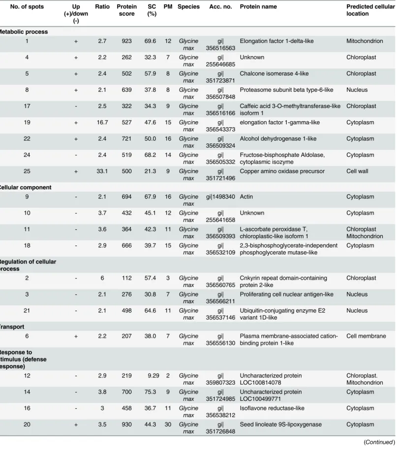
AlthoughB.maydiswas sprayed only onto the leaves of the soybean plants, proteins of the stem and roots also responded to this particular stimulus (Figs1,2,3and4). These results sug-gest that the response of soybeans toB.maydismay not be limited to the site of exposure, and that the signal of the stimulus could be transmitted to remote organs. The level of expression of several proteins was reprogrammed far from the site of inoculation. Only one pair of root and stem protein spots consisted of the same protein, e.g., S (stem) 15 and R (root) 13, an alpha-1,4-glucan-protein synthase [UDP-forming] homolog. Two pairs of stem and leaf protein spots (L17 and S24) comprised the same protein, the NDK homolog. Leaf protein spot (L34) and stem protein spot (S7) contained an oxygen-evolving enhancer protein homolog. However, there was no protein overlap between leaves and roots. These results suggest that roots, stems, and leaves of soybeans could differentially respond toB.maydischallenge on leaves, which could render a whole plant resistant toB.maydis. Thus, almost all of the metabolic proteins were triggered in almost all subcellular structures. These included the cell wall, cell membrane, cell skeleton, cytoplasm, chloroplasts, mitochondria, nucleus, Golgi apparatus, and endoplas-mic reticulum (Fig 4A). This is the first report to find evidence of a multiple-responsive NHR in soybeans againstB.maydis, a pathogenic fungus of a phylogenetically distant crop.
Table 3. (Continued)
No. of spots Up
(+)/down (-)
Ratio Protein score
SC (%)
PM Species Acc. no. Protein name Predicted cellular
location
Catalytic activity (molecular function)
13 - 3.4 366 48.9 14 Glycine
max
gi| 356495127
Alpha-1,4-glucan-protein synthase [UDP-forming]-like
Golgi apparatus
23 - 2.8 323 29.2 8 Glycine
max
gi| 356505967
Auxin-induced protein PCNT115-like isoform 2
Chloroplast
doi:10.1371/journal.pone.0141264.t003
Proteomic Analysis of the Relationship between Metabolism and NHR in Soybeans
Proteins involved in NHR in soybean overlapped with metabolic proteins
NHR is strongly associated to basic plant metabolism, and host pathogens are known to target such primary metabolic pathways to establish virulence [2]. The results of the present study showed that proteins related to metabolic processes could be co-induced in response to non-adapted pathogens. For example, the ability of soybean plants to cope withB.maydisstress depends on a number of changes in their proteins, which the present study has determined to be reprogrammed after inoculation ofB.maydison leaves, and the changes remained in effect until at least 72 hpi. Several metabolic proteins in the leaves, stems, and roots of soybeans were invoked in response toB.maydis. Most of these proteins had essential functions, either in the regulation of the response, or in the metabolic process, allowing plants to survive and recover from the stresses [17].The leaf protein RuBisCO operates as a metabolic branch point, channeling carbon either to the photosynthetic carbon fixation (Calvin–Benson) cycle or to the photorespiratory pathway. It is the most abundant protein in green plant tissues and is a bifunctional enzyme [28,29]. Its most common form (type I presents in cyanobacteria, green algae, and higher plants) is com-posed of eight chloroplast-encoded large subunits (~55 kDa) and eight nuclear gene-encoded small subunits (~15 kDa) [28,29]. Two protein spots were identified as RuBisCO large subunits, which suggested that soybean photosynthesis was reprogrammed underB. may-disstress conditions.
BAHD acyltransferase catalyzes the acylation of various plant secondary metabolites [30]. The decrease in intensity of BAHD acyltransferase DCR-like (spot 66 in leaf) protein suggests that the metabolites were inhibited underB.maydisstress.
Fig 4. Proteins responsive toB.maydisand its predicted multiple subcellular locations.A) A conceptual diagram of a systematic NHR of soybean againstB.maydis. H2O2production and callose deposition were processed upon the interaction between soybean andB.maydisstarting at the cell wall of soybean. The subcellular locations of the differentially expressed proteins were predicted. Changed proteins are shown in italics, and the organelle is written in blue. For instance, Hsp 70 was predicted to exist in chloroplasts and mitochondria. All of the major organelles responded toB.maydis. B) Venn diagram representing the differentially expressed proteins found in the leaves, stems, and roots of soybean plants. An L, S, or R character was placed before the spots (corresponding to the 2-DE pattern) corresponding to leaf, stem, and root proteins. Two pairs of leaf and stem protein spots were attributed to the same protein, L17 and S24, and L34 and S7. There was only one pair of stem and root protein spots that was attributed to the same protein, S15 and R13. However, no protein overlap between leaf and root was detected.
doi:10.1371/journal.pone.0141264.g004
Proteomic Analysis of the Relationship between Metabolism and NHR in Soybeans
The intensity of the spot attributable to 4-hydroxy-3-methylbut-2-enyl diphosphate reduc-tase (HDR) f-like protein decreased in response toB.maydis, which is also involved in a pivotal secondary metabolite process in NHR in soybeans (spot 77 inTable 1). Plant isoprenoids are involved in several key biological processes, including membrane fluidity, photosynthesis, growth regulation, cell division, communication, and defense response [31,32]. Gene knockout has demonstrated that HDR activity is not only essential toEscherichia coligrowth, but also plays a key role in plant isoprenoid biosynthesis [33–35]. Decreases in the intensity of leaf HDR and BAHD proteins suggests thatB.maydisstress inhibited the metabolic processes of soybeans or some decrease in fitness is needed for the soybeans to resistB.maydis, although no visible symptoms have been detected.
There are many families of cysteine proteases in stems. These are also known as thiol prote-ases. Among these, C1A cysteine protease is the most abundant enzyme and is responsible for protein degradation during plant senescence [36]. Dozens of lattices from different plant fami-lies are known to contain cysteine proteases [37]. Several of these plant proteases accumulate in the central vacuole, from which these can be released as programmed cell death stimuli or other stress cues [25,38]. These vacuole-localized proteases belong to the C1A family of cyste-ine proteases and are involved in both developmental and pathogen-related PCD [25]. Stem (spot 16) mitochondrial processing peptidase (MPP; EC 3.4.24.64) is a heterodimeric enzyme that plays a pivotal role in mitochondrial protein import. MPP is integrated into a protein com-plex of the respiratory chain in plants, and plays an essential role in proteolytical processing [39,40]. The increase in the intensity of spots attributable to the production of subunits of MPP might be due to the NHR of soybean.
Heat shock proteins (Hsps) are molecular chaperones that facilitate proper protein folding and function in almost all organisms. These are classified into families based on their molecular weight: Hsps include Hsps 40, 60, 70, 90, and 100 [41]. In this present study, some Hsps were detected in either the chloroplasts or mitochondria. Stress often increases the number of unfolded proteins in the cytosol. The unfolded proteins require more molecular chaperones for re-folding, which makes the stressed cell more dependent upon the Hsps than unstressed cells [41]. In the present study, Hsp 70 was detected in stem protein 19, and it showed the most pro-nounced increase in spot intensity of any protein in the mitochondria (12-fold), which sug-gested that stem proteins were involved in the response toB.maydis.
The eukaryotic translation elongation factor plays an important role in translation elonga-tion. The accumulation of maize chloroplast protein synthesis elongation factor has been reported to alleviate heat stress [42]. Elongation factor 1-delta-like protein was identified in root spot 1 (intensity increased by 2.7-fold) and elongation factor 1-gamma-like protein was found in root spot 19 (increased by 16.7-fold). Both were highly expressed in root cells, which suggested that the high level accumulation of elongation factor might be good evidence that new proteins are produced in soybean roots in response to exposure to the non-adapted patho-genB.maydis.
Protein spot 8 was attributed to root proteins related to increased protease concentration. This suggested that the protein degradation process also increased in response toB.maydis
(Table 3), which suggested that protein processing occurred in the root organs of soybeans. Some major metabolic processes such as photosynthesis, acylation of plant secondary metabolites, synthesis and degradation of proteins, and protein refolding, were determined to be involved in soybean NHR againstB.maydis. Therefore, soybean plants can undergo NHR in response to resistantB.maydisat the expense of metabolism-related proteins.
Analysis of the interactions among 64 proteins differentially expressed in the roots, stems, and leaves of soybeans in response toB.maydisprovided further bioinformatic evidence of the overlap between metabolism and stimulus response. This analysis was performed using
Proteomic Analysis of the Relationship between Metabolism and NHR in Soybeans
STRING (Fig 5). The results suggested that ~80% of proteins were involved in metabolic pro-cesses (P= 1.070e -10) and response to stimulus (P= 3.970e -10). For example, LOS2 is a cop-per ion binding/phosphopyruvate hydratase. It encodes an enolase that is involved in light-dependent cold tolerance. Its protein is tyrosine-phosphorylated and its phosphorylation state is modulated in response to ABA inArabidopsis thalianaseeds (www.string.db.org) (Fig 5). BLAST analysis using theArabidopsisDB was performed. These proteins belonged to both the response pathway and biological processes. This theoretical overlap of metabolism and defense processes was consistent with the observed overlap between metabolic and defense processes (Fig 4, Tables1–3).
Defense-related proteins in response to
B
.
maydis
In addition to co-induction of metabolic proteins againstB.maydis, NHR in soybeans also acti-vates defense-related proteins in response toB.maydis. Chitinases expressed during plant-pathogen interactions are associated with plant defense against plant-pathogens, which can catalyze polychitin molecules that are present in the cell walls of most fungi and homologues in plant-pathogen interactions [43]. Plants possess a large reservoir of protease inhibitor (PI) genes, which have been proposed to act as storage proteins, regulators of endogenous proteases, and most notably in defense against herbivores. They have potent applications in resistant crop breeding [44]. However, an Ecc licitor-induced Kunitz trypsin inhibitor gene (AtKTI1)
Fig 5.ArabidopsisDB-based BLAST analysis of the interaction network of 64 differentially expressed proteins in the roots, stems, and leaves of soybean plants in response toB.maydis.GO analysis indicated that the 38 red nodes in A might be involved in metabolic processes (P value = 1.070e
−10). Another 35 red nodes in B were involved in stimulus response (P value = 3.970e−10). Thirty proteins belonged to the both metabolic process and
response to stimulus, which suggested that the same protein could perform different functions in soybean plants in response toB.maydisstress.
doi:10.1371/journal.pone.0141264.g005
Proteomic Analysis of the Relationship between Metabolism and NHR in Soybeans
encoding a functional Kunitz type PI protein was isolated inArabidopsis, and demonstrated that the PI protein played a regulatory role in PCD antagonizing pathogen and FB1-induced cell death [44]. In the present study, the expression of the trypsin inhibitor and acidic chitinase increased in the vacuoles of soybean leaf cells, which indicated that these played key roles in NHR of soybeans toB.maydis.
Malate dehydrogenases (MDHs) often use malate or oxaloacetate as substrates to generate pyruvate and preferentially utilize NADP or NAD as cofactors. These differ in their subcellular localization among and within species [45]. The function and regulation of MDH in animals are specifically associated with cell differentiation and proliferation, ontogenic development, hormonal control, and diseases [45]. Stem protein spots 23 and 24 were identified as malate dehydrogenases (MDHs), which suggested that these might play a role in soybean NHR against
B.maydisand provide new insights into their regulatory function beyond their classical role in basic metabolism.
Isoflavone reductase is an enzyme associated with isoflavonoid biosynthesis in plants, and its overexpression of rice isoflavone reductase-like gene (OsIRL) causes tolerance to reactive oxygen species, thus indicating that rice isoflavone reductase plays an essential role in main-taining the levels of ROS [46]. Root protein spot 16 was identified as soybean isoflavone reduc-tase-like protein, which might be involved in NHR of soybean toB.maydis.
Cell-wall-associated kinases (WAKs 1–5) and their isoforms have been shown to function-ally mediate differential signals from the extracellular matrix to the cells [47]. Oxygen evolving enhancer protein 1 (OEE1) is a chloroplast protein that acts as an auxiliary component of the photosystem II manganese cluster. A previous study has shown that OEE2 interacts with and acts as a substrate for WAK1, and is phosphorylated via the action of AtGRP-3 [47]. Phosphor-ylation of OEE2 is also induced inArabidopsisby exposure to the avirulentPseudomonas syrin-gae. OEE2 is downstream ofAtGRP-3/WAK1, and may be involved in defense signaling [47,
48]. OEE1 was upregulated 11-fold in this present study (stem spot 7 and upregulated 2.5-fold in leaf protein spot 34), which suggested that strong defense signaling had been taking place in the soybean cells under theB.maydisstress conditions. In addition, Ning’s results showed that OEE played an important role in photosynthesis and stress resistance for plants [49]. Its subcel-lular location was predicted in chloroplasts.
Similarity and differences of NHR in soybean and other plants
The proteomic basis of NHR in the soybean response toB.maydis, a pathogen affecting remotely related species, was assessed. Results showed the soybean NHR response to have many similarities to that of other plants such as callose deposition and H2O2production [9]. The basicbiological processes involved in soybean NHR againstB.maydis, including some vital metabolic components for plant growth and development such as RuBisCO, BAHD, and OEE were differ-ent from the NHRs of other plants. Soybean NHR involved almost all of the organelle (Fig 4,
Table 4); this is the first work to indicate the involvement of NHR in organelles.
These results have allowed us to propose a hypothesis relating to soybean NHR, which affected almost all of the organelles in the cells of soybean plants (Fig 4andTable 4). Soybean NHR againstB.maydisis a type I nonhost resistance. Unlike host resistance that is mediated by the products of plant resistance genes (R), which involves pathogen-race- or plant-cultivar-specific interaction [9], the soybean NHR involved multiple-gene resistance, which involved multiple biological process, cellular components, and molecular functions in response to non-adapted pathogens. This may explain why a pathogen that is virulent to one plant species is nonpathogenic to another. These results could not only be used in intercropping systems involving soybeans but also in rotations involving nonhost plants in an agroecosystem.
Proteomic Analysis of the Relationship between Metabolism and NHR in Soybeans
A preliminary conclusion can be drawn from the results of the present study. Soybeans can respond toB.maydisstimulus and this involves almost all subcellular structures and major metabolic processes and multiple-gene resistance through reprogramming far from the site of inoculation. This response included multiple biological processes, cellular components, and molecular functions. Some major proteins such as RuBisCO, BAHD, NDK, and OEE were involved in NHR in soybean and overlapped with metabolic proteins. Proteins related to meta-bolic processes were co-induced in response to non-adapted pathogens, which differs from the results of previous studies of NHR [1–3; 8–10; 12–13]. Similarly, the expression of acidic chiti-nases, protease, and Kunitz type PI (protease inhibitor) proteins changed in the vacuoles of soybean leaf cells, which indicated that these played major roles in NHR of soybeans toB. may-dis. This is the first report to find evidence for a multiple-responsive NHR in soybeans against
B.maydis, a pathogenic fungus of a phylogenetically distant crop. These findings may offer insights into the microbe-mediated interactions between plant species, which play a basic role in shaping natural communities and crop yields [26,27]. However, identification of the associ-ations among the systematic responses and assessment of resistance at multiple levels in soy-beans require further investigation.
Supporting Information
S1 Fig. Cytological stress response indicated via H2O2production.H2O2in situleaves of
soybean seedlings exposed toB.maydis, DAB was allowed to react with H2O2, producing a
brown polymerization product in the presence of peroxidases. Hyphae (HY) germinated from the conidia (CO) might be delimited by H2O2production. The color mainly appeared oriented
to the position localized by the interaction between soybean leaves andB.maydis. Bars are 20μm in A and B. The microscope and software used for image handling were LEICA DM2000 and Adobe Photoshop 7.0.1.
(TIF)
S2 Fig. 2-DE map of control soybean stem proteins. (TIF)
S3 Fig. 2-DE map of treated soybean stem proteins. (TIF)
S4 Fig. 2-DE map of control soybean leaf proteins. (TIF)
S5 Fig. 2-DE map of soybean leaf proteins. (TIF)
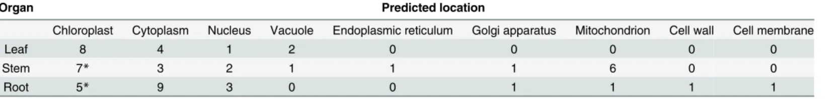
Table 4. Predicted locations of differentially expressed proteins in soybean toB.maydis.
Organ Predicted location
Chloroplast Cytoplasm Nucleus Vacuole Endoplasmic reticulum Golgi apparatus Mitochondrion Cell wall Cell membrane
Leaf 8 4 1 2 0 0 0 0 0
Stem 7* 3 2 1 1 1 6 0 0
Root 5* 9 3 0 0 1 1 1 1
Some proteins are predicted in multiple locations, which could be located in chloroplast and mitochondrion.
doi:10.1371/journal.pone.0141264.t004
Proteomic Analysis of the Relationship between Metabolism and NHR in Soybeans
Acknowledgments
We would like to thank Dr. Zhao Zhenglong of the Yunnan Agricultural University Depart-ment of Plant Protection for the preparation ofB.maydisisolates. We would also like to thank Prof. Fullen Michael of the University of Wolverhampton, U.K. and Dr. Meng Dong of China Agricultural University, China for their linguistic assistance during the preparation of this manuscript, and LetPub for linguistic assistance during the revision.
Author Contributions
Conceived and designed the experiments: CYL YYZ YMD. Performed the experiments: YMD MHP. Analyzed the data: YMD YS PY XHH MHP. Contributed reagents/materials/analysis tools: YMD YS PY MY SSZ XYM. Wrote the paper: YMD CYL.
References
1. Lee HA, Kim SY, Oh SK, Yeom SI, Kim SB, Kim MS, et al. Multiple recognition of RXLR effectors is associated with nonhost resistance of pepper against Phytophthora infestans. New Phytol. 2014; 203 (3): 926–38. doi:10.1111/nph.12861PMID:24889686
2. Senthil-Kumar M, Mysore KS. Nonhost resistance against bacterial pathogens: retrospectives and prospects. Annu Rev Phytopathol. 2013; 51:407–27. doi:10.1146/annurev-phyto-082712-102319 PMID:23725473
3. Lipka U, Fuchs R, Kuhns C, Petutschnig E, Lipka V. Live and let die-Arabidopsis nonhost resistance to powdery mildews. Eur J Cell Biol. 2010; 89(2–3): 194–9. doi:10.1016/j.ejcb.2009.11.011PMID: 19963301
4. Luo Q, Yu B, Liu Y. Differential sensitivity to chloride and sodium ions in seedlings of Glycine max and G. soja under NaCl stress. J Plant Physiol. 2005; 162: 1003–1012. PMID:16173461
5. Yang M, Zhang Y, Qi L, Mei X, Liao J, Ding X, et al. Plant-plant-microbe mechanisms involved in soil-borne disease suppression on a maize and pepper intercropping system. PLoS One. 2014; 9(12): e115052. doi:10.1371/journal.pone.0115052PMID:25551554
6. Li C, He X, Zhu S, Zhou H, Wang Y, Li Y, et al. Crop Diversity for Yield Increase PLoS ONE. 2009; 4 (11): e8049. doi:10.1371/journal.pone.0008049PMID:19956624
7. Dong YM, Pan MH, Zhao ZL, Yang M, Li CY, He XH. Callose deposition in the leaf cells associated with induced resistance of Glycine max to Bipolaris maydis. Journal of Yunnan Agricultural University. 2013; 38(1): 16–20.
8. Heath MC. Nonhost resistance and nonspecific plant defenses. Current opinion in plant biology. 2000; 3(4): 315–319. PMID:10873843
9. Cheng Y, Zhang H, Yao J, Wang X, Xu J, Han Q, et al. Characterization of non-host resistance in broad bean to the wheat stripe rust pathogen. BMC Plant Biol. 2012; 12: 96. doi:10.1186/1471-2229-12-96 PMID:22716957
10. Yun BW, Atkinson HA, Gaborit C, Greenland A, Read ND, Pallas JA, et al. Loss of actin cytoskeletal function and EDS1 activity, in combination, severely compromises non-host resistance in Arabidopsis against wheat powdery mildew. Plant J. 2003; 34(6): 768–77. PMID:12795697
11. Zheng MS, Takahashi H, Miyazaki A, Hamamoto H, Shah J, Yamaguchi I, et al. Up-regulation of Arabi-dopsis thaliana NHL10 in the hypersensitive response to cucumber mosaic virus infection and in senes-cing leaves is controlled by signalling pathways that differ in salicylate involvement. Planta. 2004; 218: 740–750. PMID:14666423
12. Zimmerli L, Stein M, Lipka V, Schulze-Lefert P, Somerville S. Host and non-host pathogens elicit differ-ent jasmonate/ethylene responses in Arabidopsis. Plant J. 2004; 40: 633–646. PMID:15546348
13. Mysore KS, Ryu CM. Nonhost resistance: how much do we know? Trends in plant science. 2004; 9(2): 97–104. PMID:15102376
14. Kim MY, Lee S, Van K, Kim TH, Jeong SC, Choi IY, et al. Whole-genome sequencing and intensive analysis of the undomesticated soybean (Glycine soja Sieb. and Zucc.) genome. Proc Natl Acad Sci USA. 2010; 107(51): 22032–7. doi:10.1073/pnas.1009526107PMID:21131573
15. Li YH, Zhou G, Ma J, Jiang W, Jin LG, Zhang Z, et al. De novo assembly of soybean wild relatives for pan-genome analysis of diversity and agronomic traits Nature Biotechnology 2014; 32: 1045–1052. doi:10.1038/nbt.2979PMID:25218520
Proteomic Analysis of the Relationship between Metabolism and NHR in Soybeans
16. Acero FJ, Carbú M, El-Akhal MR, Garrido C, González-Rodríguez VE, Cantoral JM. Development of proteomics-based fungicides: new strategies for environmentally friendly control of fungal plant dis-eases. Intern J Mol Sci. 2011; 12(1): 795–816.
17. Hakeem KR, Chandna R, Ahmad P, Iqbal M, Ozturk M. Relevance of proteomic investigations in plant abiotic stress physiology. OMICS 2012; 16(11): 621–35. doi:10.1089/omi.2012.0041PMID:23046473
18. Quach TN, Tran LS, Valliyodan B, Nguyen HT, Kumar R, Neelakandan AK, et al. Functional Analysis of Water Stress-Responsive Soybean GmNAC003 and GmNAC004 Transcription Factors in Lateral Root Development in Arabidopsis. PLoS ONE. 2014; 9(1): e84886. doi:10.1371/journal.pone.0084886 PMID:24465446
19. Wang X, Ma J, Li X, Zhao X, Lin Z, Chen J, et al. Optimization of chemical fungicide combinations tar-geting the maize fungal pathogen, Bipolaris maydis: a systematic quantitative approach. IEEE Trans Biomed Eng. 2015; 62(1): 80–7. PMID:25055377
20. Bradford MM. A rapid and sensitive method for the quantitation of microgramquantities of protein utiliz-ing the principle of protein-dye bindutiliz-ing. Anal Biochem. 1976; 72: 248–54. PMID:942051
21. Komatsu S, Makino T, Yasue H. Proteomic and biochemical analyses of the cotyledon and root of flood-ing-stressed soybean plants. PLoS One. 2013; 8(6): e65301. doi:10.1371/journal.pone.0065301 PMID:23799004
22. Hossain Z, Makino T, Komatsu S. Proteomic study ofβ-aminobutyric acid-mediated cadmium stress alleviation in soybean. J Proteomics. 2012; 75(13): 4151–64. doi:10.1016/j.jprot.2012.05.037PMID: 22652489
23. Komatsu S, Nanjo Y, Nishimura M. Proteomic analysis of the flooding tolerance mechanism in mutant soybean. J Proteomics 2013; 79: 231–50. doi:10.1016/j.jprot.2012.12.023PMID:23313221
24. Sobhanian H, Razavizadeh R, Nanjo Y, Ehsanpour AA, Jazii FR, Motamed N. Proteome analysis of soybean leaves, hypocotyls and roots under salt stress. Proteome Sci. 2010; 8:19. doi: 10.1186/1477-5956-8-19PMID:20350314
25. Van der Hoorn RA. Plant proteases: from phenotypes to molecular mechanisms. Annu. Rev. Plant Biol. 2008; 59: 191–223. doi:10.1146/annurev.arplant.59.032607.092835PMID:18257708
26. Gagliano M, Renton M, Duvdevani N, Timmins M, Mancuso S. Out of sight but not out of mind: alterna-tive means of communication in plants. PLoS One. 2012; 7(5):e37382. doi:10.1371/journal.pone. 0037382PMID:22629387
27. Gagliano M, Renton M. Love thy neighbour: facilitation through an alternative signalling modality in plants. BMC Ecol. 2013, 13:19. doi:10.1186/1472-6785-13-19PMID:23647722
28. García-Murria MJ, Karkehabadi S, Marín-Navarro J, Satagopan S, Andersson I, Spreitzer RJ, et al. Structural and functional consequences of the replacement of proximal residues Cys(172) and Cys (192) in the large subunit of ribulose-1,5-bisphosphate carboxylase/oxygenase from Chlamydomonas reinhardtii. Biochem J. 2008; 411(2): 241–7. PMID:18072944
29. Widjaja I, Naumann K, Roth U, Wolf N, Mackey D, Dangl JL, et al. Combining subproteome enrichment and Rubisco depletion enables identification of low abundance proteins differentially regulated during plant defense. Proteomics. 2009; 9 (1): 138–47. doi:10.1002/pmic.200800293PMID:19053141
30. Grienenberger E, Besseau S, Geoffroy P, Debayle D, Heintz D, Lapierre C, et al. A BAHD acyltransfer-ase is expressed in the tapetum of Arabidopsis anthers and is involved in the synthesis of hydroxycin-namoyl spermidines. Plant J. 2009; 58(2): 246–59. doi:10.1111/j.1365-313X.2008.03773.xPMID: 19077165
31. Bouvier F, Rahier A, Camara B. Biogenesis, molecular regulation and function of plant isoprenoids. Prog Lipid Res. 2005; 44: 357–429. PMID:16289312
32. Ramos AA, Marques AR, Rodrigues M, Henriques N, Baumgartner A, Castilho R, et al. Molecular and functional characterization of a cDNA encoding 4-hydroxy-3-methylbut-2-enyl diphosphate reductase from Dunaliella salina. J Plant Physiol. 2009; 166(9): 968–77. doi:10.1016/j.jplph.2008.11.008PMID: 19155093
33. Cunningham FX Jr, Lafond TP, Gantt E. Evidence of a role for LytB in the nonmevalonate path way of isoprenoid biosynthesis. J Bacteriol. 2000; 182: 5841–8. PMID:11004185
34. Altincicek B, Kollas A, Eberl M, Wiesner J, Sanderbrand S, Hintz M, et al. LytB, a novel gene of the 2-C-methyl-D-erythritol 4-phosphate pathway of isoprenoid biosynthesis in Escherichia coli. FEBS Lett 2001; 499: 37–40. PMID:11418107
35. Page JE, Hause G, Raschke M, Gao W, Schmidt J, Zenk MH, et al. Functional analysis of the final steps of the 1-deoxy-D-xylulose 5-phosphate (DXP) pathway to isoprenoids in plants using virus-induced gene silencing. Plant Physiol. 2004; 134: 1401–13. PMID:15064370
Proteomic Analysis of the Relationship between Metabolism and NHR in Soybeans
36. Díaz-Mendoza M, Velasco-Arroyo B, González-Melendi P, Martínez M, Díaz I. C1A cysteine protease-cystatin interactions in leaf senescence. J Exp Bot. 2014; 65(14): 3825–3833. doi:10.1093/jxb/eru043 PMID:24600023
37. Domsalla A, Melzig MF. Occurrence and properties of proteases in plant latices. Planta Med. 2008; 74 (7): 699–711. doi:10.1055/s-2008-1074530PMID:18496785
38. Van Doorn WG, Beers EP, Dangl JL, Franklin-Tong VE, Gallois P, Hara-Nishimura I, et al. Morphologi-cal classification of plant cell deaths. Cell Death Differ. 2011; 18: 1241–1246. doi:10.1038/cdd.2011. 36PMID:21494263
39. Braun HP, Schmitz UK. The mitochondrial processing peptidase. Int J Biochem Cell Biol. 1997; 29(8–
9):1043–5. PMID:9415998
40. Ito A. Mitochondrial processing peptidase: multiple-site recognition of precursor proteins. Biochem Bio-phys Res Commun. 1999; 265(3):611–6. PMID:10600469
41. McConnell JR., McAlpine SR. Heat shock proteins 27, 40, and 70 as combinational and dual therapeu-tic cancer targets. Bioorg Med Chem Lett. 2013; 23(7): 1923–8. doi:10.1016/j.bmcl.2013.02.014 PMID:23453837
42. Momcilovic I, Ristic Z. Expression of chloroplast protein synthesis elongation factor, EF-Tu, in two lines of maize with contrasting tolerance to heat stress during early stages of plant development. J Plant Phy-siol. 2007; 164(1): 90–9. PMID:16542752
43. Su Y, Xu L, Fu Z, Yang Y, Guo J, Wang S, et al. ScChi, encoding an acidic class III chitinase of garcane, confers positive responses to biotic and abiotic stresses in sugarcane. Int J Mol Sci. 2014: 15(2): 2738–60. doi:10.3390/ijms15022738PMID:24552874
44. Li J, Brader G, Palva ET. Kunitz trypsin inhibitor: an antagonist of cell death triggered by phytopatho-gens and fumonisin b1 in Arabidopsis. Mol Plant. 2008; 1(3):482–95. doi:10.1093/mp/ssn013PMID: 19825555
45. Danis P, Farkas R. Hormone-dependent and hormone-independent control of metabolic and develop-mental functions of malate dehydrogenase—review. Endocr Regul. 2009; 43(1): 39–52. PMID: 19309237
46. Kim SG, Kim ST, Wang Y, Kim SK, Lee CH, Kim KK, et al. Overexpression of rice isoflavone reduc-tase-like gene (OsIRL) confers tolerance to reactive oxygen species. Physiol Plant. 2010; 138(1): 1–9. doi:10.1111/j.1399-3054.2009.01290.xPMID:19825006
47. Yang EJ, Oh YA, Lee ES, Park AR, Cho SK, Yoo YJ, et al. Oxygen-evolving enhancer protein 2 is phos-phorylated by glycine-rich protein 3/wall-associated kinase 1 in Arabidopsis. Biochem Biophys Res Commun. 2003; 305(4): 862–8. PMID:12767910
48. Heide H, Kalisz HM, Follmann H. The oxygen evolving enhancer protein 1 (OEE) of photosystem II in green algae exhibits thioredoxin activity. J Plant Physiol. 2014; 161(2): 139–49.
49. Ning CJ, Ding N, Wu GL, Meng HJ, Wang YN, Wang QH. Proteomics research on the effects of apply-ing selenium to apple leaves on photosynthesis. Plant Physiol Biochem. 2013; 70: 1–6. doi:10.1016/j. plaphy.2013.05.008PMID:23770588
Proteomic Analysis of the Relationship between Metabolism and NHR in Soybeans