UNIVERSIDADE DE LISBOA FACULDADE DE
CIÊNCIAS
DEPARTAMENTO DE BIOLOGIA ANIMAL
The role of Dmrt2 in the establishment of Left-Right
asymmetry in the Chick embryo.
Henrique Caldevilla Sentieiro Lyon de Castro
DISSERTAÇÃO MESTRADO EM BIOLOGIA EVOLUTIVA E DO
DESENVOLVIMENTO 2015
UNIVERSIDADE DE LISBOA FACULDADE DE
CIÊNCIAS
DEPARTAMENTO DE BIOLOGIA ANIMAL
The role of Dmrt2 in the establishment of Left-Right
asymmetry in the Chick embryo.
Henrique Caldevilla Sentieiro Lyon de Castro
DISSERTAÇÃO MESTRADO EM BIOLOGIA EVOLUTIVA E DO
DESENVOLVIMENTO 2015
Dissertação de Mestrado orientada por:
Prof. Doutora Maria Leonor Tavares Saúde (iMM Lisboa / FMUL) Prof. Doutora Sólveig Thorsteinsdóttir (DBA/FCUL)
Acknowledgments
“Fifty percent of what we know is wrong. The problem is that we do not know which 50% it is.”
- Timothy Noakes, PhD -
I would like to thank Dra. Leonor Saúde for giving me the opportunity to work in her lab, for the constant support during this past hard working year, and most importantly for believing in my work. I couldn´t have asked for a better supervisor. To all members of LSaúde Lab for the help and advice they gave me throughout this year. I know that I’m not the easiest person to work with so I realy do appretiate the support given.
To Professor Solveig Thorsteinsdottir for being my internal supervisor.
To all of my friends, for making my life better by being there and making me laught when I was down. You are the best!
And last but not least, to my family for the unconditional love and support that they gave me all of these years. I love you guys!!!
Resumo:
Durante o desenvolvimento, diferentes eventos acontecem ao longo dos eixos de um embrião, para que um individuo adulto seja formado. Estes eventos podem dividir-se em dois tipos: (1) Eventos simétricos; (2) Eventos assimétricos.
Um dos eventos simétricos chave é a somitogénese. A somitogénese é o processo pelo qual temos iniciação da segmentação do sistema músculo-esquelético, através da formação dos sómitos, segmentos mesodérmicos transitórios. Este processo de segmentação ocorre progressivamente na mesoderme pré-somítica, numa direcção anterior para posterior. Os pares de sómitos formam-se ao mesmo tempo dos dois lados e sempre com a mesma periodicidade segundo o modelo, proposto em 1976 por Cooke and Zeeman, “Clock and Wavefront model”. Este modelo supõe a existência de uma interacção de factores, cuja produção é cíclica, que marca o “tempo” que as células passam numa zona indeterminada da mesoderme paraxial. Associada a esta ciclicidade de factores encontra-se-ía ainda um gradiente de um outro factor, distinto dos primeiros, que seria responsável por determinar o espaço onde se dá a maturação das células. Quando as células atingem uma determinada região do gradiente, estas interpretariam a combinação de ambos os sinais como informação para se diferenciarem e aglomerarem, formando um novo sómito.
Um conjunto de eventos que se enquadra no ponto (2), é o da formação e organização dos órgãos internos. Um exemplo clássico é o da formação do coração, que está localizado no lado esquerdo do organismo (no direito no caso da galinha) e que sofre uma torção de maneira a que as aurículas fiquem em cima e os ventrículos em baixo. Outro exemplo é o da formação das vísceras que, de maneira a ficarem alojadas correctamente na cavidade abdominal, têm que sofrer uma série de rotações.
Todos estes processos assimétricos têm como base um conjunto de mecanismos que distribui, assimetricamente várias moléculas sinalizadoras, entre eles Shh, Wnts e FGFs, que vão controlar uma cadeia genética altamente conservada entre espécies. Hoje em dia sabe-se que este conjunto de mecanismos, iniciadores de assimetria, são variados e que apresentam algum grau de conservação entre espécies, actuando em diferentes tempos do desenvolvimento. Alguns destes mecanismos são: (1) A distribuição assimétrica de iões Ca2+, antes da gastrulação; (2) Estabelecimento de
um gradiente assimétrico de determinantes via cílios, presentes no organizador, durante a gastrulação; (3) Expressão diferencial de uma cadeia de genes durante a formação de sómitos. Esta cadeia é a cadeia de Nodal-Pitx2, que se sabe ser assimétrica, tendo expressão no lado esquerdo do embrião.
Sabe-se no entanto que os processos de simetria e assimetria, que formam um individuo adulto, ocorrem ao mesmo tempo, pelo que é necessário um, ou vários, mediadores para que a informação seja transmitida corretamente para as diferentes vias. Uma incorreta transmissão desta informação induz graves problemas no correcto posicionamento das diferentes estruturas que constituem um organismo, pelo que, uma boa compreensão dos eventos que coordenam as diferentes vias, é necessária.
Um dos mediadores é o ácido retinóico (RA), que é a versão oxidada e activa da vitamina A. Estudos revelaram que de facto o RA tem um papel importante na protecção dos sómitos contra sinais assimétricos, uma vez que a sua inibição leva a graves problemas na formação dos sómitos, observando-se uma distribuição assimétrica de expressão génica. Um destes genes é o lunatic fringe (Lfng), que faz parte do “relógio molecular” da somitogénese, e está envolvido na diferenciação dos sómitos.
Mais recentemente foi descoberto um novo efector nesta via de protecção. Este efector é o factor de transcrição Dmrt2. Este factor de transcrição, embora pertença a uma família maioritariamente associada à determinação sexual, não só é necessário para a formação simétrica dos sómitos, como é para o correcto estabelecimento da assimetria esquerda-direita. O seu papel exacto nas vias responsáveis por estes eventos, ainda é vastamente desconhecido, pelo que um estudo detalhado sobre este factor de transcrição é necessário.
O objectivo deste projeto seria aprofundar o nosso conhecimento relativamente ao papel do Dmrt2 na formação de estruturas simétricas e assimétricas ao longo do desenvolvimento. Para que tal fosse possível iria ser utilizado o embrião de galinha como modelo, uma vez que este é o modelo em que menos se conhece o papel do
Dmrt2, pelo que a importância de estudos neste organismo é maior.
Para que estas funções sejam desvendadas, iriam ser feitas experiências de ganho de função, através da electroporação do Dmrt2, clonado no vector de expressão pGAGGS, em embriões de galinha no estádio HH3+ e em cultura New, para perceber
qual o seu papel na padronização esquerda-direita. Tal não foi possível pois durante o processo de clonagem percebemos que a região a 5’ do gene não estava bem anotada, pelo que o desenho de primers forward era impossível. Ainda foi tentada uma 5’-RACE (técnica de amplificação de extremidades de DNA), para obter a região 5’, mas sem sucesso.
Para perceber quais os genes que estariam a regular a sua atividade, partimos para uma abordagem de “gene candidato”. Para tal foram feitas manipulações na sinalização de sonic hedgehog (Shh), uma vez que este gene tem a mesma expressão que Dmrt2 mas mais cedo no desenvolvimento. Estas manipulações foram realizadas pondo uma solução de ciclopamina, inibidor da via de sinalização de Shh, in ovo, em embriões por volta do estádio HH4.
Para ver se a expressão de Dmrt2 era afectada, realizamos hibridações in situ para o
Dmrt2, com uma sonda produzida por nós em laboratório, através de plasmídeos
infectados com uma porção do Dmrt2, que já tinha sido isolada, no âmbito de um projecto anterior. Apesar de se terem utilizado concentrações diferentes de droga, não se notou diferenças significativas na expressão entre embriões manipulados
versus controlo.
Tendo em conta estes resultados propomos que a abordagem futura a seguir, será a de optimizar os protocolos usados, nomeadamente o da 5’-RACE, e o da inibição de sinalização hedgehog, via ciclopamina. Deveram ainda ser feitas experiências adicionais, após realizar a experiência de ganho de função, tais como ver a expressão de genes candidatos, que estejam a ser regulados via Dmrt2, e o registo do fenótipo observado. Este registo pode ter como parâmetros a posição de órgãos com disposição assimétrica, como por exemplo o coração.
Abstract:
In order to form a well-developed body, different events need to happen. These events can be symmetric or asymmetric. An example of a symmetric event is the formation of the axial skeleton and it’s associated muscles. An asymmetric event is for instance the establishment of organ morphology and situs. It is known that these events happen in the same time window and in embryonic territories closely located. A well-coordinated set of pathways is therefore needed, otherwise an incorrect transfer of information occurs, leading to serious problems in both somite and organ formation and positioning.
It was shown that in zebrafish, Dmrt2a, a transcription factor of the DM domain family, protects the somites from asymmetric signals and conveys asymmetric signals to place the heart. It is known that zebrafish dmrt2a/terra is expressed in the anterior region of the PSM and somites. In addition, our lab detected
dmrt2a/terra transcripts in the zebrafish KV from the 3-somite stage until the
10-somite stage, showing that this transcription factor has a function in the transition of information from the KV to the LPM.
This transcription factor was also shown to have an asymmetric expression in the chicken’s Hensen’s node and a symmetric expression in the somites, although its function in this embryo has not been accessed..
In this study we tried to assess the role of the Dmrt2 in the establishment of left-right asymmetry in the chick embryo. We tried to clone the full length of dmrt2, by standard PCR and by 5’-RACE, in order to do experiments of gain-of-function, but without success. After this we tried to understand if Sonic hedgehog (Shh) is regulating the asymmetric expression of Dmrt2 in the chicken node, by inhibiting Hedgehog signalling, with different concentrations of cyclopamine, a drug that
inhibits Patched. In situ hybridizations were done in order to understand if Dmrt2 expression was downregulated when Hedgehog signaling was inhibited. No conclusive results were obtained.
The lack of results and the technical hurdles will be discussed and future experiments and expected results will be presented.
Table of Contents:
1. Introduction... 13
1.1. Symmetry and its pathways……….….…….13
1.1.1. Somitogenesis………..……….13
1.1.1.1. The Segmentation Clock……….15
1.1.1.2. The wavefront……….17
1.1.2. Formation of the axial skeleton and it’s associated muscles………18
1.2. Asymmetry and its pathways………...20
1.3. Protection of symmetric events……….……….…24
1.3.1. Retinoic acid……….……….….25
1.3.2. Dmrt2……….26
1.4. Aim of this project………..………..27
2. Materials and Methods ... 28
2.1. Eggs and embryos...28
2.2. Total RNA extraction from chicken embryos...28
2.3. Reverse transcription to obtain cDNA... 29
2.4. Primer design ...29
2.5. Cloning and sequencing…... 30
2.5.1. Standard PCR using chicken cDNA... 30
2.5.2. Optimization PCR using chicken cDNA...30
2.5.3. 5’-RACE using chicken cDNA... 31
2.5.4.1. Gel electrophoresis………...………...32
2.5.4.1. Band extraction………..………..32
2.5.5. Cloning………...………...………...33
2.5.6. Plasmid DNA isolation and purification………..34
2.5.6.1. Lysis of bacterial cells……….……..………....34
2.5.6.2. Purification of Plasmid DNA………..…..….….35
2.5.6.3. STETS (Dirty-Preps) protocol (only used for 5’-RACE plasmids)……..35
2.5.7. DNA sequencing……….…36
2.6. Whole Mount In situ Hybridization………...….…36
2.6.1. Plasmid DNA linearization………...……….…36
2.6.2. Anti-sense RNA probe preparation for in situ hybridization…..………….….37
2.6.2.1. RNA transcription……….………37
2.6.2.3. RNA precipitation……….……….……37
2.6.2.4. RNA probe hydrolysis……….……38
2.6.3. Whole-mount in situ hybridization protocol for chicken embryos……...…38
2.6.4. Image recording and analysis………...……….…..…………40
2.7. New culture………..……..…………..…40
2.8. Electroporation of chicken embryos……….……41
2.9. Drug treatment………..………41
3. Results...48
3.1. Attempts to clone the chicken dmrt2 full-length gene………...………...48
3.1.1. Standard PCR approach to clone the full-length dmrt2 ... 49
3.1.3. Attempt to amplify the 5’ end of dmrt2 using 5’-RACE technique...53
3.2. Shh signalling, a possible upstream regulator of dmrt2 expression...57
4. Discussion ... 60
1. Introduction
In order to form a well-developed body, different events need to happen. These events can be symmetric or asymmetric. An example of a symmetric event is the formation of the bilaterally symmetric structures known as somites. An asymmetric event is for instance the positioning of the organs inside the body cavities.
It is known that these events happen in the same time window and in embryonic territories closely located. A well-coordinated set of pathways is needed, otherwise an incorrect transfer of information would occur.
When we have an interruption in one of the components of these pathways we have severe developmental abnormalities such has, situs inversus, atrophic musculature, and skeletal malformations. Due to these problems, studies in this area of investigation are crucial. To have a better comprehension of the events behind this we first have to look to each one separately.
1.1. Symmetry and its pathways
1.1.1. Somitogenesis
An example of a symmetric event is somitogenesis. Somitogenesis is a fundamental process that initiates segmentation in developing embryos, through the formation of somites. These transient metameric and epithelial structures are located symmetrically on either side of the axial structures (i.e. the notochord and the neural tube) and will give rise to our axial skeleton, skeletal musculature, dermis and the tendons. Temporal and spatial regulation is key to proper somite development.
14
(PSM) in a rostral-to-caudal direction [1,2].
As somites bud off from the anterior end of the PSM, the posterior end of the PSM, is constantly replenished by cells entering from the tail bud [3].
The time required to form a new pair of somites as well as the total number of somites formed is constant and species-specific depending on the temperature. In the chick embryo, at 37oC, a somite pair is formed every 90 minutes, in a total of 50
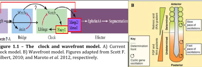
somite pairs formed. In the human, a new pair of somites is formed every 4–5 hours, in the mouse every 120 minutes and in the zebrafish every 30 minutes [4]. This process of differentiation of the PSM to somites is done in a time and space dependent manner, which can be explained by the clock and wavefront model proposed by Cooke and Zeeman, in 1976. This model postulates that a biochemical oscillator – clock – is operating synchronously is PSM cells, while a gradient of maturation – wavefront – sweeps the embryos along the rostral-to-caudal axis (A-P axis), determing the size of each pair of somites (Figure 1.1). This model has been proposed to be translating temporal information into positional information in PSM cells [5].
FCUL – 2013 Marta Sofia Rodrigues, nº. 40097
TEÓRICA 21 – 06.05.2013
Diferenciação de sómitos em vertebrados
Segmentação em Drosophila e em vertebrados
Temos um gene de segmentação em comum - o gene hairy, mas muitos diferentes.
A acção de hairy no sincício de Drosophila é diferente da expressão cíclica de hairy que existe em vertebrados e alguns gastrópodes. Em Drosophila, o
hairy não está envolvido na sinalização notch, pois é expresso antes de
haver sequer celularização do embrião (nestes o hairy é apenas um factor de transcrição que regionaliza no sincício a existência dos 14 segmentos), enquanto que nos vertebrados a expressão cícilica de hairy está envolvido num ciclo de notch que leva a um loop entre células.
Estabelecimento dos eixos
Em Drosophila, o passo seguinte à celularização é a expressão simultânea dos genes de polaridade segmentar e dos genes homeóticos, que vão levar à expressão de genes efectores.
A
Figure 1.1 – The clock and wavefront model. A) Current clock model. B) Wavefront model. Figures adapted from Scott F. Gilbert, 2010; and Maroto et al. 2012, respectively.
1.1.1.1. The segmentation clock
The segmentation clock is responsible for the dynamic and periodic expression of mRNA of a number of “clock” genes across the PSM in a posterior to anterior fashion, with a periodicity that matches somite formation (Figure 1.1. A).
The molecular evidence for the existence of this segmentation clock came with the discovery of the first cyclic gene, the avian basic-helix-loop-helix (bHLH) transcription factor hairy1. The chick hairy1 gene shows a dynamic and reiterated expression pattern in the PSM with the exact same periodicity of somite formation [6]. These hairy1 messenger RNA (mRNA) oscillations occur autonomously in PSM cells and because they are synchronized with adjacent cells, describe a wave of expression starting at the posterior PSM and moving towards the anterior PSM, where it slows down and eventually stops, concomitant with somite formation (Figure 1.1. A). Therefore, PSM cells undergo several periodic oscillations of hairy1 gene expression before they incorporate into the next somite [6].
Hairy1 is a bHLH transcription factor downstream of the Notch-Delta pathway. Other bHLH transcription factors with a cyclic behaviour were next found namely, Hairy2 in chick, Hairy and enhancer of split 1 (Hes1) and Hes7 in mouse, Hairy and enhancer of split-related 1 (Her1) and Her7 in zebrafish, just to name a few.
Indeed, the analysis of mouse and zebrafish mutants for several components of the Notch pathway revealed that cyclic gene expression and somite boundary formation were disrupted to varying degrees. Nevertheless, the anterior somites developed normally and only the posterior ones were affected in the Notch mutants [7][8][9]. These findings showed that Notch signalling is not entirely necessary for somite formation but instead suggested that its failure leads to a gradual perturbation in somite segmentation.
both Lunatic fringe (Lfng) [10][11] and Hairy1 [6], in chicken, that exert inhibitory action upon their own promoters, thus leading to a negative feedback loop.
Lfng is activated, by Notch intracellular domain (NICD), acting post-translationally
in the notch receptor, altering its sensibility to Delta, leading to a decrease in notch-delta signaling [5]. Hairy1 expression, also activated by NICD, will then be responsible for the inhibition of Lfng and, when its protein expression levels get high enough, its own. When the concentration of Hairy 1 is low enough the notch-delta signaling becomes active again and a new cycle is started.
Recent studies showed that components of the Wnt pathway have a cyclic expression in the same phase with Notch pathway components, and have a role in somitogenesis [12] [13]. In fact, mutants for Wnt3a showed a downregulation of the expression of Lfng and Dll1 in the tailbud [13]. This function of Wnt signalling is regulated by a negative feedback loop of Axin2 in the PSM [12] [14]. This Axin2 is a critical component of the Wnt signaling pathway that acts as a scaffold for the β-catenin destruction complex.
Although Wnt signalling is implicated in the somitogenesis process, components of the FGF pathway, like snail1 and snail2, have also been showed to have cyclic expression. The determining agent in linking FGF signaling to Notch-related cyclic gene expression and, ultimately, proper somite segmentation in the zebrafish embryo is Hairy/Enhancer-of-Split molecule, Her13.2. Expression of her13.2 is induced by soaked beads and decreased by an FGF signalling inhibitor. FGF-induced Her13.2 acts as a dimerization partner for Her1, and the formation of this heterodimer is required for transcriptional feedback repression on Her1 promoter, ensuring its cyclic expression, and for somitic border formation throughout the entire A-P axis [15] [16]. Another study described cyclic expression of snail1 and
pathways and was Notch-independent. This link of SNAIL proteins and the FGF pathway is observed when FGF signalling is inhibited. snail1 expression is lost in the PSM of mouse mutants for FGFR1 [17]. When the activity of FGFR1 is blocked, snail2 was found to be downregulated in the PSM, whereas it was upregulated in the neural tube and the lateral plate. Overexpression of FGF8 by electroporation blocked somite formation, but no ectopic expression of snail2 was observed in the paraxial mesoderm of FGF8-overexpressing embryos. These experiments suggest that FGF signalling is necessary, but not sufficient, for snail2 expression in the chick PSM. Nevertheless, snail2 misexpression ceased lfng oscillations and impaired epithelial somite formation [18].
In summary, FGF and Notch clusters of cycling genes are activated in phase, while genes belonging to the Wnt cluster present an opposite cycling phase to the Notch/FGF cluster.
1.1.1.2. The wavefront
The wavefront component of the model states that between the determined and undetermined region of a developing embryo is a determination front, corresponding to the intersection of FGF8 and retinoic acid (RA) gradients, the first going from caudal to rostral, and the second going rostral to caudal (Figure 1.1. B) [19]. Once the PSM cells cross this front, the somitogenic program is activated and deposition of extracellular matrix (ECM) starts the individualization of somites [20]. The posterior-to-anterior gradient of fgf8 mRNA in the PSM was the first one to be described in several vertebrate embryos (mouse, zebrafish and chick) [21][22][23]. In fact, displacing the position of the determination front by altering the extent of the Fgf8 gradient results in the shift of the somite boundary position [21]. This is because Fgf8 is responsible for maintaining the PSM cells in an undifferentiated
state.
What we see is a more intense RNA gradient, in the posterior part of the embryo, fading out as we reach the anterior part of the PSM. This more intense RNA gradient is because fgf8 mRNA is transcribed in PSM progenitors but not in the PSM itself. This fading out of the gradient is established by the cells movements, which are occurring when cells leave the tailbud, and also due to the long half-life of fgf8. As the mRNA gets more mature the expression of FGF8 becomes more fade-out, because of the degradation of the more mature mRNA. This diminishing of protein expression leads to a more permissive environment for cells to differentiate. The precise regulation between undifferentiated and differentiated fronts is done by the counteracting gradient of RA. Once the cells contact with this determination front of low FGF8 and increasingly RA concentration they start the differentiation process, which culminates inamesenchymal-to-epithelial transition (MET).
1.1.2. Formation of the axial skeleton and it’s associated muscles
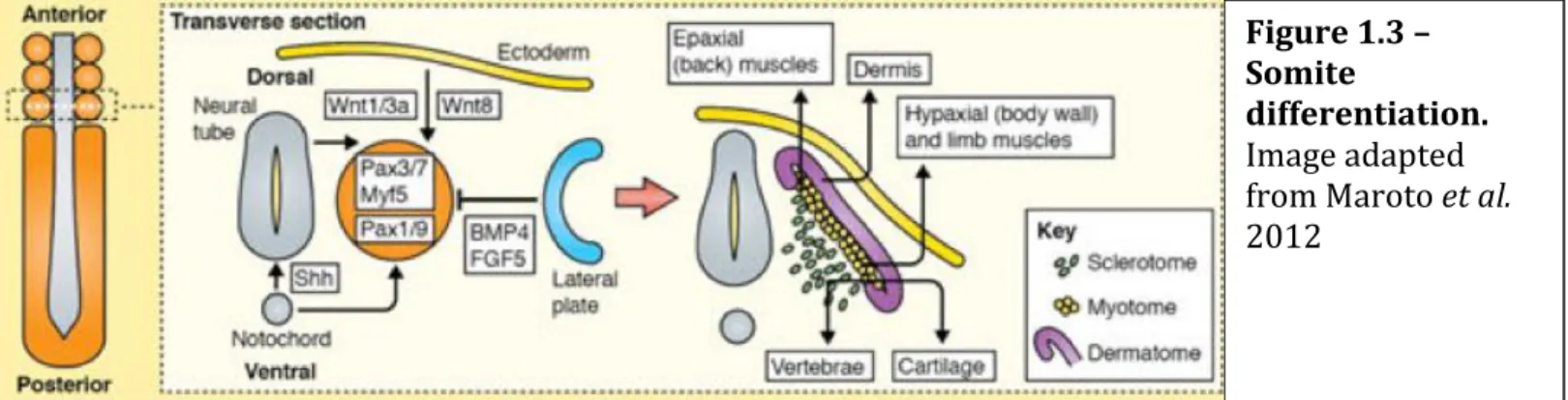
After the individualization of the somite, a maturation process begins. Two major compartments will form within the somite: the sclerotome and the dermamyotome (this will then divide to from the myotome and the dermatome) (Figure 1.3.) [22].
The sclerotome is formed when cells from the ventral part of the somite
de-Figure 1.3 – Somite
differentiation. Image adapted from Maroto et al. 2012
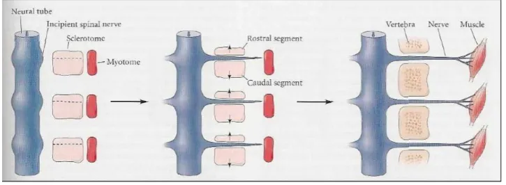
epithelized and turn into mesenchymal cells. The ventromedial portion of the somite is induced to become the sclerotome by paracrine factors, especially Noggin and Sonic hedgehog, which are secreted from the notochord [23][24]. This paracrine factors will induce the expression of Pax1, which is required for their epithelial-to-mesenchymal transition and subsequent differentiation into cartilage [25]. The sclerotome will then give rise to the vertebrae and rib cartilage, and the syndetome (tendon precursor) in a process called somite resegmentation. In this resegmentation we will have the division of the sclerotome in a rostral and caudal segment (Figure 1.4.). Each caudal segment combines with the next anterior sclerotome, forming the vertebral rudiment. Meanwhile the motor neurons, that will innervate the muscles, migrate trough the anterior part of the sclerotome dividing it in half, helping in this way the resegmentation process.
Figure 1.4 - Resegmentation of the sclerotome. Each caudal segment combines with the next anterior sclerotome, forming the vertebral rudiment. Meanwhile the motor neurons, that will enervate the muscles, migrate trough the anterior part of the sclerotome dividing it in half. Image adapted from Scott F. Gilbret, 2010.
The dorsal cells of the somite maintain the epithelial identity forming the dermamyotome (precursor of skeletal musculature and dermis of the dorsal skin). This dermamyotome will then be divided in myotome and dermatome.
Myotome formation involves two sequential steps: first, cells from the dorsomedial edge of the dermomyotome move underneath and form the epaxial myotome (musculature of the back and intercostals); in a second phase the central dermomyotome cells de-epithelialize and form the hypaxial musculature and the dorsal dermis of the trunk. This differentiation is due to Wnt signalling, namely Wnt1/3a from the neural tube, Wnt7a and Wnt8c from the ectoderm, that participate in the induction of the myogenic program in the epaxial and hypaxial lips respectively [26][27][1]. This myogenic program starts with the expression of the myogenic factors Myf5 (myogenic factor 5) and MyoD (myogenic differentiation 1), in Pax3/7 positive cells, eventually generating the epaxial and hypaxial muscles and also part of the dermis of the back.
The dermatome, the precursor of dermis tissue, is formed through signals via Neurotrophin 3 (NTF3) from the neural tube [27].
1.2. Asymmetry and its pathways
Although symmetric developmental processes are crucial, asymmetric developmental events are also needed in order to have a fully developed body.
One of the main mechanisms for the establishment of Left-Right asymmetry is the Nodal cascade, which takes place during somitogenesis, and is present in all vertebrate model systems and some invertebrates. Nodal is expressed on the left side of the node and the lateral plate mesoderm (LPM) [28][29], and it was shown that inhibition of Nodal will lead to right isomerism (formation of two right sides)
[30], making it a left-side determinant.
Other genes in this cascade are the Lefty gene family, composed by lefty1 and lefty2 [31], which are antagonists of Nodal signalling, thus creating a regulatory network between Nodal and the Lefty genes. So, when Nodal protein reaches a cell in the left side, it will activate Nodal in the left LPM (Lateral Plate Mesoderm), that when Nodal protein reaches a certain concentration, activates Lefty1 and 2. Lefty1 will be expressed in the midline inhibiting the expression of Nodal in the right side. Lefty2 will restrict the domain of Nodal on the left LPM [32][33].
This expression of nodal in the left LPM will lead also to the expression of the homeobox transcription factor pitx2. Pitx2 will promote the asymmetric establishment of the internal organs [33]. The mechanism by which Pitx2 contributes to this left-right asymmetry is not yet fully understood.
This conserved cascade can be activated differentially according to the organism. In the mouse and chick we have the activation of Nodal cascade, in the node, by Notch-Delta signalling [34][35]. Although Notch-Notch-Delta activates the Nodal cascade in the mouse and chick, in zebrafish we have the activation of Charon, an inhibitor of Nodal, wich is expressed in Kupffer’s vesicle, the node homologous LR organ [36]. Another molecule that is responsible for the asymmetric expression of nodal is Fgf8. The gene fgf8 is expressed in the chick, mouse and zebrafish, but has a different role in one of each organism. In mouse fgf8 acts as a positive regulator of nodal, therefore acting as a left determinant [37]. In the chick fgf8 acts has a repressor of
nodal, having its expression in the right side of the Hensen’s node, thus acting like a
right determinant [38]. In zebrafish, the function of fgf8 has not been determinated, although fgf8 mutants have LR abnormalities [39].
One molecule that is also responsible for the expression of nodal in the left side of the perinodal region, is Shh (Sonic hedgehog) that has its expression restricted to
the left side around stage HH4+ in chick [28][29]. Studies showed that the
asymmetrical shh expression at stage HH5 is considered to be itself upstream of asymmetric nodal expression, because right-sided implantation of Shh producing cells induces ectopic nodal expression in right LPM [28], whereas Shh antibody administration at stage HH5 effectively suppresses initiation of nodal expression in the left LPM [40].
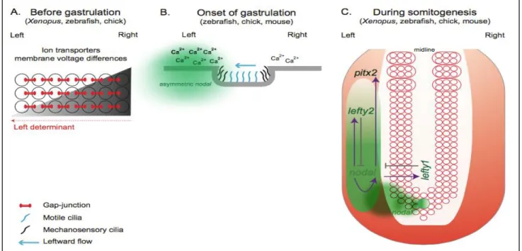
One question that remained unanswered for a long time was how is this differential gene expression established. The ways by which these asymmetric processes are established are diverse, and can be through three distinguish mechanisms that act in different developmental times (Figure 1.8)[33].
Figure 1.8 - Set of events that might culminate with the establishment of the left-right patterning in different vertebrates. A) Prior to gastrulation in Xenopus, zebrafish and
chick, ion transporters asymmetrically distributed in the embryo generate differences in membrane voltage potential between the left and right side. B) In mouse, it is though that mechanosensory cilia present in the node epithelia sense the leftward fluid flow created by motile cilia and as a consequence trigger an asymmetric Ca2+ release which will induce an asymmetric nodal expression around the node. C) A conserved Nodal cascade is activated at the onset of gastrulation in Xenopus, zebrafish, chick and mouse. Nodal is asymmetrically
One of these mechanisms that are observed in chick, zebrafish and Xenopus, prior to gastrulation, is the establishment of a differential membrane potential due to an asymmetric distribution of ions done by channels present in the cell membrane. As a consequence, we will have an asymmetric distribution of extracellular Ca2+, via
gap-junction communication channels (GJC). If we have GJC inhibition, LR patterning problems will appear [41][42]. Also Ca2+ accumulation was shown to induce an
asymmetric activation of Notch on the left side of the node that then translates this differential activity into asymmetric nodal expression. Perturbing this early asymmetric ion flux, will lead to randomized gene expression and organ heterotaxia [43].
Another mechanism, that happens during gastrulation, is the presence of motile and mechanosensory cilia [33][34]. One theory states that a morphogen is transported by the leftward flow created by the motile cilia, present in the node cells, thus breaking the symmetry. This leftward flow is possible because the cilia are posteriorly tilted [32]. The mechanism by which mechanosensory cilia break symmetry is due to an asymmetric intracellular Ca2+ flux, which will induce
asymmetric nodal expression [45]. This asymmetric Ca2+ is generated by
polycystin-2 (PKD) calcium activated channel [46]. This cilia mechanism of breaking symmetry can be found on the mouse node, zebrafish Kupffer’s vesicle, and Xenopus gastrocoel roof plate [33].
There are no motile cilia in chicken embryos so the way of breaking symmetry in this organism is by leftward cell movements, downstream of the H+/K+-ATPase
pump activity. This alternative strategy was seen in chick embryos at stage HH4 [47]. These cell movements stopped at stage HH5 by a N-cadherin dependent process, that is localized in the right side of the node, leading to asymmetric expression of nodal and fgf8 [48].
1.3. Protection of symmetric events
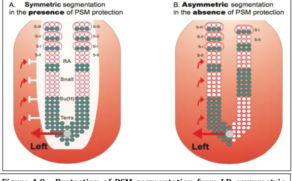
As we saw, in order to have a well developed body a coordinated set of events, either symmetric or asymmetric, need to occur. Since these contradictory processes occur in the same timeline and in close territories the problem of how these events are protected from each other arises. Studies in the area showed that a set of conserved mediators are responsible for this protective barrier. Some of these mediators are RA, Snail and Dmrt2 (former Terra) (Figure 1.9).
Figure 1.9 - Protection of PSM segmentation from LR asymmetric patterning cues. A) PSM is protected from LR signals that come from
the node and are implicated in left-right patterning (red arrows). This protection consists of a “shield” (white) which so far has been shown to be composed by RA, Snail, Su(H) and Terra. In its presence, cyclic gene expression (blue) and somite formation are symmetric between the left and right sides. B) In the absence of this protection, cyclic gene expression becomes desynchronised between both sides. Consequently, somite formation proceeds in an asymmetric way, with the left side exhibiting more somites than the right (this biased asymmetry towards the right side is seen in mouse and fish embryos, while in chick
asymmetries are biased to the left side). Figure adopted from Lourenço
1.3.1. Retinoic acid
Retinoic acid (RA) is a morphogen derived from retinol (vitamin A) that plays important roles in cell growth, differentiation, and organogenesis [49]. It has been shown that RA acts has a buffer, during somitogenesis, preventing LR signals from disrupting symmetric signals that will form the somites [50]. When we have a blocking in the production of RA, in chicken, mouse and zebrafish embryos a desynchronization of somite formation between the two embryonic sides is observed [51][52]. This desynchronization is accompanied, in chicken embryos, by an expression domain of Lfng extend more anteriorly on the left than on the right embryonic side, in disulphiram-treated embryos (disulphiram inhibits raldh2, the enzyme responsible for converting retinol into RA). No asymmetric expression was seen for fgf8 in the chicken embryos [52]. In mouse expression of the cyclic genes
hes7 and lfng are out of phase between the left and right sides. The same erratic
expression pattern is observed in deltaC, her1 and her7 in zebrafish. Also consistent with the somite phenotype, is the anterior displacement of the wavefront seen by the anterior expansion of fgf8 on the right side of the PSM. These observations showed that the primary target of the uncoordination process is the oscillations of the clock [52][53].
One question that arises is: what are the mechanisms by which RA is protecting the symmetric signals from the asymmetric ones? One of the molecules of this RA pathway is Snail, namely snail1 that is transiently expressed in the right LPM, in both chick and mouse embryos, having a role in organ lateralization. It was seen that this transient expression of snail1 corresponds to the period of RA expression in the PSM. If we have absence of RA signalling, during that period, snail1 expression is affected leading to its expression on the right anterior PSM. This erratic expression of snail1 leads to asymmetric expression of the cyclic genes snail2 e lfng leading to
asynchronous somitogenesis [33].
1.3.2. Dmrt2
Recently the role of a new mediator in the crosstalk between symmetry and asymmetry was discovered. This mediator is Dmrt2, a transcription factor of the DM domain family, that is a family previously involved in sex determination [54]. Despite being mainly expressed in developing gonads and associated with sex differentiation, not all the vertebrate dmrt genes are associated exclusively with this function. So far, from the eight known dmrt genes, five of them have already been implicated in other developmental processes other than sex differentiation. dmrt genes have been detected in the central nervous system, nasal placodes and in the somites [55].
Dmrt2 in particular has a role in somitogenesis, more specifically in somite
differentiation in mice and zebrafish, having its expression in the dermomyotome of developing vertebrate somites [56][57][58]. Mouse dmrt2-/- mutants die due to abnormal rib and sternal development, having also defects in the expression pattern of dermomyotomal and myotomal transcription factors [57].
In zebrafish, we have Dmrt2a and Dmrt2b, due to the genome duplication event, with different functions. Zebrafish injected with Dmrt2a-morpholino, displayed a randomization of clock-specific genes, such has deltaC, her1, her7 and left dterminants like pitx2 and spaw; a randomization of the heart position was also seen [59]. The dmrt2b gene is functionally divergent from dmrt2a regarding its role during somite formation. Instead of being necessary for symmetric somite formation, it is in fact involved in the regulation of somite differentiation at the level of slow muscle development through the regulation of the Hedgehog pathway [60].
Dmrt2a and dmrt2b share the same expression pattern in the anterior region of the
patterns, it was showed that dmrt2a is also expressed in the KV, in the 3-somite stage until the 10-somite stage, from where dmrt2b is in fact absent [33]. This observations lead to the suspicion that Dmrt2a is acting at the level of the KV to regulate both pathways, by doing the transition of information from the KV to the LPM, and that its expression in the anterior PSM region and somites is probably involved in somites differentiation. If this is true, it may help to understand why
dmrt2b is not necessary for symmetry somite formation and only somite
differentiation. A plausible explanation for the fact that dmrt2b is involved in the regulation of the LR asymmetry pathway, despite being absent from the KV, is that
dmrt2b is regulating the Hedgehog signalling at the level of the midline, whose
integrity is necessary for the correct establishment of the LR asymmetry pathway. The role of the dmrt2 in the chick embryo is still unknown; a role in left-right patterning, similar to dmrt2a in zebrafish, is a plausible hypothesis, due to the asymmetric expression in the left side of Hensen’s node in HH4+ stage embryos [59],
that is similar to the expression pattern of other genes that are known to have a role in LR patterning, like shh and nodal [28]. Dmrt2 is also expressed in the somites [59]. Although the expression of Dmrt2 in chick is known, its function is not. Revealing this function can lead to major breakthroughs in this area, thus giving a more complete vision of the processes behind LR patterning, and consequently to the problems that are behind LR defects, such as situs inversus.
1.4. Aim of this project
The aim of this project was to evaluate the function of Dmrt2 in LR patterning in the chicken embryo and uncover potential upstream regulators. In order to do so our initial idea was to do gain-of-function experiments and perform drug treatment experiments respectevly.
2. Materials and Methods
2.1. Eggs and embryos
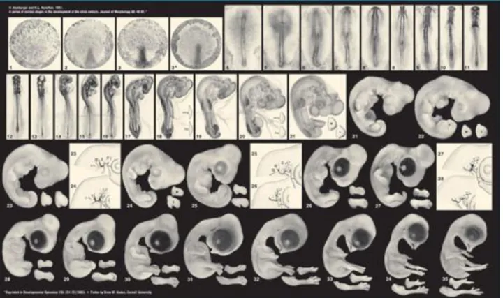
Fertilized chicken (Gallus gallus) eggs were obtained from commercial sources (Sociedade Agrícola Quinta da Freiria, Portugal) and incubated at 37°C in a 17% humidified incubator. Embryos were staged according to the Hamburger and Hamilton development table (Figure 2.1.) [61].
2.2. Total RNA extraction from chicken embryos
Total RNA was isolated from entire embryos at stage HH10 and from the Hensen’s node of stage HH4 embryos. To disrupt the cells and dissolve cellular components, maintaining the integrity of the RNA, 1 ml of Trizol reagent (Gibco-BRL) was added to the tubes per each 50-100 mg of tissue, which was thoroughly ressuspended. Next, the mixture was incubated for 5 minutes at 30°C, to allow the complete dissociation of protein complexes. This was followed by a dilution with 0,2 ml of Chloroform per each 1 ml of Trizol reagent initially used, a shaking for 30 seconds and an incubation for 3 minutes at 30°C. Then, the mixture was separated into three phases through a centrifugation at 4°C for 30 minutes at 6.000 g. This centrifugation places the red phenol-chloroform phase lower, followed by an interphase and finally a colourless upper aqueous phase where the RNA remains. The aqueous phase was transferred to another eppendorf tube. To precipitate the RNA, 0,5 ml of Isopropyl Alcohol were added per each 1 ml of Trizol reagent initially used, followed by an incubation of 10 minutes at 30°C. The RNA was then centrifuged at 4°C during 15 minutes at 10.000 g. The supernatant was discarded, the pellet washed once with 1 ml of 75% EtOH, per each 1 ml of Trizol, and centrifuged at 4°C during 15 minutes at 10.000 g. Finally, the RNA pellet was allowed to air dry, dissolved in 30 µl of RNase
free water and stored at -80°C.
2.3. Reverse transcription to obtain cDNA
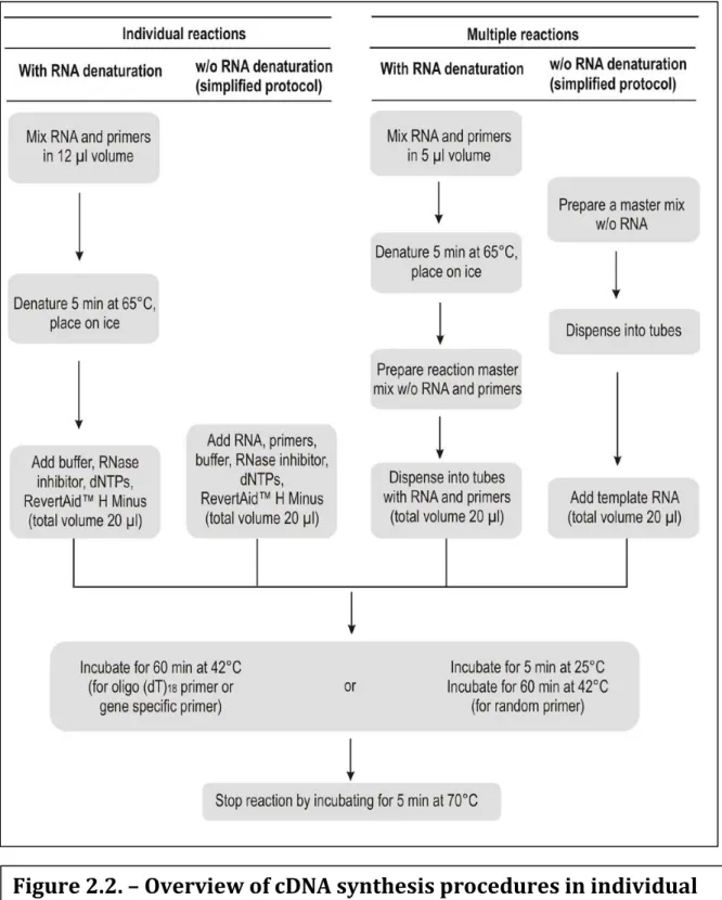
The reverse transcription is a method that allows obtaining complementary DNA (cDNA), from a RNA sample. This reaction is done by an enzyme, which is found in viruses, called reverse transcriptase. This technique functions as a standard PCR, with the difference that the template is RNA, not DNA. With this technique we are cloning expressed genes by reverse transcribing the RNA of interest, not simply generating copies of a gene. For the general process see Figure 2.2.
For the reverse transcription we used the Thermo Scientific RevertAid H Minus First Strand cDNA Synthesis Kit #K1631, #K1632 (Thermo Scientific). For this reaction we used a random hexamer primer provided by the kit. The reason why we used random primers is because they initiate cDNA synthesis from the total RNA population (rRNA and mRNA), instead of the oligo(dT) that give us exclusively the mRNA population. Therefore, using random primers for first strand synthesis results in a greater complexity of the generated cDNA (since we started with a bigger pool of RNAs) compared with the oligo(dT) (exclusively the mRNA population).
2.4. Primer design
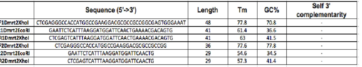
Sets of gene specific primers for dmrt2 were design. For the sake of simplicity we have grouped the primers into 3 types. Type 1 (Table 2.1.) and Type 2 (Table 2.2.) primers were design using Primer BLAST from NCBI. Type 3 Primers (Table 2.3.) were designed using Primer3. The type 1 primers have restriction sites added to its 5’ and 3’ ends, for vector cloning. The type 2 primers differ from the type 1 simply because they are missing the restrictions sites. In the type 3 primers we designed
only reverse primers using specific characteristics such as, length (23-28 nt), melting temperature (Tm≥65°C), GC% (50-70%) and not complementary to the
3’-end of the Universal Primer Mix (For the specific numbers consult table 2.3.).
2.5. Cloning and sequencing
2.5.1. Standard PCR using chicken cDNA
For the standard PCR using type 1 primers, four master mixes of solutions were prepared. Mix 1 was done with forward and reverse primers with a Xho1 restriction site; Mix 2 was done with forward primer and reverse primers with a EcoR1 restriction site; Mix 3 was done with a forward primer with a Xho1 restriction site and a reverse primer with a EcoR1 restriction site; Mix4 was done with a forward primer with a EcoR1 restriction site and a reverse primer with a Xho1 restriction site. The PCR program was the one described in Table 5. The polymerase used for this PCR was Phusion High-Fidelity DNA polymerase (#F-530L, Thermo Scientific).
2.5.2. Optimization PCR using chicken cDNA
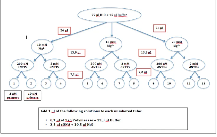
An optimization PCR protocol was done using the different combinations of Type 2 Primers (Table 2.2.), and different annealing temperatures (obtained by subtracting 50C of the Tm of primer), in a one-degree interval (one bellow and one higher of the
calculated temperature). The polymerase used in these reactions was Taq DNA Polymerase (recombinant) (#EP0402, Thermo Scientific). The optimization protocol is described in Figure 2.3.
2.5.3. 5’-RACE using chicken cDNA
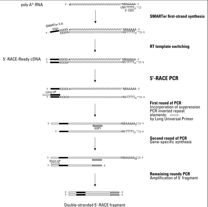
5’-RACE is a method for performing 5’-rapid amplification of cDNA ends (RACE). The way that this is done is by producing a cDNA sample that has an additional SMARTer sequence (3–5 modified bases that anneal to the extended cDNA tail). After this a two rounds PCR is done with different primers in order to have a Double-stranded 5'-RACE fragment (for the mechanism in detail consult Figure 2.4.) The 5’-RACE was done using the SMARTer RACE cDNA Amplification Kit (PT4096-1, Clontech), starting with 1 µg of chicken total RNA (for the RNA sampling see section 2.2.). For our gene-specific primers (GSP) we used the type 3 primers (Table 2.3.).
2.5.4. Gel electrophoresis and band extraction
2.5.4.1. Gel electrophoresis
The success of the amplification was determined by an agarose gel electrophoresis. The gel was prepared by dissolving agarose (Gibco-BRL) in 1x TAE buffer (40 mM Tris-acetate; 2,0 mM EDTA pH 8,5), to a final concentration of 2% (for Standard and Optimized PCR), and 1,2% (for 5’-RACE). To dissolve the agarose, the mixture was heated until a transparent solution was obtained and, when cooled, Red Safe 20 000X (Intron Biotechnology). This is a substance that intercalates with DNA bases, becoming fluorescent when exposed to ultraviolet (UV) light, allowing visualization of DNA molecules. The agarose mixture was then transferred to a gel mould with the appropriate comb in place. The gel was allowed to polymerize and was then covered with 1x TAE buffer. In eppendorf tubes, the following mixtures were made: 50 µl of PCR/5’-RACE product + 5 µl of 6x Loading Buffer (6 mM EDTA; 0,5% Bromophenol Blue; 40% Sucrose); 5 µl of Smart Ladder (5 μl/lane) (Eurogentec molecular weight
marker that allows the determination of the DNA fragment size). The samples were briefly centrifuged and loaded in the gel wells (25 µl of product/well). An electric field of 100V was applied until the appropriate resolution was achieved. Since the nucleic acids are negatively charged, the DNA runs from the negative pole (black) towards the positively charged pole (red) of the electrophoresis apparatus. In the end, the gel was placed on a UV light box and a photograph was taken using the ImageLab program.
2.5.4.2. Band extraction
After photographed, the gel was placed under UV light and a band with approximately 1,4 Kb size (obtained from Standard and Optimized PCRs) and a band with 200/450 bp size (obtained from 5’-RACE), were excised with a clean sharp scalpel and placed in a eppendorf tube. The gel slice containing the DNA fragment was weighted and the DNA extraction from the agarose gel was carried out using the QIAQuick gel extraction kit. 3 volumes of Buffer QG (solubilization and binding buffer) were added for each volume of gel (100 mg ~ 100 µl), being the maximum amount of gel slice per QIAQuick column 400 mg (for gel slices > 400 mg more than one QIAQuick column was used). The tube was then incubated at 50°C for 10 minutes, until the gel slice was completely dissolved (to help dissolving the gel, the tube was vortexed every 2 minutes during the incubation). After that, one gel volume of Isopropanol was added to the tube. To collect the DNA, the content of the tube was transferred into a QIAQuick spin column (which was inserted in a 2 ml collection tube) and centrifuged for 1 minute at RT (the maximum volume of the column was 800 µl). The flow through was discarded, the column placed back in the same collection tube and 0,5 ml of Buffer QG were added, followed by a centrifugation of 1 minute at RT. To wash, 0,75 ml of Buffer PE were added to the
column, which was left stand 2-5 minutes before centrifugation of 1 minute at RT. The flow through was once again discarded and the column centrifuged for an additional minute at RT. The column was then placed into a sterile 1,5 ml eppendorf tube and the DNA eluted with 30µl of RNase free water, being centrifuged for 1 minute at maximum speed and RT. The DNA was then stored at -20°C. Quantification of purified DNA was done using a NanoDrop 2000, a full-spectrum (220-275 nm) spectrophotometer that measures 1 μl of samples with high accuracy.
2.5.5. Cloning
As our cloning vector, we used pGEM-T and pGEM-T Easy Vector Systems (Promega). The reason for using pGEM-T, was because this vector can be sequenced using universal primers making the process of sequencing much less laborious. Since the pGEM-T vector has a polyT tail, the transformation is also easier, since we only need to add a polyA to our sequence in order to successfully clone our vector. The best-fit ratio for the pGEM-T transformation was calculated by using the ligation calculator of NEBioCalculator. The produced plasmids were introduced into Subcloning Efficiency™ DH5α™ Competent Cells (18265-017, Thermo Fisher Scientific) and the transformed bacteria were cultured in LB medium with 100µg/ml of Ampicillin (A9518-SG, Sigma). A background control, with the plasmid alone, was done, in order to evaluate the success of the tranformation. In order to identify the bacteria colonies that are effenciatly transformed we performed a blue-white selection test using XGal-4 99,5% 1g (Bio-37035, BIOLINE), at 20mg/ml in DMF.
2.5.6. Plasmid DNA isolation and purification
2.5.6.1. Lysis of bacterial cells
Purification of plasmid DNA was done using the Promega’s Wizard® Plus SV
Minipreps DNA Purification System. A 4 ml bacterial culture was pelleted by centrifugation at room temperature (RT) for 5 minutes, at 16.000 g (centrifugal force). The supernatant was discarded and the tube was inverted on a paper towel in order to remove the excess media. Next, the bacterial pellet was thoroughly resuspended in 250 µl of Cell Resuspension Solution (50 mM Tris-HCl pH 7,5; 10 mM EDTA; 100 μl/ml RNase A), followed by the addition of 250 µl of cell lysis solution (0,2 M NaOH; 1% SDS). The tube was inverted 4 times and incubated at RT for 5 minutes, until the cell suspension cleared. Then, in order to inactivate endonucleases and other proteins released during the lysis of bacterial cells (that can affect the quality of the isolated DNA), 10 µl of Alkaline Protease Solution (approximately 250 μg per sample) were added. The tube was inverted 4 times and incubated at RT for 5 minutes. Finally, 350 µl of Neutralization Solution (4,09 M guanidine hydrochloride; 0,759 M potassium acetate; 2,12 M glacial acetic acid) were added and the tube inverted 4 times, followed by a centrifugation of the bacterial lysate at RT for 10 minutes, at 16.000 g.
2.5.6.2. Purification of Plasmid DNA
The supernatant obtained in 2.5.6. was transferred to a spin column (inserted in a 2 ml collection tube), avoiding the transfer of any of the white precipitate, and centrifuged at RT for 1 minute at 16.000 g. The spin column was then removed from
the collection tube and the flow through was discarded. This was followed by the reinsertion of the spin column into the collection tube and the addition of 750 µl of column Wash Solution (162,8 mM potassium acetate; 22,6 mM Tris-HCl pH 7,5; 0,109 mM EDTA pH 8,0). The centrifugation occurred at RT for 1 minute, at 16.000 g. The spin column was once more removed from the collection tube and the flow through discarded. The column was reinserted into the collection tube, and the wash procedure repeated using 250 µl of column Wash Solution. This was centrifuged at RT for 2 minutes, at 16.000 g. The spin column was transferred to a new sterile 1,5 ml microcentrifuge tube and the plasmid DNA eluted in 30 µl of nuclease-free water, followed by a centrifugation at RT for 1 minute at 16.000 g. After eluting the DNA, the spin column was discarded. Before storing the DNA at -20°C (where it remains stable), 1 μl of the sample was collected in order to quantify the DNA. The concentration of the purified DNA was determined spectrophotometricaly using the Nanodrop 2000.
2.5.6.3. STETS (Dirty-Preps) protocol (only used for 5’-RACE
plasmids)
After growing our colonies, 12 to 24 were inoculated into a 15 ml falcon with 5 ml of LB medium with Ampicillin (A9518-SG, Sigma). Colonies were lefted to grow O/N with agitation at 37°C. Next day the falcons were centrifuged for 30 seconds to 1 minute at 13000 rpm. After centrifuged the flowthrough was discarded. Next the pellet was ressuspended in 750 μl of STET (5% Triton, 5% Sucralose, 50 mM EDTA, 50 mM TrisHCl pH 8), plus Lysozime. The solution was vortexed and boild for 2 minutes at 100°C in a dry bath. Then we centrifuged at 13000 rpm for 10 minutes, and removed de pellet with a sterile toothpick. After removing the pellet, 750 μl of Isopropanol was added to the flowthrough followed by brief vortexing. Next the
solution was centrifuge for 10 minutes at 13000 rpm and washed with 500 μl of ethanol at 70%, and the falcons were centrifuged for an additional 2 minutes. After centrifuged the flowthrough was discarted and the pellet was left to dry. After all the ethanol evaporated the pellet was ressuspended in 30 µl of Diethylpyrocarbonate (DEPC, alkylating agent that destroys the enzymatic activity of RNase) treated water.
2.5.7. DNA sequencing
The DNA minipreps we sent to StabVida Company for sequencing. The sequences were analysed using Nucleotide BLAST from NCBI to confirm the degree of similiatity with the gene dmrt2.
2.6. Whole Mount In situ Hybridization
In situ hybridization is a technique that allows the identification and localization of a
particular nucleic acid sequence in preserved embryonic tissues or sections. It consists in the annealing of a labelled probe to complementary sequences (in this case, the annealing between an antisense mRNA probe and its complementary sense mRNA) in fixed tissues, followed by its detection with a specific antibody conjugated with an enzyme that catalysis a colorimetric reaction.
2.6.1. Plasmid DNA linearization
The different plasmids, pGEM-T; TERRA 1, 2, 3 and 4 (plasmids with the chicken EST 465h15, already available in our lab as a glicerol stock at -800C) used in this work
were linearized with the appropriate restriction enzyme (see Table 2.4.). We have chosen a restriction site located beyond the 3’ prime end of the sense strand to
allow the RNA polymerase to transcribe the entire gene, without transcribing plasmid sequences. The plasmid linearization was done in a 20 µl reaction mix containing the following components: 5 µg of plasmid DNA; 1x Restriction Enzyme Buffer, specific for each enzyme; 10 units of the appropriate Restriction Enzyme (see Table 2.4.). The tube was briefly centrifuged, followed by an incubation at 37°C for 2,5 hours. After that time, it was briefly centrifuged and kept on ice.
2.6.2. Anti-sense RNA probe preparation for in situ hybridization
2.6.2.1. RNA transcription
Single-stranded mRNA probes were in vitro synthesised in RNase free conditions. The 20 µl reaction mixture contained: 1µg of linearized DNA template; 1x Transcription Buffer, specific for each RNA polymerase; 1x Digoxigenin (DIG)-RNA Labelling Mix; 40 units of Ribonucleases Inhibitor; 20 units of the appropriate RNA Polymerase (see Table 2.4.), which synthesizes RNA complementary to a DNA template. The mixture was then vortexed and incubated at 37°C for 4 hours. In the commercially available DIG-RNA labelling mix approximately 1/3 of the uridine oligonucleotides are labelled with DIG. DIG is a steroid only found in Digitalis plants, to which a specific antibody has been raised. In this way, it is ensured that the anti-DIG antibody does not bind to other biological material. Using this specific antibody is possible to detect the incorporated DIG labelled nucleotides and precisely detect the RNA probe. After the 4 hours incubation, 2 units of DNAse (RNAse free) were added to the tube, which was incubated at 37°C for 30 minutes. The tube was then briefly centrifuged and kept on ice, followed by visualization of the RNA by an agarose gel electrophoresis (see section 2.5.4.1. Material and Methods).
2.6.2.2. RNA precipitation
To precipitate the RNA, after transcription, the following solutions were added to the tube: 2,5 µl of 4 M LiCl; 1µl of 0,5 M EDTA; 75 µl of cold 100% EtOH. The RNA was left to precipitate at –80°C for 40 minutes or at –20°C O/N. Finally, to obtain the RNA probe, the tube was centrifuged at 4°C during 30 minutes, at 16.000 g. The supernatant was removed, the pellet washed with 500 µl of cold 70% EtOH and centrifuged at 4°C during 10 minutes, at 16.000 g. The supernatant was discarded, the pellet allowed to air dry and ressuspended in 20 µl of DEPC treated water. An agarose gel electrophoresis was used (see section 2.5.4.1. Material and Methods) to check the integrity of the RNA probe, which was then stored at -20°C.
2.6.2.3. RNA probe hidrolysis
After RNA precipitation, 5 µl of sodium carbonate 0,6 M and 5 µl of sodium bicarbonate 0,4 M were added. The solution was heated at 600C during 34 minutes.
After this step, 200 µl of DEPC H2O, 25 µl sodium acetate 3 M and 600 µl of ethanol
100% at -200C were added to the solution. The solution was placed at -200C O/N to
percipitate the RNA. The next day the solution was centrifuged at 40C during 30
minutes, at 12.000 g. The supernatant was removed, the pellet washed with 1 ml of cold 70% EtOH and centrifuged at 40C during 15 minutes, at 12.000 g. The
supernatant was discarded, the pellet allowed to air dry and ressuspended in 30 µl of DEPC treated water.
2.6.3. Whole-mount in situ hybridization protocol for chicken
embryos
The embryos were fixed O/N in PFA 4% fixative. After O/N fixation, embryos were dehydrated in methanol (50% first, then 2x times 100% washes, 5 minutes each) and left at -20°C, at least O/N (and no more than 3 months).
On day 1 of the in situ hybridization protocol embryos were rehydrated in a 75%-50%-25% methanol series. Embryos were then permeabilized with Proteinase K (Roche #3115879001) at 10μg/mL. Exposure times to Proteinase K depended on embryo stage (one minute per stage, i.e HH1=1 minute, HH10=10 minutes and so on). After that the embryos were post-fixed in 4% formaldehyde and 0.1% glutaraldehyde (Sigma #G5882) in PBT, to inactivate the Proteinase K and prevent deterioration of the tissues. Finally, embryos were passed through Hybmix by rinsing in a 1:1 PBT:Hybmix solution, and then left to incubate in Hybmix at 65°C/70°C for at least an hour. Instead of the incubation time, some embryos were put in Hybmix and stored at -20°C for future hybridization. After the 1h incubation they were submerged in 500μL of probe (diluted in Hybmix in a concentration of 1 μg/mL). The RNA probes used were as followed: nodal (positive control) and dmrt2. On day 2, the probe was recollected from the embryos and stored at -20°C to be reused. After that embryos were washed, for 30 minutes, twice in Hybridization Mix at 65°C/70°C, and then transitioned to TBST 1X by washing in a 1:1 TBST: Hybmix solution for 10 minutes at 65°C/70°C, and then twice in TBST 1X. The embryos were then washed for one hour in in 2% Blocking, 20% Sheep Serum solution in TBST 1X, at room temperature (RT) and with agitation, in 2% Blocking, 20% Sheep Serum solution in TBST 1X. Finally, the embryos were incubated O/N, at 4°C, with Anti-Digoxigenin-AP Fab fragments (11093274910, Roche), diluted 1:2000 in 2% Blocking, 20% Sheep Serum solution in TBST 1X.
On day 3, depending on how clean (less background) the probe’s development is, embryos were either washed for ≈3h (often more) in TBST 1X, or washed all day, several times, in MABT. In the last case, they were then stored O/N, at 4°C, in TBST 1X, and the rest of the protocol happened on day 4. Embryos were then washed twice, for 10 minutes, at RT, in Alkaline Phosphatase (AP) Buffer (NTMT, 100mM MgCl2, 100mM Tris-HCl pH9,5, 100mM NaCl and 1% Tween, in H2O), and then developed in a 0.5ml BM Purple (11442074001, Roche) solution per well.
To photograph, the AP reaction was either temporarily paused in TBST 1X, or completely stopped in PBT. If paused in TBST 1X, the reaction was continued incubating again in NTMT followed by BM Purple. After stopping the reaction in PBT and photographing the embryos, they were placed in 1% Sodium Azide in PBT at stored at 4°C.
2.6.4. Image recording and analysis
Photographs of the embryos, in a Petri dish with agar 1% (1g Bactoagar : 100ml dH2O), were taken using a LEICA DFC320 colour camera coupled to a LEICA M2 16 FA stereomicroscope and to a computer. Images were treated using the Photoshop programme and ImageJ 1.48u4.
2.7. New culture
The New culture is a technique for in vitro culture of early avian embryos. Eggs were incubated until stage HH3+ of developmental was reached. The egg was opened and
the albumin was removed using a plastic Pasteur pipette and kept aside. The yolk was transferred into a bowl containing 1X PBS (1X Phosphate Buffer Saline : 1x PBS: 136 mM NaCl; 2,7 mM KCl; 8,0 mM Na2HPO4.H2O; 1,5 mM KH2PO4 pH 7,4-7,6) with
of the yolk. Since early stage embryos are weakly attached to the vitelline membrane, this was peeled very slowly using a pair of forceps. The vitelline membrane, with the embryo still attached and with its ventral side up, was transferred with a spoon to a Petri dish and washed with PBS with 1% penicillin/streptomycin (15140122/15140163, Gibco). The vitelline membrane was then stretched around a 26 mm diameter glass ring, making sure that the embryo stays in the centre of the ring. When the periphery of the area opaca reaches the glass ring the development of the embryo slows down, so the use of larger rings recommended. The ring with the vitelline membrane and embryo attached was then lifted and transferred into a small plastic Petri dish, filled with egg albumin which is good bacteriostatic culture medium. (the strong bacteriostatic properties of the albumin made it possible to dispense strict sterile techniques).
2.8. Electroporation of chicken embryos
New culture embryos (Section 2.7 of Materials and Methods) were transferred to a silicon pool with a 2 mm2 cathode (CUY701P2E electrode; Nepa Gene). The embryos were covered with Hank’s balanced salt solution (GIBCO) and injected with the DNA solution (1 ml DNA; 0.1% Fast Green) in the prospective node territory. pCAGGS-green fluorescent protein (GFP) sample was used at a concentration of 1 mg/ml. Electroporation was performed with five pulses of 5 V for 50 ms at 500 ms intervals using an Electro Square Porator ECM830 (BTX) (Figure 2.5.).
2.9. Drug treatment
A hole was made on the round end of the chicken eggshell of embryos at the appropriate developmental stage, arround HH3+ and HH4. After opening the shell,
(5 µM or 10 µM) was administrated. As a control we used DMSO (D2650, Sigma) dilluted in 1X PBS in the same concentrations has the cyclopamine solutions. After drug and DMSO administration, the embrios were sealed and re-incubated for a period of approximately 6 h. When embryos reached stage HH5 they were collected from the egg and transferred to a Petri dish containing 1X PBS and then any adherent yolk or vitelline membrane was removed. The embryo was transferred with a spoon to a Petri dish, which had been previously coated with charcoal-containing resin (rhodorsyl, Rhône-Poulenc, France). This dark surface allows a better visualisation of the embryo, since it contrasts with the transparent embryo. Embryos were examined for any phenotype abnormalites, and were then fixed O/N in 4% PFA in PBS and dehydrated the next day, for in situ hybridization (see section 2.6.2. of Materials and Methods chapter).
Figure 2.1. - Hamburger and Hamilton development table. Image adapted
The table below describes the main steps of the first strand cDNA synthesis protocol. A detailed protocol is provided on p.6. The protocol describes the set-up of an individual reaction that includes an RNA denaturation step. Other reaction layouts use same reagent amounts per 20 µl reaction, but require different order of reagent assembly (see table below). Table 1. Overview of cDNA synthesis procedures in individual and multiple reactions.
Figure 2.2. – Overview of cDNA synthesis procedures in individual and multiple reactions. Image adopted from Thermo scientific.
Table 2.1. - Type 1 Primers. Used in standard PCR with Phusion polymerase,
with the restriction sites for XhoI and EcoRI. (Primers designed by Rita Pinto, PhD student in our lab)
Table 2.2. - Type 2 Primers. Used in the optimization protocol for standard
PCR with Taq polymerase. Primers rearranged in the different combinations used for forward (pF) and reversed (pR) primers.
Figure 2.3.- Optimization protocol for PCR. Red boxes are the volume to add
Figure 2.4. - Detailed mechanism of the 5’-RACE reactions. Image adopted
Plasmid Linearization site RNA Polymerase Terra1,2,3,4 (Chicken) SacI-HF T3
pGEM-T NcoI/SpeI -
Table 2.4.- Appropriate restriction enzyme and RNA polymerase for each probe
Figure 2.5. - Detailed set-up of the Electroporation. Image adopted from Cui et