IN ST IT U TO D E C IÊ N C IA S B IO M ÉD IC A S A BE L S A LA Z A R FA C U LD A D E D E CI ÊN CI A S FA C U LD A DE DE M ED IC IN A
G
on
ça
lo
R
odr
ig
ue
s. D
efi
nin
g t
he r
ole o
f t
um
or
-d
er
ive
d
ex
os
om
es i
n b
ra
in m
eta
sta
sis
D
efi
ni
ng t
he ro
le o
f t
um
or
-d
er
iv
ed
ex
os
om
es i
n b
ra
in m
et
as
ta
sis
G
onç
alo
R
odr
igue
s
Defining the role of tumor-derived
exosomes in brain metastasis
Gonçalo Rodrigues
D
2020D
.ICB
AS
2020
E A D M IN IST R AT IV A PROGRAMA DOUTORAL BIOLOGIA BÁSICA E APLICADAi
GONÇALO MARTINS DA COSTA PEREIRA RODRIGUES
DEFINING THE ROLE OF TUMOR-DERIVED EXOSOMES IN BRAIN
METASTASIS
Tese de Candidatura ao grau de Doutor em Biologia Básica e Aplicada submetida ao Instituto de Ciências Biomédicas Abel Salazar da Universidade do Porto.
Orientador – Doutor David Lyden
Categoria – Professor of Pediatrics, and of Cell and Developmental Biology
Afiliação – Weill Cornell Medical College, Cornell University
Co-orientador – Doutora Maria de Sousa Categoria – Professora Catedrática
Afiliação – Instituto de Ciências Biomédicas Abel Salazar da Universidade do Porto.
iii
Os artigos científicos publicados no âmbito desta tese, e que serviram para a escrita da mesma, foram:
Tumour exosomal CEMIP protein promotes cancer cell colonization in brain metastasis.
Rodrigues G*, Hoshino A*, Kenific CM*, Matei I*, Steiner L, Freitas D, Kim HS, Oxley PR,
Scandariato I, Casanova-Salas I, Dai J, Badwe CR, Gril B, Mark MT, Dill BD, Molina H, Zhang H, Benito-Martin A, Bojmar L, Ararso Y, Offer K, LaPlant Q, Buehring W, Wang H, Jiang X, Lu TM, Liu Y, Sabari JK, Shin SJ, Narula N, Ginter PS, Rajasekhar VK, Healey JH, Meylan E, Costa-Silva B, Wang SE, Rafii S, Altorki NK, Rudin CM, Jones DR, Steeg PS, Peinado H, Ghajar CM, Bromberg J, De Sousa M#, Pisapia D#, Lyden D#.
Nat Cell Biol. 2019 Nov;21(11):1403-1412. doi: 10.1038/s41556-019-0404-4. Epub 2019 Nov 4.
PMID: 31685984
Tumour vesicular micromachinery uncovered.
Rodrigues G, Zhang H, Lyden D.
Nat Cell Biol. 2019 Jul;21(7):795-797. doi: 10.1038/s41556-019-0351-0.
PMID: 31235937
Pre-metastatic niches: organ-specific homes for metastases.
Peinado H*, Zhang H*, Matei IR*, Costa-Silva B, Hoshino A, Rodrigues G, Psaila B, Kaplan RN, Bromberg JF, Kang Y, Bissell MJ, Cox TR, Giaccia AJ, Erler JT, Hiratsuka S, Ghajar CM, Lyden D.
Nat Rev Cancer. 2017 May;17(5):302-317. doi: 10.1038/nrc.2017.6. Epub 2017 Mar 17. Review.
PMID: 28303905
Tumour exosome integrins determine organotropic metastasis.
Hoshino A*, Costa-Silva B*, Shen TL*, Rodrigues G, Hashimoto A, Tesic Mark M, Molina H, Kohsaka S, Di Giannatale A, Ceder S, Singh S, Williams C, Soplop N, Uryu K, Pharmer L, King T, Bojmar L, Davies AE, Ararso Y, Zhang T, Zhang H, Hernandez J, Weiss JM, Dumont-Cole VD, Kramer K, Wexler LH, Narendran A, Schwartz GK, Healey JH, Sandstrom P, Labori KJ, Kure EH, Grandgenett PM, Hollingsworth MA, de Sousa M, Kaur S, Jain M, Mallya K, Batra SK, Jarnagin WR, Brady MS, Fodstad O, Muller V, Pantel K, Minn AJ, Bissell MJ, Garcia BA, Kang Y, Rajasekhar VK, Ghajar CM, Matei I, Peinado H, Bromberg J, Lyden D.
Nature. 2015 Nov 19;527(7578):329-35. doi: 10.1038/nature15756. Epub 2015 Oct 28. PMID:
26524530
Pancreatic cancer exosomes initiate pre-metastatic niche formation in the liver.
Costa-Silva B, Aiello NM, Ocean AJ, Singh S, Zhang H, Thakur BK, Becker A, Hoshino A, Mark MT, Molina H, Xiang J, Zhang T, Theilen TM, García-Santos G, Williams C, Ararso Y, Huang Y,
Rodrigues G, Shen TL, Labori KJ, Lothe IM, Kure EH, Hernandez J, Doussot A, Ebbesen SH,
Grandgenett PM, Hollingsworth MA, Jain M, Mallya K, Batra SK, Jarnagin WR, Schwartz RE, Matei I, Peinado H, Stanger BZ, Bromberg J, Lyden D.
Nat Cell Biol. 2015 Jun;17(6):816-26. doi: 10.1038/ncb3169. Epub 2015 May 18. PMID:
v
Dedicado à minha mãe e em memória do meu pai,
The saddest aspect of life right now is that science gathers knowledge faster than society gathers wisdom
vii
ix
My experience as a PhD student ended up cementing the idyllic idea I had when I started doing research, that the uncharted and challenging paths are indeed the ones worth pursuing in science. However, one should not disregard lightly the risks and setbacks that accompany it. In these paths, mistakes and wrong turns easily remove you from the right track and as time goes by they become increasingly costly, sometimes leading to dismal scenarios of frustration. Luckily, during this same path I also had the privilege to encounter amazing people, from scientific colleagues to friends, who helped me to steer my boat in the right direction so I could reach my scientific destination and the conclusion of the thesis work presented here. To all of you, I whole-heartedly dedicate the following paragraphs.
“If I have seen further it is by standing on the shoulders of Giants.” – Isaac Newton
To begin with, this experience would have never been possible without the amazing work of David Lyden, whose ideas captivated me so much that they made me move one ocean apart from the place I called home, so I could learn in first-hand more about them. For that alone, and for giving me the opportunity of integrating his lab, I owe an enormous thank you to David. Besides being a brilliant scientist and dedicated doctor, David mentored me in my scientific approach, and thought me how to sharpen my scientific mind, go after my objectives, and not be afraid of aiming high at challenging new ideas. Thank you for giving me the liberty to pursue my own path and believing in my capabilities.
A colossal thank you to Professor Maria de Sousa, for accepting me as her student, and more importantly, for keeping me as so throughout this time and not giving up on me, although the though must have occurred more than a handful of times. Jokes aside, without the guidance and help of Professor Maria de Sousa I would have been lost in this journey from the start. She wasn’t always the supporter that I wanted, but she always was the mentor that I needed. In addition to the extremely important contributions in shaping my thesis work, Professor Maria always provided me with an insightful, honest and direct perspective on matters of science and life. I will forever cherish the coffee place scientific discussions we shared about different and intriguing scientific questions, especially the one surrounding that weirdly named molecule – KIAA1199, of which there was very little known about, and that would eventually become the center piece of this thesis work.
A huge thank you to Irina Matei, one that words cannot really describe. Besides her crucial and direct contributions for this thesis work, Irina was one of the best persons I had the privilege to cross paths with. Irina helped me in every single obstacle I encountered, even the ones not related to my work. Irina was a lighthouse for me in critical periods and I know for sure that I would have not been able to carry out this project without her. In addition to her friendliness and helping nature, Irina’s extended scientific knowledge, common interest in brain metastasis, and exquisite scientific writing skills, helped me to grow as a scientist and provided me with the precious tools needed to finish my thesis and for use my future.
There are many more wonderful people who supported me greatly and were key in materializing this thesis, such as Ayuko Hoshino, who was by my side since the first time I stepped into the lab and that thought me everything about science on a day-to-day basis. I thank Ayuko not only for all the teachings and skills I learned from her but above all, for so openly sharing her scientific journey with me and believing in myself when I wouldn’t. Hector Peinado, who pioneered the study of exosomes in our lab and received me with open arms when I first arrived, mentoring me during my initial forming years. Hector’s door was always open, regardless of the issue. Besides teaching me a lot of what I know today, he also thought me that you can deal with anything in life with a positive attitude. Bruno Costa-Silva, with who I had the pleasure to share the bench, and the language, and who thought me the importance of perseverance and determination to become
x
successful in science. Candia Kenific and my other fellow lab members who were extremely involved in this project, giving critical contributions for the accomplishment of this work. Namely, my student Loic Steiner, who will become in his own right a great PhD student, for all the maturity shown and the hard work he had in helping me with experiments and putting up with me. I would also like to further extend my sincere thank you to everyone else not mentioned here that was involved in this project. The work presented here results from the effort of many more people, and without each of them this project would have been even harder, and very likely, not as good.
Obviously an enormous thanks to the GABBA program, which was the catalyst for all of this. Specially for allowing me to pursue what I was truly interested in and giving me the privilege to do a PhD in such a wonderful lab abroad. I thank the current and former directors, as well as Catarina and the office members of ICBAS for all their help and support throughout my thesis.
My PhD experience would have not been the same if I would have not met and been with my fellow GABBAs 15th, in special Joao, Joana, Mafalda and Diana. I became great friends with them and knew I could always count on tem if in need of friendly support or to spend a great time just being silly all around. Thank you for all the beautiful moments and memories I so fondly remember while writing this section.
To Porto and the friends I made there for accepting me how I am and embracing me as one of their own, and to New York for opening me the doors to the world and forcing me to become a better person by so bluntly exposing my flaws and weak points. Thanks to my friends of always from home, Alex, Angelo, Miguel and Nuno, for always supporting me and remaining close in spite of the distance. And, Yonathan, of course, who cannot go unmentioned, for being the truest of friends during my stay in the US, and an inspiration on hard work and dedication.
Obrigado a minha mae e avo, pelo apoio constante e amor incondicional que me deram, e por me deixarem seguir os meus sonhos, independentemente dos seus desejos proprios. Obrigado tambem pelos bons conselhos que me dao todos os dias, as qualidades que tenho e a razao de estar hoje aqui, a fechar com sucesso mais um capitulo da minha vida, sao resultado da vossa boa influencia em mim.
Finally, a very loving and special thanks to Irene, my partner, who joined me in this ride during a tormented time and was a ray of sunshine that helped me see life in brighter colors, not only on rainy days but every day we have shared together. Thank you for balancing me, reminding me of what is important in life and continuously contributing in my strive to become a better person. That the completion of this thesis marks not only a new chapter in my life, but in ours.
xi
xiii
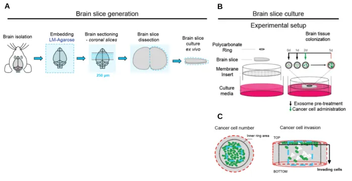
Despite advances in our knowledge of the molecular determinants that drive the overall process of metastasis, metastatic entry and adaptation to specific organs remain poorly understood. This is particularly true for brain metastasis, as evidenced by its growing incidence, now ten times higher than that of all primary brain tumors combined. Brain metastases most commonly arise from cancers of the lung and breast and constitute one of the most pernicious outcomes of cancer, characterized by neurological complications, poor prognosis and high mortality, with no effective therapies available. Hence, discovery of new drivers of brain metastasis with potential to become therapeutic and preventive targets is required to advance the care of brain metastasis patients as well as cancer patients at risk of metastatic spread to the brain. The major challenges facing brain metastasis research stem not only from our incomplete understanding of the molecular mechanisms regulating metastasis to this organ, but also from the limited number of pre-clinical models available that mimic human disease and enable the study of the complex interactions between tumor cells and the brain microenvironment. Brain metastatic colonization underlies acquisition of key adaptations by metastatic tumor cells and is determined by both tumor-intrinsic properties and the crosstalk between tumor cells and stromal cells in the brain. The low metastatic efficiency observed for this organ results from its particular microenvironment and resident cells, which constitute a hostile “soil” for seeding tumor cells to colonize. Ultimately, it is the ability of tumor cells to remodel the brain microenvironment and create a supportive niche for metastatic tumor cell survival and outgrowth that determines successful metastatic colonization. Among tumor-secreted factors, which are recognized as major contributors to the formation of pre-metastatic and pre-metastatic niches, tumor-derived exosomes have recently arisen as crucial players in cell-to-cell communication and in the remodeling of distant microenvironments that favor organ-specific metastasis. Therefore, we sought to determine the role of tumor-derived exosomes in the modulation of the brain microenvironment and their specific contribution to supporting metastatic colonization of the brain. For that, we used in vivo pre-clinical models of brain metastasis and adapted an organotypic brain slice ex vivo model for the study of tumor-derived exosomes in brain metastasis, allowing us to assess their role in metastatic colonization and tumor outgrowth.
xiv
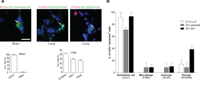
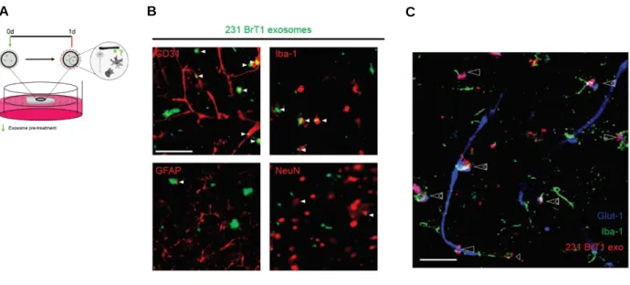
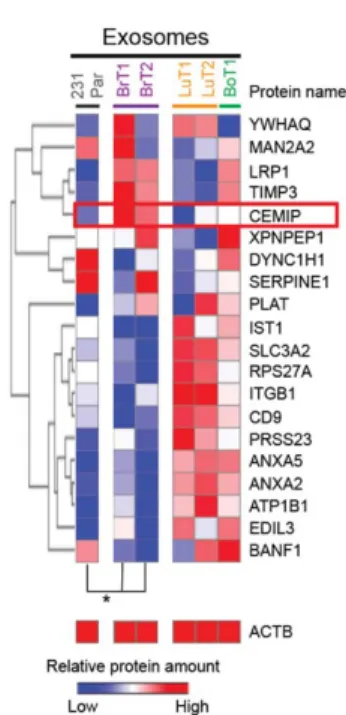
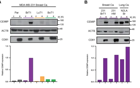
Our characterization of the organ distribution of tumor exosomes revealed that circulating exosomes of brain metastatic cell origin preferentially localize to the brain, where they interact predominantly with endothelial cells. Interestingly, exosome-positive vessels in the brain showed indication of vascular dysfunction, suggesting that their interaction with the brain microenvironment, and the vascular niche in particular, can lead to pro-metastatic effects of relevance for brain metastasis. Using our optimized ex vivo brain slice model we observed that pre-conditioning the brain microenvironment with exosomes from brain metastatic cells enhances cancer cell colonization of the brain. We then set to investigate which factor present in brain metastatic exosomes could be responsible for their pro-metastatic. Given the relevance of the exosome protein content in previously documented pro-metastatic functions of tumor exosomes we decided to pursue the characterization of the specific exosomal protein composition of exosomes from brain metastatic models. Our unbiased proteomic analysis of exosomes isolated from tumor cell models of common origin but different metastatic organotropisms identified cell migration-inducing and hyaluronan-binding protein (CEMIP, KIAA1199) as a protein distinctively enriched in exosomes from brain metastatic breast and lung tumor cell models. We now show that CEMIP, a Wnt-related protein involved in cancer and inflammation, mediates brain metastasis and promotes a pro-metastatic environment conducive to tumor cell-vascular association. Mechanistically, exosomal CEMIP stimulated a pro-inflammatory state in the brain vascular niche, namely through its effect on brain endothelial cells and microglia, characterized by the upregulation of diverse cytokines and chemokines known to promote brain tissue colonization (e.g. Ptgs2, Tnf, and Ccl/Cxcl family genes). Importantly, CEMIP was identified in exosomes secreted from surgically resected live human brain metastatic tissue samples obtained from breast and lung cancer patients. Moreover, high CEMIP expression in human primary and metastatic tumor tissue samples significantly associated with faster brain metastasis progression and poor survival.
Our work defining the contribution of CEMIP to brain metastasis brings new insight into the molecular mechanisms through which tumor-secreted exosomes promote metastasis to the brain and highlights the importance of the trophic properties of the brain vascular niche in this process.
xv
xvii
Apesar de avanços no nosso conhecimento relativamente aos determinantes moleculares que governam o processo geral de metastização, a entrada e a adaptação de células metastáticas em órgãos específicos permanece pouco compreendida. Esta afirmação é particularmente verdadeira para o caso das metástases cerebrais, como é evidenciado pela sua crescente incidência, agora dez vezes maior que a de todos os tumores cerebrais primários combinados. As metástases cerebrais surgem normalmente de cancros do pulmão e de mama e são umas das consequencias mais perniciosas da metastização do cancro, sendo caracterizadas por complicações neurológicas, mau prognóstico e alta mortalidade, padecendo actualmente de terapias eficazes disponíveis para o seu tratamento. Por este motivo, é necessário compreender melhor os processos que estão na origem da formação de metástases cerebrais e descobrir novos mecanismos envolvidos nestes processos que tenham potencial para se tornarem alvos terapêuticos e preventivos. Desta forma, poderemos avançar o tratamento de pacientes com metástases cerebrais e pacientes oncológicos em risco de disseminação metastática para o cérebro. Os principais desafios enfrentados pela pesquisa em metástases cerebrais não são apenas fruto da nossa compreensão incompleta dos mecanismos moleculares que o regulam este processo, mas também do número limitado de modelos pré-clínicos disponíveis capazes de mimetizar a progressão doença em humanos e que também permitam o estudo das complexas interações entre as células tumorais e as células do microambiente cerebral. A colonização metastática do cérebro está subjacente à aquisição de adaptações cruciais pelas células tumorais metastáticas e é determinada pelas propriedades intrínsecas do tumor e pela comunicação entre as células tumorais e as células estromais do cérebro. A baixa eficiência metastática observada para este órgão resulta do seu microambiente e células residentes, que constituem um "solo" hostil para a colonização das células tumorais, que o semeiam. Como resultado, é a capacidade que as células tumorais têm de remodelar o microambiente cerebral e criar um nicho que promova a sobrevivência e crescimento das mesmas que determina uma colonização metastática bem-sucedida no cérebro. Entre os factores secretados por tumores, reconhecidos como os principais contribuintes na formação de nichos pré-metastáticos e metastáticos, os exossomas surgiram recentemente como actores críticos para a comunicação célula-a-célula e para a
xviii
remodelação de microambientes distantes que favoreçam a metastização específica para distintos órgãos. Por estes motivos, procurámos determinar o papel dos exossomas derivados de tumores na modulação do microambiente cerebral e sua contribuição específica no suporte da colonização metastática do cérebro. Para isso, usámos modelos pré-clinicos de metástases cerebrais in vivo e adaptámos um modelo organotípico de secções cerebrais ex vivo para o estudo de exossomas derivados de tumores metastáticos cerebrais, permitindo desta forma avaliar seu papel específico na colonização metastática e no crescimento tumoral.
A nossa caracterização da distribuição de exossomas tumorais em diferentes órgãos revelou que os exossomas circulantes de origem celular metastática cerebral localizam-se preferencialmente no cérebro, onde interagem predominantemente com células endoteliais. Curiosamente, os vasos sanguíneos encontrados no cérebro que interagiram com exosomas tumorais demonstraram indicações de disfunção vascular, sugerindo que a interação de exossomas com o microambiente cerebral, e o nicho vascular em particular, pode levar a efeitos pró-metastáticos relevantes para a formação metástases cerebrais. Utilizando o nosso modelo optimizado de secções cerebrais ex vivo, observamos que o pré-condicionamento do microambiente cerebral com exossomas de células metastáticas cerebrais promove a colonização de células cancerígenas no cérebro. Em seguida, decidimos investigar a identidade do fator presente em exossomas metastáticos cerebrais que poderia ser responsável pelos efeitos pró-metastáticos observados. Dada a relevância do conteúdo proteico de exossomas tumorais em funções pró-metastáticas previamente documentadas, decidimos prosseguir com a caracterização da composição proteica específica dos exossomas derivados de modelos metastáticos cerebrais. Utilizando uma abordagem exploratória imparcial, a análise proteómica de exossomas isolados de modelos tumorais celulares com origem comum mas predilecção metastática para diferentes orgãos, identificou a cell migration-inducing
and hyaluronan-binding protein, ou proteína inductora de migração celular e de ligação
ao ácido hialurónico (CEMIP, KIAA1199), como sendo uma proteína enriquecida distintamente em exossomas derivadas de células metastáticas cerebrais de cancro de da mama e de pulmão. Neste trabalho, demonstramos que a CEMIP, uma proteína relacionada com a sinalização Wnt, e previamente envolvida na progressão de diferentes
xix
cancros e em processos de inflamação, medeia a formação de metástases cerebrais e promove um ambiente pró-metastático, propício à associação de células tumorais com a vasculatura cerebral. Mecanisticamente, a CEMIP exossomal estimulou um estado pró-inflamatório no nicho vascular do cérebro, através do seu efeito em células endoteliais do cérebro e micróglia, que é caracterizado pela promoção da expressão génica de diversas citocinas conhecidas por promover a colonização do tecido cerebral (ex.: Ptgs2,
Tnf, e genes da família Ccl/Cxcl). É importante salientar que o CEMIP também foi
identificado em exossomas secretados a partir de amostras de tecido metastático cerebral humano, obtidas a partir da resecção cirúrgica de pacientes com cancro da mama e do pulmão. Além disso, a expressão elevada de CEMIP em amostras de tecidos tumorais primários e metastáticos humanos associou-se significativamente a uma progressão mais rápida da formação de metástases cerebrais e a baixas taxas de sobrevivência.
O nosso trabalho, define desta forma a contribuição do CEMIP para metástases cerebrais, trazendo uma nova visão dos mecanismos moleculares através dos quais os exossomas secretados por tumores promovem metástases para o cérebro, destacando também a importância das propriedades tróficas do nicho vascular cerebral neste processo.
xx
xxii
α Alpha
β Beta
3D Tri-dimensional
ACA Anterior cerebral artery ACTB Beta-actin
BA Basilar artery BBB Blood brain barrier BCA Bicinchoninic acid
BMDC Bone-marrow derived cell BoT Bone tropic
BrEC Brain endothelial cell BrM Brain metastasis BrT Brain tropic
c Cells
CC Cell compartment
CCR7 C-C chemokine receptor type 7
CEMIP Cell migration-inducing and hyaluronan-binding protein Cm Conditioned media
CNS Central nervous system COX2 Cyclooxygenase 2
Cp Cytoplasm
Cs Cytoskeleton CSF Cerebrospinal fluid CTC Circulating tumor cell
CXCR4 C-X-C chemokine receptor type 4 DTC Disseminated tumor cell
e Exosomes
exo Exosomes
EC Endothelial cell ECM Extracellular matrix EGFR Epidermal growth factor
xxiii
ER Estrogen receptor
ESCRT Endosomal sorting complex required for transport ETF Endothelial tube formation
EV Extracellular vesicle
FACS Fluorescence-activated cell sorting FBS Fetal bovine serum
FDR False discovery rate
Fig Figure
FOV Field of view
GFP Green fluorescent protein gRNA Guide RNA
h Hours
HA Hyaluronic acid
H&E Hematoxylin and eosin
IA Intussusceptive angiogenesis IC Internal carotid
IHC Immunohistochemistry ITG Integrin
ILV Intra-luminal vesicle
IPA Ingenuity Pathway analysis
KO Knock-out
LFQ Label-free quantification LFU Last follow-up
LuT Lung tropic m Molecular marker Mb Membrane Met Metastasis MG Microglia MMP Metalloproteinase MT Metastatic tumor MVB Multivesicular bodies
xxiv
Nc Nuclear
NSLC Non-small cell lung cancer OE Overexpression
PC Polycarbonate
PCA Principal component analysis PCR Polymerase chain reaction PMN Pre-metastatic niche PR Progesterone receptor p/s Photons per second PT Primary tumor RNA Ribonucleic acid
SCA Superior cerebellar artery SEM Standard error of mean SPC Surfactant protein C Sup Supplementary
TEM Transmission electron microscope VA Ventral artery
VEGF Vascular endothelial growth factor
WC Whole cell
xxv
xxvii Section Page ACKNOWLEDGEMENTS vii ABSTRACT xi RESUMO xv LIST OF ABBREVIATIONS xx TABLE OF CONTENTS xxv CHAPTER I: INTRODUCTION 1
Section 1: Mechanisms of Cancer Metastasis 3
Section 2: Brain Metastasis Biology 11
Section 3: Exosomes in Cancer progression and Metastasis 18
Section 4: Background and Research Aims 23
CHAPTER II: MATERIALS AND METHODS 26
- Cancer cell lines and cell culture 28
- Exosome purification, labelling, characterization and analysis 28
- Brain slice assay 29
- Proteomics methods and data analysis 32
- Western blot analysis 33
- OptiPrep™ density gradient (ODG) exosome isolation 34
- Mouse studies 34
- Tissue processing and immunostaining 37
- Generation of CEMIP knockout and overexpression cell lines 38 - Proliferation and invasion in vitro assays 40 - Brain endothelial cell isolation, culture and assays 40
- FACS analysis 42
- RNA preparation, sequencing and data analysis 43
- Human studies 44
xxviii
CHAPTER III: BIODISTRIBUTION AND VASCULAR INTERACTION OF
METASTATIC TUMOR EXOSOMES IN THE BRAIN 48
CHAPTER IV: EX VIVO MODELING OF BRAIN METASTATIC COLONIZATION
FOR TUMOR EXOSOME STUDIES 59
CHAPTER V: CEMIP, A TUMOR EXOSOME PROTEIN WITH FUNCTIONS IN
BRAIN METASTASIS 69
CHAPTER VI: EXOSOMAL CEMIP-DEPENDENT MECHANISMS UNDERLYING
BRAIN VASCULAR NICHE RESHAPING 84
CHAPTER VII: CEMIP AS A BIOMARKER OF BRAIN METASTASIS 97
CHAPTER VIII: DISCUSSION 106
1
CHAPTER I
3
Section 1
4
Section 1 – Mechanisms of Cancer Metastasis
Despite continuous research efforts to understand the molecular drivers of cancer for the past 70 years, metastasis remains a mystery and the primary cause of cancer-related death. Metastasis encompasses the spread of cancer from its primary organ site to other organs. Development of secondary malignant tumor lesions in distant organs disturbs physiological homeostasis and can provoke physical alterations in tissue architecture which can lead to loss of organ function and death. It is now well established that organs of future metastasis are not passive receivers of circulating tumor cells, but are instead selectively and actively modified by the primary tumor before metastatic spread has even occurred. Sowing the ‘seeds’ of metastasis requires the action of tumor-secreted factors and tumor-shed extracellular vesicles that enable the ‘soil’ at distant metastatic sites to encourage the outgrowth of incoming cancer cells [1].
1.1 – Tumor progression and evolution
Primary tumors were initially viewed as homogeneous masses of cancer cells; however, work in the past 30 years has shown that primary tumors are composed of a heterogeneous mixture of cancer cells, with diverse genetic profiles, as well as stromal cells and infiltrating immune cells [2]. Recent work has demonstrated that clonal evolution occurs in primary tumors, driving the emergence of distinct cancer cell clones with different characteristics and contributions at the tumor cell population level. Genetic tumor heterogeneity, either intrinsic or developed as a result of selection due to environmental stress, may confer tumor cells properties that allow them to become more malignant. Genetic alterations and epigenetic regulation of genes impacting biological pathways such as cell migration and remodeling of surrounding extracellular matrixes, are likely to impact the ability of tumor cells to disseminate and conquer new territories, one of the hallmarks of metastasis.
Alterations in key pro-metastatic drivers synergize, rendering tumor cells metastatic and (e.g. EMT) allowing them to escape the primary tumor and generate secondary lesions in distinct organs [3]. Metastasis is a multi-step process, and rather inefficient, as tumor cells encounter challenges at every step [4]. Tumor cells first need to escape the primary tumor
5
mass by intravasating into the vasculature that supplies the primary tumor and this way spread into circulation to reach distant organs. Depending on the type of vasculature present in the tumor, metastatic spread can occur through the haematogenous or lymphatic systems, via the blood vessels or lymphatic vessels, respectively. After exiting the primary tumor, tumor cells have to survive in circulation, avoiding destruction by the immune system and eventually arresting in capillaries of distant organs where they could give rise to secondary tumor lesions [2]. Due to all these requirements, the metastatic process is extremely inefficient and only a very small percentage of disseminated tumor cells ever becomes a metastatic lesion [4]. Tumor cell arrest within the vasculature is a complex process which depends on vascular architecture and the physical properties of the blood/lymph flow, contributions from the clotting system, and expression of cell membrane adhesion molecules by tumor cells. After intravascular arrest tumor cells extravasate into the tissues of the new organ. For extravasation, tumor cells need to overcome the vascular barrier, usually by disrupting it, so they can access the parenchyma of the target organ. Interaction with endothelial cells of distinct organs is a common feature of all metastasis [5]. Cancer cells closely interact with endothelium during initial arrest and seeding, subsequent extravasation, and then later during angiogenic remodeling that occurs in metastatic growth phase. Extravasation is a critical step which can severely limit tumor cell entry and subsequent metastases formation. Once this barrier is surpassed, the ability of tumor cells to survive within their new environment and remodel it in support of their growth fully determines their true potential to generate metastases.
In addition to the intrinsic properties of tumor cells that confer them motility and metastatic potential, it is their unique ability to remodel their surrounding microenvironment that determines successful metastatic colonization [3].
1.2 – Metastatic tumor colonization
The first mechanisms proposed to explain organ-specific metastasis focused mainly on tumor cell intrinsic properties, such as metabolic or extracellular matrix (ECM)-interacting/remodeling adaptations, which lead to increased cancer cell survival at
6
metastatic sites and successful metastatic colonization. During the initial phase of metastatic colonization, tumor cells rely on ligand/receptor interactions between cancer cell membrane proteins and ECM components of the vasculature and microenvironment of distinct organs during metastatic seeding (e.g. tumor integrin interactions with vascular adhesion and ECM molecules). These and other observations underscore the importance and contribution of the colonizing organ microenvironment to metastasis as a major driver of tumor malignancy and progression [6, 7].
The understanding that the microenvironment is a critical determinant of tumor growth as well as metastatic outcome, led to a shift towards a new, non-tumor centric perspective, leading to the identification of tumor cell extrinsic drivers of metastasis. Thus, it was established that receptor/ligand interactions, paracrine signaling and secreted factors mediate the crosstalk between tumor and stromal cells in the metastatic organ microenvironment. The tumor and surrounding environment (blood vessels, stromal cells, immune cells, ECM and signaling molecules) constantly interact and co-evolve [2]. Interestingly, tumor-secreted factors, such as cytokines, chemokines, metabolites, and extracellular vesicles/particles can not only mediate crosstalk in the local tumor microenvironment, but can also have systemic effects, influencing the behavior of stromal and immune cells at distant sites [8]. This discovery brought tumor secreted factors into the spotlight, and they became a major focus of metastasis research in the past decade. More importantly, tumor-secreted factors can affect several distinct steps of the metastatic cascade by: a) inducing thrombotic events and adhesive properties in the endothelium during cancer cell arrest phase that allow cancer cell attachment ; b) altering the permeability of vessels during extravasation phase to facilitate cell entry ; c) promoting cancer cell invasion and tissue colonization through basement membrane processing and ECM deposition by stromal cells via inflammatory signaling ; d) promoting stromal cell recruitment and induction of pro-survival signaling in stroma by release of chemotactic signals during metastatic growth ; and e) inhibiting immune cell function [3].
7
1.3 – Tumor-type specific organ metastatic patterns
A pivotal discovery by Stephen Paget in 1889 [9] postulated that metastasis is dependent on the interactions between ‘seeds’ (or the cancer cells) and the ‘soil’ (or the host microenvironment). Paget’s theory was challenged in the 1930s by James Ewing [10], who advocated that metastatic dissemination could be explained solely by the dynamics of haematogenous flow. Ewing’s perspective became the prevalent viewpoint until Isaiah Fidler’s research in the 1970s [11] demonstrated that, although the mechanical properties of blood flow were important, successful metastatic colonization could occur only at certain organ sites and that distinct tumor-types displayed specific metastatic organ patterns that could not solely be explained by the mechanic flow properties.
In experimental metastasis assays, Fidler et al. demonstrated that cancer cells derived from a certain metastatic site displayed enhanced abilities to metastasize to that specific organ, providing support for Paget’s organ-specific metastasis theory [12].
In addition to strengthening Paget’s theory, Fidler’s findings reignited interest in the question that first captivated Paget: why do tumor cells emerge only as disseminated tumor cells (DTCs) within specific organs? Is metastatic seeding monoclonal or polyclonal in nature? Moreover, does metastatic seeding occur only directly from the primary tumor or is secondary seeding from one metastatic organ to another also a biologically relevant event? This organ specificity observed in metastasis is known as organotropism and remains one of the most intriguing unanswered questions in cancer research [13].
Despite Stephen Paget’s 131-year-old “seed-and-soil” hypothesis [9], insufficient progress has been made towards decoding the mechanisms governing organ-specific metastasis. Subsequent studies investigating organ-specific metastasis focused largely on the role of intrinsic cancer cell properties, such as genes and pathways regulating colonization, in directing organotropism. Breast cancer cells express chemokine receptors, such as C-X-C motif receptor 4 (CXCR4) and C-C motif receptor 7 (CCR7), which partner with chemokine ligands expressed in lymph nodes (CXCL12) and lung (CCL21), thus guiding metastasis [14]. Tumor-secreted factors can also increase metastasis by inducing vascular leakiness, promoting the recruitment of pro-angiogenic immune cells , and influencing organotropism [15]. Furthermore, the ability of breast
8
cancer to form osteolytic lesions depends on osteoclast stimulating growth factors (for example, PTHRP and GM-CSF) released into the bone microenvironment [16]. Therefore, previous observations by the Lyden laboratory that metastatic melanoma-derived factors dictate organotropism are not surprising [17]. We found that medium conditioned by highly metastatic melanoma cells was sufficient to expand the metastatic repertoire of lung carcinoma cells that would typically only metastasize to the lung itself. This particular observation suggested that the formation of niches permissive for tumor outgrowth at future sites of metastasis could overcome the tumor cell’s intrinsic metastatic properties and more importantly, that particular tumor-secreted factors could account for niche formation in distinct organs and this may determine metastatic organotropism.
1.4 – Pre-metastatic niches and organotropic metastasis
Fundamental discoveries revealed that tumors induce the formation of microenvironments in distant organs that are conducive to the survival and outgrowth of tumor cells before their arrival at these sites. These predetermined microenvironments are termed ‘pre-metastatic niches’ (PMNs) [18]. Although congruent with both Paget’s and Ewing’s theories, the concept of the PMN is unique as it proposes that the primary tumor preconditions specific organ sites for future metastatic disease (that is, before CTC arrival) via tumor-derived factors. Therefore, in contrast to the metastatic niche, which is initiated and shaped upon CTC arrival, the PMN represents an abnormal, tumor growth-favoring microenvironment devoid of cancer cells.
Since the existence of the PMN was first demonstrated, numerous studies have identified various molecules that regulate its stepwise evolution, highlighting the complex molecular and cellular changes that occur in the PMN to support future metastatic tumor growth [19]. PMNs are the result of the combined systemic effects of tumor-secreted factors and tumor-shed extracellular vesicles (EVs) that promote a temporal sequence of events during the evolution of PMNs. Vascular leakiness is the earliest event in this sequence, followed by the alteration of local resident cells, such as fibroblasts, and the recruitment of non-resident cells, such as bone marrow-derived cells (BMDCs), to these PMNs, subsequently attracting circulating tumor cells (CTCs) [19, 20]. Pre-metastatic niche formation requires S100 proteins and ECM deposition, such as fibronectin upregulation
9
by lung resident cells, as well as the recruitment of bone-marrow-derived myeloid cells in response to tumor-secreted factors [17]. It has become clear that vascular niche reshaping and extracellular matrix (ECM) remodeling is crucial for establishing the PMN. Moreover, PMNs are probable sites of immune deregulation, owing to the presence of a pro-tumorigenic, inflammatory milieu induced by tumor-secreted factors, which creates immunosuppression and coagulation disorders [5, 21] These events synergize to establish a favorable microenvironment that promotes the growth of disseminated tumor cells upon their arrival.
Although the mechanisms of lung PMN generation have started to be described, the cellular and ECM constitution and underlying molecular mechanisms of PMN formation in other metastatic organs remains to be characterized. Since distinct challenges are faced by cancer cells during metastatic colonization depending on the organ of metastasis, specific needs regarding the constitution of pro-tumoral microenvironments are likely to differ as well. This implies that the organ-specific mechanisms of metastatic colonization can offer potential targets for organ-specific metastatic therapy. The specific contribution of tumor-secreted factors and tumor-shed extracellular vesicles and exosomes is also not fully explored and requires deeper analysis. Action of these tumor agents is of particular interest in the context of brain metastasis, an organ in which metastases develop late but associate with poor outcome. This implies that brain metastasis usually happens late after primary tumor formation, with a long pre-metastatic phase during which tumor-secreted factors could affect the brain microenvironment and generate a PMN in the brain. The brain also poses particular challenges to metastasis that are unique to this organ, such as the presence of a restrictive vascular barrier structure, the blood-brain-barrier (BBB), that is much less permissive than other vascular beds as a result of their multi-cellular and complex composition and tight endothelial cell junctions. In addition, the brain microenvironment is suppressive and requires tumor cells to acquire additional survival mechanisms and specific signaling to colonize it. Due to the complexity of the brain metastatic process and limitations of the available pre-clinical models of brain metastasis, the development of therapeutic approaches for this dreadful type of metastasis is still lacking in comparison to other organs. Therefore, further understanding of the specific mechanisms involved in the formation of hospitable and
10
conducive environments to enhance tumor colonization in organs such as the brain, and improvements on the current research models, are required for development of effective anti-metastatic therapies.
11
Section 2
12
Section 2 – Brain Metastasis Biology
Metastatic spread to the brain remains one of the least understood and most detrimental outcomes of cancer. Brain metastases are estimated to occur in approximately 20% of all cancer patients and overshadow the incidence of primary brain tumors by more than ten-fold [22, 23]. Brain metastases present a unique set of clinical challenges and their presence is associated with poor survival, development of severe neurological issues, and ultimately death [24]. The brain microenvironment is vastly different from other organ environments, presenting complex anatomical structures, unique cell types, and particular immune and metabolic properties. Thus, the brain microenvironment imposes a very distinct and strong selective pressure on tumor cells that shapes the metastatic process and therapy responses. Importantly, the brain vasculature has emerged as a key determinant factor in brain metastasis, not only by due to its role in the initial seeding of circulating tumor cells and promoting vascular-associated tumor growth, but also due to its capacity to limit the access of systemic therapies to the brain [5]. The major limiting factor for the study of brain metastasis has been the scarcity of robust preclinical models that recapitulate the full process of metastatic spread to the brain and/or that allow to dissect out the key relationships between tumor cells and the brain microenvironment which allow for successful colonization of this organ [25]. Therefore, the study of brain metastasis and further characterization of their interaction with the brain microenvironment, and in particular the brain vascular niche, could unveil new therapeutic targets to address this unmet clinical need.
2.1 – Epidemiology of brain metastasis
Although the true prevalence of brain metastasis is not certain, epidemiological population-based studies estimate that their incidence ranges from 8.3 to 14.4 per 100,000 [26]. However, this value is likely an underestimate given that the majority of the epidemiological studies pre-date modern imaging technologies and no further information on disease course or subsequent involvement of metastatic sites was available for patients not presenting with neurological symptoms or brain metastasis at time of initial diagnosis. In support of this claim, autopsy studies suggest a higher incidence of
13
intracranial metastasis, detected in up to 40% of cancer patients [27]. Furthermore, it is also believed that while the improvement in cancer detection, patient care, and systemic therapies for extra-cranial metastasis implemented over the past decades, while having extended the life span of cancer patients, may account for and inversely correlate with the rising incidence of brain metastasis.
The incidence of brain metastasis largely depends on factors such as primary tumor site, stage of disease, subtype of cancer, and other key prognostic factors [22, 28]. Although any type of cancer has the ability to metastasize to the brain, the most common primary tumors associated with brain metastasis are lung (20 – 56% of patients), breast (5 – 20%), and melanoma (7 – 16%)[22, 26], followed by colorectal and renal cancers [22]. Within these, the type of primary tumor and its molecular subtype further impact the risk of developing brain metastasis. This is particularly well established in breast cancer, where tumors presenting triple-negative hormone-receptor status molecular subtype [i.e. estrogen receptor (ER)-negative, progesterone receptor (PR)-negative and normal human epidermal growth factor receptor 2 (ERBB2; also known as HER2) levels] or with HER2 amplification, have higher risk of developing brain metastases [29]. In lung cancer, patients with non-small cell lung cancer (NSCLC) or small-cell lung cancer (SCLC) primary tumor types are the most afflicted with brain metastases (15 – 26%, and 24% respectively) [30], with NSCLC molecular subtype with ALK rearrangements shown to be at increased risk [31].
In addition to these specific cancers and molecular subtypes, the risk of developing brain metastases also increases with more advanced primary disease [30], and can also be influenced by additional factors such as sex, age and ethnicity. Regardless, despite the factors described above and efforts to develop data-driven tools to specifically prognosticate brain metastasis patients earlier and more accurately (such as the graded prognostic assessment index – GPA, which takes into account performance of daily tasks by patients, treatment status, number of metastases, among others), their clinical value remains limited, as illustrated by the very short and poor survival estimates for brain metastasis patients [32].
14
Diagnosis of brain metastasis has improved during the last decades due to advancements in imaging technologies that allow earlier detection. However, current treatments for brain metastasis, such as whole-brain radiation therapy or local radiation therapy carry many toxic outcomes given that this is not a targeted therapy and therefore affects healthy normal brain cells as well. Due to the shortcomings of radiation, systemic treatments with efficacy in the CNS that target only tumor cells are needed. Thus, a better understanding of the molecular mechanisms driving brain metastasis are necessary for the development of novel and improved therapeutic approaches.
2.2 – Molecular mechanisms of brain metastatic colonization
A better understanding of the metastatic spread to the brain and the underlying molecular mechanisms governing it is necessary to address the dismal prognosis that brain metastasis patients currently face. From the analysis of preclinical models of brain metastasis, it has become apparent that cancer cells require two critical competences in order to successfully metastasize to the brain. First, they need to be able to reach and surpass the blood-brain-barrier (BBB) in order to enter the brain, and later they need to adapt to and remodel the brain milieu so they can survive and thrive within this new environment. The BBB is a multi-cellular and complex barrier structure that protects and insulates the brain and its neuronal signaling from systemic insults [24]. Alongside the blood-cerebrospinal fluid (CSF) barrier, the BBB comprises the most important functional barrier in the brain that tumor cells need to overcome to enter the brain. This structure consists of endothelial cells that are connected by strong tight junctions and present low transcytosis rates and expression of several efflux pumps. Furthermore, in addition to endothelial cells and their basement membrane, the BBB is composed by bordering pericytes and terminal processes of astrocytes, which further contribute for this barrier’s functions and selective permeability. Due to the properties of the BBB, only small uncharged compounds are able to freely diffuse through it under normal conditions. However, inflammation occurring in the context of infections and autoimmunity, will open the BBB to immune cells. Through mechanisms which are still not completely elucidated, cancer cells can also overcome the BBB and gain access to the CNS [33].
15
Cancer cells are thought to arrive to the brain and arrest within the vasculature either as single cells or emboli. Their arrest is thought to be promoted by a combination of factors, ranging from aberrant sizes of cancer cells, slower blood flow movement at capillary branch points and activation of coagulation by cancer cells and the factors they secrete [34, 35]. Interestingly, several studies have shown that only a very small proportion of cells that arrest within the brain microvasculature form metastases, highlighting the importance of subsequent steps for successful brain metastatic colonization [35]. After arrest, the tumor cells’ potential to extravasate into the brain and colonize the brain is mostly mediated by their ability to interact with brain endothelial cells. Successful interaction between tumor cells and brain endothelial cells is mainly determined by expression of adhesion factors, such as the membrane glycosyltransferase ST6GALNAC5, shown to specifically mediate circulating cancer cell adhesion to the endothelium of the brain [36]. During extravasation, tumor cells express factors that induce vascular permeability, promoting migration across the BBB, such as VEGF, COX2, and HBEGF. Additionally, recruitment of immune cells and activation of glial cells of the BBB by extravasating tumor cells results in VEGF signaling activation and release of proteinases such as cathepsin S and matrix metalloproteinases (MMPs), such as MMP9 [25]. The later are able to further contribute to this process by cleaving junctional adhesion molecules of the endothelial cell barrier and leading to focal extracellular matrix degradation, respectively. Once extravasation is completed, different cell fates might await tumor cells. They might undergo cell death due to the conditions of the brain microenvironment [24], remain in a dormant state with slow cell-cycling [36, 37] or survive and give rise to metastases.
Once within the brain, thriving tumor cells experience complex interactions with the brain microenvironment that govern their outgrowth. Although this process is not completely understood, the insights gathered so from pre-clinical models and analysis of patient samples indicates that the colonization process is characterized by induction of neuroinflammatory cascades and mechanisms of vascular niche reshaping. A particularly interesting observation is that surviving cancer cells appear to remain in proximity to the perivascular niche [35, 38]. This particular localization of tumor cells allows easy access to oxygen and nutrients from blood circulation, a settling contact point for attachment
16
within the basal lamina of vessels and exquisite access to angiocrine factors produced by endothelial cells that support tumor growth. Furthermore, brain metastatic tumor cells are known to exploit the vasculature of the brain rather than generate their own vascular supply system, through the process of angiogenesis, as commonly observed for extracranial metastases [24]. In the brain, tumor cells typically interact with the pre-existing vasculature and grow along the abluminal side, through a process known as vascular co-option [39]. Later, they reshape the architecture of the vasculature with which they associate in order to integrate it into the growing metastatic mass, a phenomenon typically observed in brain metastases and referred to as angiocentric growth. This mainly occurs through a process termed intussusceptive angiogenesis, where new vessels are formed by the remodeling and splitting of previously existing ones [40-43]. Adhesion molecules involved in vascular co-option include L1CAM and β1 integrin [38, 44]. Interestingly, another integrin family member, the αvβ3 integrin heterodimer, has been shown to promote cell adhesion to the endothelium and key signaling for initial colonization. In addition to their pivotal interaction with brain endothelial cells, tumor cells are exposed to and interact with other stromal cells of the brain microenvironment that can determine the success of their colonization. Of particular importance, tumor cells induce a prominent neuroinflammatory response, activating astrocytes and microglia. Astrocytes can influence tumor cell growth through pro-metastatic transfer of metabolites such as cGAMP thorough gap-junctions and secretion of inflammatory chemokines such as interferon-α and TNF, as well as anti-metastatic secretion of cell-death inducing plasmin [25]. In contrast, the complex role of microglia is equally acknowledged, but less explored. While microglia can hinder brain metastasis through cytotoxic effects which can be mitigated by tumor cell expression of neurotrophin-3 [45], they can also promote brain metastasis by producing pro-tumorigenic cytokines, such as CCL2 and other members of the CCL/CXCL family [46]. Finally, additional properties and adaptations by tumor cells that have been shown to assist in brain colonization include metabolic adaptations, impacting processes such as glucose metabolism or reactive oxygen species production [47]. As described in this section, the incomplete understanding of the roles that different brain stromal cells play in metastatic colonization, and the dependence of some of the mechanisms described to particular tumor types or circumstances, underscores the need
17
for improved brain metastasis pre-clinical models that allow the study of the interactions of tumor cells with the brain microenvironment and respective outcome for metastasis. Use of alternative models such as 3D organotypic brain slice ex vivo cultures [38, 48], or 3D multi-cellular cultures composed of different brain stromal cells in vitro, might be the key to help to bridge this gap.
18
Section 3
19
Section 3 – Exosomes in cancer progression and metastasis
Among tumor-secreted factors, which are recognized as major contributors to the formation of pre-metastatic and metastatic niches, tumor-derived exosomes have recently arisen as crucial players in cell-to-cell communication and in the remodeling of distant microenvironments that favor organ-specific metastasis.
3.1 – Extracellular vesicle biology and exosome biogenesis
Exosomes are a class of extracellular vesicles (EVs) ranging in size between 30 –150 nm that derive from the multivesicular endosomal pathway [49]. This class of vesicles are secreted into circulation (blood, lymphatics fluid, saliva, urine, etc.) by both normal and tumor cells. In particular, tumor-secreted exosomes are critical mediators of intercellular communication between tumor cells and stromal cells in local and distant microenvironments, and therefore, are key modulators of tumor progression and metastasis.
EVs were first described in 1967 by work from Peter Wolf, who first demonstrated a role for platelet-secreted vesicles during blood coagulation [50]. In 1980, Trams
et al. [51] uncovered the essential role that EVs play in intercellular transport of trophic substances or nutrients. In the 1980s, several groups described the role of secretory vesicles in reticulocyte maturation through recycling of transferrin and its receptor [52-54]. In addition, pioneer studies by Raposo et al. [55] further demonstrated the importance of EVs in immune modulation. Since then, many studies, including those from our group [17, 56, 57], have further demonstrated the crucial role of EVs in immune modulation, cell signaling, and onset and progression of disease.
EVs are classified on the basis of their size and origin. Microvesicles (>150 nm) are formed at the cell surface through a budding mechanism while exosomes (30–150 nm) are produced in a regulated manner by the endolysosomal and multivesicular body compartments for release outside the cell [1]. The biogenesis of exosomes starts with the invagination of endosomal limiting membranes which leads to the formation of intraluminal vesicles (ILVs) contained within the endosome. This compartment, termed a multivesicular body (MVB), fuses with the plasma membrane, culminating in the extracellular release of ILVs as exosomes. The machineries involved in the release of
20
exosomes, their docking and fusion during both physiological and pathological processes are still being deciphered. However, there is no doubt that exosome biogenesis is a multistep process which requires the coordinated efforts of several protein networks in the cell. Among these known actors are: 1) Rab GTPases, proteins which control endosomal trafficking; 2) endosomal sorting complexes required for transport (ESCRT), multiple protein complexes that regulate ILV formation; 3) tetraspanins, transmembrane proteins that induce membrane curvatures enabling vesicle formation; and 4) lipid-modifying enzymes (i.e. sphingomyelinase), that generate ceramides involved in promoting vesicle formation [58].
It has been shown that tumor cells are able to package and secrete, through exosomes, proteins (receptors, transcription factors, enzymes, extracellular matrix proteins) as well as nucleic acids (DNA, mRNA, microRNA [miRNA], and other non-coding RNAs), metabolites and lipids [59]. This exosome cargo becomes even more relevant once these vesicles are in circulation and taken up by other cells. This cargo can be transferred to recipient cells (tumor cells, stromal cells, immune cells, etc.) and influences their phenotype and/or function [1]. Therefore, exosomes are essential mediators of cell-cell communication. Remarkably, in addition to local signaling within the primary tumor microenvironment, tumors also signal over long distances to sites of future metastases to promote formation of a hospitable, pre-metastatic niche (PMN) that will foster growth of disseminated tumor cells upon their arrival [17, 56, 57].
Tumor-secreted exosomes can promote angiogenesis and coagulation, modulate the immune system, and remodel surrounding parenchymal tissue, which together support tumor progression [1]. Clinically, circulating exosomes and microvesicles isolated from cancer patients have been associated with metastasis or relapse, and therefore could serve as important diagnostic and prognostic markers as well as therapeutic targets [59].
3.2 – Exosome-mediated biological functions
Through their cargo, exosomes mediate several biological functions that cooperate to promote tumor progression and metastasis. Tumor-derived exosomes can transfer
21
oncogenic molecules as cargo between tumor cells within the primary tumor or distant metastatic sites [60]. A large number of studies have now demonstrated that exosomal proteins varies between different cancer types and even between cancers with different metastatic potential but with a similar origin. These unique patterns of exosomal protein cargo seem to also influence the metastatic capabilities of their cells of origin since exosomes from more aggressive tumor cells are more efficient at promoting metastasis than exosomes derived from less metastatic cells [17]. Furthermore, this differential and selective packaging also occurs in cancer with specific metastatic tropism (organotropism). For instance, packaging of distinct integrins in exosomes from different cancer types has a role in determining the organs that take up tumor exosomes [56] As tumors form and progress, they modify the stroma through soluble factors, exosomes, and direct cell-cell interaction. Therefore, tumor-derived exosomes exert also complex effects on neighboring stromal cells, such as endothelial cells and fibroblasts. Conversely, exosomes secreted by tumor stroma can also influence tumor progression [61]. Therefore, exosomes mediate bidirectional communication between tumor cells and their environment and are central effectors of a feedforward signaling loop that shapes the ever-evolving tumor microenvironment.
The properties of cancer exosomes in metastatic tumor progression as well as their presence in numerous fluids and tissues have made them a promising new source of biomarkers for cancer progression and as novel targets for future anticancer therapies.
3.3. – Regulation of metastasis by tumor exosomes
Tumor cell-derived EVs fuse with resident cells in both PMNs and tumors, transferring their cargo, including genetic material (DNA, mRNA and miRNA), metabolites (lipids and small metabolites) and protein. Pioneering studies demonstrated that platelet-derived EVs (particularly microparticles) induced angiogenesis and metastasis in lung and breast cancers [62, 63]; however, the relevance of EVs in PMN formation was not evaluated in these studies. Since then, EVs have been shown to contribute to the recruitment and
transfer of material to other stromal cell types, including those populating PMNs [64, 65].
Tumor-derived exosomal miRNA and proteins reprogramme or educate target cells that take up exosomes towards a pro-metastatic and pro-inflammatory phenotype, creating
22
the PMN [17, 57]. Education of stromal cells depends on the cancer type and pre-metastatic organ analysed. In melanoma, the hepatocyte growth factor receptor, MET,
which is secreted in B16-F10 melanoma-derived exosomes, reinforces the expression of
tyrosine kinase with immunoglobulin (Ig) and epidermal growth factor (EGF) homology domains 2 (TIE2) and MET in blood-circulating BMDCs, and promotes pro-vasculogenic and pro-migratory properties in these cells. These re-educated BMDCs then exit the bone marrow and contribute to the formation of PMNs in lungs [17]. Importantly, exosomes
secreted by B16-F10 melanoma cells promote vascular leakiness in lung PMNs, inducing
a pro-inflammatory response by increasing the expression of cytokines and chemokines such as TNF, S100A8 and S100A9, which, in turn, recruit BMDCs to these PMNs [17]. However, the source of S100 proteins was not defined in this study. Breast and pancreatic cancer cell-secreted exosomes express integrins on their surface, which promotes their
homing to PMNs [56]. Expression of the integrin α6β4 heterodimer on the surface of
tumor-derived exosomes promotes their homing to lung PMNs, whereas αvβ5 targets
them to liver PMNs. The homing of exosomes to these organs caused an increase in pro-inflammatory S100 family proteins in the PMN, generating a supportive microenvironment for subsequent metastasis [56].
Regarding liver PMNs, pancreatic tumor-derived exosomes expressing MIF promote TGFβ secretion in Kupffer cells, stimulating hepatic stellate cells to secrete fibronectin and promoting the recruitment of BMDCs [57].
Interestingly, genomic content (that is, miRNAs) is also packaged selectively within EVs and is involved in PMN formation [66, 67]
Recently, RNAs packaged within primary tumor-derived exosomes were found to be involved in the activation of TLR3-dependent signalling in lung epithelial cells, inducing chemokine secretion in the lung and promoting PMN formation by recruiting neutrophils [68]. These findings only encompass the beginning of our understanding on how tumor-derived EVs are involved in metastasis and opens the door for the pursuit of their roles in PMN BrM formation, which were remained completely unexplored at the starting time of this thesis.
23
Section 4
24
Section 4 – Background and Research Aims
Despite increased research interest in the molecular mechanisms driving BrM, there have been few advances in the early diagnosis and therapeutic targeting of this disease. The role of tumor-derived exosomes, emerging players in the interaction between tumor cells and the host microenvironment, remains widely unexplored in the brain metastatic process. Hence, gaining insight into the mechanisms of BrM and the specific contribution of tumor-derived exosomes to this process may open new avenues of investigation with potential clinical applications. Due to the aforementioned functions of tumor exosomes in regulating cancer metastasis, we aimed to determine the role of tumor-derived exosomes in the remodeling of the brain microenvironment and their specific contribution to supporting metastatic colonization of the brain.
The main objectives of the work presented in this manuscript are the ensuing:
- Characterize the brain biodistribution of tumor exosomes and their interaction with brain cells (Chapter III)
- Evaluate if tumor exosome uptake by the brain microenvironment generates a pro-metastatic niche supportive of pro-metastatic colonization (Chapter IV)
- Identify candidate exosomal molecule(s) with functions in brain metastasis and characterize their biological mechanisms of action (Chapter V and VI)
- Address the potential significance of candidate exosomal molecule(s) for brain metastasis in cancer patients (Chapter VII)
The findings documented for each of these objectives will be discussed further on
Chapter VIII. We expect that our understanding of the role of tumor-derived
exosomes in the reshaping of the brain microenvironment for successful metastatic colonization can provide significant advances to the brain metastasis research field. Furthermore, we contemplate that new brain metastasis targets may be identified from this study and potentially applied to the diagnostic and therapeutic management of brain metastasis in cancer patients.
26
CHAPTER II
28
Cancer cell lines and cell culture. All cancer cell lines used in this study are of human
breast or lung cancer origin. The breast cancer cell lines were provided as follows: brain-tropic 231BR cells by P. Steeg (NCI); brain-brain-tropic MDA-MB-231-HM cells [69] by S. Wang (UC San Diego); MDA-MD-231 organotropic 831 (brain-tropic), 4175 (lung-tropic) and 1833 (bone-tropic) cells by J. Massagué (MSKCC); and lung-tropic 4173 cells by A. Minn (University of Pennsylvania). The brain metastatic derivative N2LA-BR of the lung cancer cell line N2LA (N2LA-JHH-VKR) was generated from a patient with metastatic lung cancer by V. Rajasekhar (MSKCC). The breast cancer cell line MDA-MB-231 (parental) was purchased from American Type Culture Collection (ATCC). Breast cancer cells were cultured in Dulbecco’s Modified Eagle Medium (DMEM) and lung cancer cells in RPMI, both supplemented with 10% fetal bovine serum (FBS) (Gibco, Thermo Scientific), L-glutamine (1mM), and Penicillin/Streptomycin (100 IU/mL and 100 µg/mL). For exosome isolation from tissue culture supernatants, cells were cultured in exosome-depleted FBS. Depletion of endogenous serum exosomes was achieved by FBS ultracentrifugation at 100,000xg for 70 min. All cancer cells were maintained in a humidified incubator with 5% CO2 at 37 ºC. All cell lines used were routinely tested for mycoplasma (Universal Mycoplasma Detection Kit, ATCC) and found to be negative.
Exosome purification, labelling, characterization and analysis. Exosomes from cell
lines were purified by sequential ultracentrifugation as previously described [56]. Briefly, the fresh cell culture supernatant of a 3-day confluent culture was centrifuged at 500xg for 10 min, to remove any cells, and then at 12,000xg for 20 min, in order to clear away any cell debris or apoptotic bodies that might be present in the culture media. Exosomes were then collected from the latter supernatant by an ultracentrifugation step of 100,000xg for 70 min. Exosome collection was followed by a washing step through resuspension of the pellet in 1xPBS followed by centrifugation at 100,000xg for 70 min. For exosome imaging purposes, purified exosomes were fluorescently-labelled using the PKH67 (green) or PKH26 (red) lipophilic membrane dyes (Sigma), or the CellVue Maroonkit(far red) (Polysciences). Labelled exosomes were washed in 20 mL of 1xPBS and subjected to an additional round of ultracentrifugation at 100,000xg for 70 min. The final exosome