Darío Estraviz López
Graduado em BiologíaQuaternary fossil vertebrates from continental
Portugal: Paleobiodiversity, revision of
specimens and new localities
Dissertação para obtenção do Grau de Mestre em Paleontologia
Orientador: Prof. Associado Octávio Mateus, Faculdade de Ciências e
Tecnologia da Universidade Nova de Lisboa
Júri:
Presidente: Prof. Auxiliar Paulo Alexandre Rodrigues Roque Legoinha (FCT-UNL)
Arguente: Prof. Catedrático João Luís Cunha Cardoso (Universidade Aberta) Vogal: Prof. Associado Octávio João Madeira Mateus (FCT-UNL)
Júri:
i
Darío Estraviz López
Graduado em Biología
Quaternary fossil vertebrates
from continental Portugal:
Paleobiodiversity, revision of
specimens and new localities
Dissertação para obtenção do Grau de Mestre em
Paleontologia
Orientador: Prof. Associado Octávio Mateus, Faculdade de
Ciências e Tecnologia da Universidade Nova de Lisboa
© Darío Estraviz López, FCT/UNL e UNL
A Faculdade de Ciências e Tecnologia e a Universidade Nova de Lisboa tem o direito, perpétuo e sem limites geográficos, de arquivar e publicar esta dissertação através de exemplares impressos
reproduzidos em papel ou de forma digital, ou por qualquer outro meio conhecido ou que venha a ser inventado, e de a divulgar através de repositórios científicos e de admitir a sua cópia e distribuição com objetivos educacionais ou de investigação, não comerciais, desde que seja dado crédito ao autor e editor.
ii
“In vertebrate paleontology, increasing knowledge leads to triumphant loss of clarity.” Alfred Romer
iii
ACKNOWLEDGEMENTS
It is a strange feeling to finally write this. I have been working on this master thesis for one and a half years and at the same time that I am happy about finishing it I feel like this is the end of an era: the end of the straight road that took me from the preschool to this master, always with paleontology in mind. I could have never reached this stage without the people that have accompanied me and for them are these words:
First I have to say thanks to my family. This dream of mine is only possible because of their tireless support. Thanks to my grandparents (Mariví, Josefa and Nicolás). Infinite thanks to my father Antonio for understanding my passion for paleontology and caring for me while I am in other country, if the computer where I am writing this is still working is thanks to you. Infinite thanks to my mother Silvia and my stepfather Suso, I do not have enough words to tell how much I appreciate that you come every few months to help me at my home in Portugal. All of you are the best family of the world and I am so lucky of having you by my side. You all make me happy in every possible way.
I want to thank my advisor professor Octávio Mateus. He has given me an immense freedom to follow the research path that I felt that was the most appropriate for each topic. The freedom to do my own failures (and his help to get out of them) has made me a better scientist.
I would like to say thanks to Miguel Moreno Azanza, that have really acted a lot of time as a de facto co-advisor, even when he had to take care of his real students. I won´t forget it. Also I would like to thank Eduardo Puértolas Pascual for his comments on the thesis and his help to overcome concrete problems during it.
Thanks to my fellow master students at the Lourinhã PaleoTeam for their companionship and support, Hugo Campos (And his family that opened me their house in Algarve), João Pratas, André Saleiro, Cátia Ribeiro, Francisco Costa, Vincent Cheng and Alexandra Fernandes; especially to my classmates of the fifth master edition Filippo Maria Rotatoria and Alexandre Guillaume. I won´t forget the dinners on Thursday nights in Almada!
Big thanks to the personnel of the Museum da Lourinhã Alexandre Audigane, Bruno Pereira, Carla Alexandra Tomás, Carla Abreu, Jorge Beja and Raquel Furtado. Thanks for letting me use the museum facilities and also for the paraffin heater to prevented me to freeze during winter while working.
Thanks to all the other people that stood by my side in Lourinhã: Isabel Mateus, Marta Mateus, Simão Mateus, Silvia Batista, Ana Luz, Micael Martinho, João Marinheiro, João Russo, Marco Marzola, João Muchagata, Marta, Isabel and all the English class, my training partners at “Miga fight team” and everyone else; you all complete my “second family” in Portugal and you make me feel at home in a foreign country.
But this thesis couldn´t have been done without many people that work outside Lourinhã.
I would like to thank the entire GPS (Grupo Proteção Sicó) especially to Sérgio Medeiros and all the other speleologists that have helped me several times to descend to the Algar do Vale da Pena, for their disinterested help. I owe you my life and that is not an exaggeration!
Thanks to my former advisor at University of A Coruña, Aurora Grandal d´Anglade, that has helped me immensely during the process of doing the chemical analysis of my samples. I promise I will get something interesting for ancient DNA soon.
iv
Many thanks to the paleontologist that helped me with the identification of the fauna of Santa Margarida: Pedro Callapez, Raquel Moyá Costa, Pavlos Piskoulis and Juan Manuel López García. I really hope to be able to keep working with you in further studies about this locality in the future. Thanks to Miguel Ramalho, Jorge Sequeira and all the personnel of the Geological Museum in Lisbon, for letting me to visit their collections on short notice twice. Thanks to Jorge Graça, the Algarvian photographer that contacted Octávio Mateus and discovered the Santa Margarida locality.
Also thanks to Alexandra Elbakyan, without her work I could have never done this thesis.
Finally thanks to the students of other editions, personnel of the Department of Earth Sciences of the FCT-UNL and everyone that has helped me one way or the other and could not see his/her name on this thesis, I thank you a lot.
v
ABSTRACT
The Quaternary fossil record of Portugal is important for our understanding of the paleobiodiversity in Iberia. In the present master thesis a series of studies augment our knowledge about this topic.
A census of Quaternary paleobiodiversity is carried out in order to test how reliable the fossil record is for detecting living species, resulting in that ~38% of living terrestrial tetrapods are recognized in the fossil record for Portugal, although the number of species recognized varies between groups.
The body mass of a Portuguese proboscidean (Palaeoloxodon antiquus) is calculated via numerical methods for the first time (11metric tons) and morphometric comparisons of this species with Mammuthus primigenius are presented using an extensive Proboscidean sample.
A new fossil brown bear (Ursus arctos) locality, Algar do Vale da Pena, with numerous claw mark in the walls of the cave (the first of this type of marks described in Portugal) is presented and the fossil bear remains identified and compared to a sample from NW Spain. The bears from Algar do Vale da Pena contrast with other previously known Portuguese brown bear specimens by relative small size. A new microvertebrate locality from Algarve, Santa Margarida, is presented. It is an extraordinary rich site with one fossil for every two grams of sediment selected and processed. The locality provided the first record of two arvicoline taxa in Portugal (Iberomys huescarensis and Victoriamys chalinei), which allows giving a minimal age of around 800.000 YBP for at least part of it. This makes Santa Margarida one of the oldest three localities in the Pleistocene of Portugal.
KEYWORDS
vii
RESUMO
O registro fóssil Quaternário de Portugal é importante para nossa compreensão da paleobiodiversidade na Ibéria. Na presente tese de mestrado, uma série de estudos aumenta o nosso conhecimento sobre a matéria.
Um censo de paleobiodiversidade do Quaternário é realizado para testar a fiabilidade do registro fóssil para a detecção de espécies vivas, resultando em que 38% dos tetrápodes terrestres vivos são reconhecidos no registro fóssil de Portugal, ainda que este número varie entre grupos.
A massa corporal de um proboscídeo português (Palaeoloxodon antiquus) é calculada através de métodos numéricos pela primeira vez (11 toneladas) e comparações morfométricas desta espécie com Mammuthus primigenius são apresentadas, utilizando uma extensive amostra de proboscídeos.
É apresentada uma nova localidade de urso pardo (Ursus arctos); o Algar do Vale da Pena, com numerosas marcas de garras nas paredes da gruta (as primeiras deste tipo de marcas descritas em Portugal) e os restos fósseis de ursos são identificados e comparados com uma amostra procedente do Noroeste de Espanha. Os ursos do Algar do Vale da Pena contrastam com outros exemplares portugueses fósseis de urso pardo pelo seu tamanho relativamente pequeno.
Uma nova localidade de microvertebrados do Algarve, Santa Margarida, é apresentada. É um local extraordinariamente rico, com um fóssil por cada dois gramas de sedimentos selecionados e processados. A localidade forneceu o primeiro registro de dois taxa de Arvicolinae em Portugal (Iberomys huescarensis e Victoriamys chalinei), o que permite determinar uma idade mínima de cerca de 800.000 YBP para, pelo menos, parte da jazida. Isto faz de Santa Margarida uma das três localidades mais antigas do Plistocénico de Portugal.
PALAVRAS-CHAVE
ix
xi
INDEX OF MATERIALS
1. INTRODUCTION ... 1
1.1 History of the study of Quaternary fossil vertebrates from continental Portugal ... 1
1.2 Vertebrates in the Quaternary of Portugal ... 7
1.3 Aim, context and objectives of the present thesis... 22
2. PALEOBIODIVERSITY OF THE QUATERNARY FOSSIL TETRAPODS IN CONTINENTAL PORTUGAL ... 23
2.1 Introduction and methods ... 23
2.2 Lists of localities and fossil tetrapods of the Quaternary of Portugal ... 23
2.3 Results ... 36
2.4 Discussion ... 41
2.5 Conclusions ... 42
3.THE PALAEOLOXODON ANTIQUUS OF SANTO ANTÃO DO TOJAL ... 43
3.1 Introduction and methods ... 43
3.2 Geographical and geological settings ... 43
3.3 The material ... 44
3.4 Measurements of femur and tibia ... 48
3.5 Body mass estimation ... 52
3.6 Analysis of metrical data ... 54
3.7 Conclusions ... 66
4. ALGAR DO VALE DA PENA: A NEW FOSSIL BEAR SITE FROM PORTUGAL ... 67
4.1 Overview ... 67
4.2 Introduction ... 67
4.3 Methods, expeditions to the cave and preparation of the material ... 73
4.4 The bone assemblages ... 77
4.5 Results: The recovered bone remains ... 80
4.6 The claw marks... 118
4.7 Discussion ... 119
4.8 Conclusions ... 121
4.9 Further work ... 122
5. SANTA MARGARIDA: A NEW MICROVERTEBRATE LOCALITY FROM ALGARVE ... 123
5.1 Overview ... 123
5.2 Introduction: Location, discovery, geological settings and nature of the site ... 123
5.3 Preparation methods ... 126
xii
5.5 Discussion ... 143
5.6 Conclusions ... 146
6. CONCLUSIONS ... 147
Chapter 2: Paleobiodiversity of the Quaternary fossil tetrapods in continental Portugal ... 147
Chapter 3: The Palaeoloxodon antiquus of Santo Antão do Tojal ... 157
Chapter 4: Algar do Vale Da Pena: A new fossil bear site from Portugal ... 148
Chapter 5: Santa Margarida: A new microvertebrate locality from Algarve. ... 148
xiii
INDEX OF FIGURES
Figure 1.1: Bust of Carlos Ribeiro at the Geological Museum, Lisbon (Photo by Octávio Mateus). ... 1
Figure 1.2: Picture of Nery Delgado at exposition in the Geological Museum, Lisbon. ... 2
Figure 1.3: Hyena coprolite from Furninha cave recovered by Nery Delgado (MG). Note the glued identification.Geological Museum (Lisbon) ... 2
Figure 1.4: Picture of Paul Choffat at exposition in the Geological Museum, Lisbon. ... 3
Figure 1.5: Georges Zbyszewski (Picture by unknown author). ... 4
Figure 1.6: Miguel Telles Antunes in 2009 (Photo by Natacha Cardoso). ... 5
Figure 1.7: João Luis Cardoso in 2017 (Photo by Octávio Mateus). ... 6
Figure 1.8: “Contribuição para o conhecimento dos grandes mamíferos do Plistocénico Superior de Portugal” ... 6
Figure 1.9: Vertebra of Salamandra salamandra from Figueira Brava cave in dorsal view; scale bar 1mm. Catalog number of the specimen not provided in the original (Crespo et al., 2000). ... 8
Figure 1.10: Mandible from an indeterminate lacertid recovered at the site of Santa Margarida (Portugal). FCT-UNL. ... 8
Figure 1.11: Right dentary of Blanus cinereus from Figueira Brava in lingual view, scale bar 1 mm. Catalog number of the specimen not provided in the original (Crespo et al., 2000). ... 9
Figure 1.12: Hyoplastron of Testudo hermanni from Figueira Brava in ventral view; scale bar 1 cm. Catalog number of the specimen not provided in the original (Lapparent de Broin & Antunes, 2000). 9 Figure 1.13: Right femur of Bubo bubo (Linnaeus, 1758) from Furninha cave in anterior view. Geological Museum, Lisbon. ... 10
Figure 1.14: Right humerus of Cygnus olor from Furninha cave in anterior view. Geological Museum, Lisbon. ... 10
Figure 1.15: Pinguinus impennis proximal humerus from Furninha in lateral view. Geological Museum, Lisbon. ... 12
Figure 1.16: Right hemimandible of Erinaceus europaeus from Furninha in lingual view. Geological Museum, Lisbon. ... 13
Figure 1.17: Rodenia incisor recovered at the site of Santa Margarida (Portugal). FCT-UNL. ... 13
Figure 1.18: Molar of Palaeoloxodon antiquus (MG 3561) recovered by Paul Choffat in Condeixa during the XIX century in lateral view. ... 15
Figure 1.19: Partial molar of Stephanorhinus hemitoechus from Gruta da Serra de Molianos. Geological Museum, Lisbon. ... 16
Figure 1.20: Type specimen of Equus ferus antunesi in lateral view (MG). Geological Museum, Lisbon. ... 16
Figure 1.21: Anterior mandibular fragment of Hippopotamus incognitus from Condeixa in dorsal view. Geological Museum, Lisbon. ... 17
Figure 1.22: Skull of female Cervus elaphus from Gruta das Fontainhas in lateral view. Geological Museum, Lisbon. ... 18
xiv
Figure 1.23: Distal part of a humerus of Capra pyrenaica in anterior view from Caldeirão cave. Catalog number and scale of the specimen not provided in the original (Davis, 2002). ... 19 Figure 1.24: Fragment of left hemimandible of Cuon alpinus with P4 and fragment of M1 from
Escoural cave in oclusal (left) and labial (right) views. Catalog number of the specimen or not
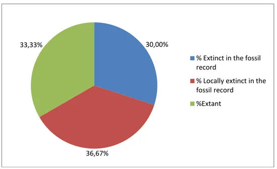
provided in the original, scale do not appear in the image (Cardoso, 1993a). ... 20 Figure 1.25: Left Ursus arctos mandible from Furninha cave in lingual view. Geological Museum, Lisbon. ... 20 Figure 1.26: Skull of Hyaena hyaena prisca from Furninha cave in dorsal view. Geological Museum, Lisbon. ... 21 Figure 1.27: The leopard (Panthera pardus) skull from Algar da Manga Larga in anterolateral view. Geological Museum, Lisbon. ... 22 Figure 2.2: Percentages of different parameters about the paleodiversity of the Quaternary in
Portugal. ... 37 Figure 2.3: Status of the different big mammals that appear in the fossil record in Portugal. ... 38 Figure 2.4: Percentage of species from each group in extant, fossil and fossil species (without cave deposits). ... 41 Figure 3.1: The location of the Santo Antão do Tojal Palaeoloxodon antiquus is marked by the
pushpin, in the Lisboa Peninsula (left) and at local level (right). Credit Google Earth. ... 43 Figure 3.2: General view of the Palaeoloxodon antiquus femur of Santo Antão do Tojal (MG 5788) at display in the geological museum in posterior view. Scale bar 7 cm. The chalk mark signals a cut in the bone where a stone tool was found, being it regarded by Zbyszewski as a proof that the animal was butchered by Neanderthals. ... 45 Figure 3.3: Right tibia of the Palaeoloxodon antiquus of Santo Antão do Tojal (MG 5793) at display in the Geological Museum in anterior view. Scale bar 7 cm. ... 46 Figure 3.4: Proboscidean tibia of Santo Antão do Tojal (MG 5793) in left, anterior and right proximal views. The principal anatomical features are marked, in white the tibial depression and in red the medial articular surface. Scale bar 7 cm. ... 46 Figure 3.5: The spinal process of the proboscidean of Santo Antão do Tojal (MG 8081) in A) anterior and B) posterior views. ... 47 Figure 3.6: The phalanx from the proboscidean of Santo Antão do Tojal in A) dorsal; B) lateral; C) ventral and D) lateral views. ... 48 Figure 3.7: Measurements of the femur of mammals by Von den Driesch, 1976. ... 49 Figure 3.8: Measurements of the tibia of mammals by Von den Driesch, 1976. ... 51 Figure 3.9: Plot of means for the previously selected measurements of the femur of Mammuthus primigenius (left) and Palaeoloxodon antiquus (right) with an interval of confidence of 95%. All measurements are in millimeters. ... 57 Figure 3.10: Plot of means for the previously selected measurements of the tibia of Mammuthus primigenius (left) and Palaeoloxodon antiquus (right) with an interval of confidence of 95%. All measurements are in millimeters. ... 58
xv
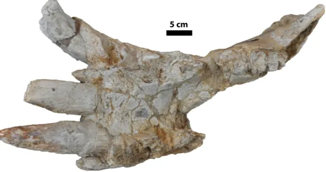
Figure 3.11: Measurements of Santo Antão do Tojal specimen (in red) compared to the means for them in Mammuthus primigenius (left) and Palaeoloxodon antiquus (right) with a 95% confidence interval. ... 59 Figure 3.12: Estimation of density for maximum length and minimum diaphysis width of the femur of Mammuthus primigenius and Palaeoloxodon antiquus. ... 60 Figure 3.13: Estimation of density for maximum length and minimum diaphysis width of the tibia of Mammuthus primigenius and Palaeoloxodon antiquus ... 61 Figure 3.14: From upper to bottom: Percentage of variation accounted by the first three Principal Component of the femur; PC1, PC2 and PC3. In PC1 and PC2 the highest contribution comes from the maximum length and the minimum diaphysis circumference respectively. ... 63 Figure 3.15: PC1 vs PC2 of the femur of selected proboscideans. The Santo Antão do Tojal specimen is marked with the red square, Mammuthus primigenius in blue and Palaeoloxodon antiquus in black. ... 63 Figure 3.16: From upper to bottom: Percentage of variation accounted by the first three Principal Component of the tibia; PC1, PC2 and PC3. In PC1 and PC2 the highest contribution comes from the maximum length and the minimum diaphysis circumference respectively. ... 65 Figure 3.17: PC1 vs PC2 of the tibia of selected proboscideans. The Santo Antão do Tojal specimen is marked with the red square, Mammuthus primigenius in blue and Palaeoloxodon antiquus in black. 65 Figure 4.1: Location of the Algar do Vale da Pena. Upper left, at peninsular level, it is situated in the Portuguese Estremadura. Bottom, view at regional level, the closest city is Alcobaça. Upper right, view at local level. Credit Google Earth. ... 67 Figure 4.2: Litostratigraphic log of the “Maciço Calcário Estremenho”, with the Candeeiros
Formation marked (Carvalho, 1996). ... 68 Figure 4.3: Location of the cave (red and orange dot) in the corresponding sheet (26 D, Caldas da Rainha) of the geologic map (1:50000) of Portugal, including legend (Zbyszewski & de Matos, 1959). ... 68 Figure 4.4: Topography of the Algar do Vale da Pena. In black, some landmarks of the cave are marked and in red, the bone findings (Modified from Amendoeira et al., 2015). ... 70 Figure 4.5: A) Entrance of the cave showing the first ramp and the top of two meters step. B) Picture taken from the base of the two meters step, showing the second ramp. C) Final vertical well of the entrance from the bottom. D) Picture from the bottom of the cave showing the final well (white line), the second ramp (green line), the two meter step (red line) and part of the firs ramp (blue line).
Pictures by Carlos Ferreira. ... 71 Figure 4.6: Claw marks (left) and dog skeleton (right) in the small conduct at the right of the cave. .. 72 Figure 4.7: Thick deposit of sands below the calcareous crust (left) and claw marks (right). ... 72 Figure 4.8: Equipment needed to descend (in black) and climb out of the cave again (in red). Left; A) Shunt, B) Descensor, C) Climbing fist. Right; D) Ventral croll and security knot to stop descending, note how the fingers secure the shunt. ... 73 Figure 4.9: First excavation around the skull of “bear number one” in October 2016. ... 74 Figure 4.10: The main block as in October 2017, during the second expedition, no new diggings where carried out this time. The skull is near the scale of seven centimeters, ... 74
xvi
Figure 4.11: A) Digging of the main block of “bear one” (note the camera of Rui Guerra). B) First layer of plaster after the block was cut. C) Putting the block in the net. D) Moving the block with the tree trunk. E) Final plastering of the block. F) Current state of the block. Pictures by Carlos Ferreira. ... 75 Figure 4.12: Before ( left) and after( right) for some ursid remains recovered in Algar do Vale da Pena: a) distal radius, b) mandibular ramus and c) basicranium. ... 76 Figure 4.13: The main block of “bear” one during the second expedition. Scapula and protruding long bones are marked with black arrows. In red, line where the block was cut, main skull with the scale of 7 cm and muddy area with numerous smaller bone remains. ... 77 Figure 4.14: Remains of the “bear number two” before (left) and after (right) removal of the block that was hiding them. ... 78 Figure 4.15: Muddy sediment below the calcareous crust, during the preparation of the hemimandible (ADVP 2.1). Scale 7 cm. ... 78 Figure 4.16: Material of bear number four encased in the calcareous crust. Left, humerus; right, femur and pelvis. ... 79 Figure 4.17: Distal humerus of the bear number five. ... 79 Figure 4.18: Damaged scapula (ADVP 1.1) from “bear number one” (Ursus arctos) of Algar do Vale da Pena. ... 81 Figure 4.19: Right scapholunate bone (ADVP 1.3) from “bear number one” (Ursus arctos) of Algar do Vale da Pena in A) proximal; B) distal; C) anterior and D) exterior views. ... 81 Figure 4.20: Measurements for the scapholunate bone in ursids according to the system of Tsoukala & Grandal-d’Anglade (2002). All of them could be recorded for the specimen. ... 82 Figure 4.21: Transverse diameter vs anteroposterior diameter of the scapholunate of brown bears (García Vázquez, 2015) as brown dots and cave bears (unpublished own data from Cova da Ceza population) as green squares; plus the scapholunate ADVP 1.3, marked as a yellow star. ... 83 Figure 4.22: Transverse diameter vs anteroposterior diameter of the scapholunate of brown bears (data from García Vázquez, (2015)) as brown dots; plus the scapholunate ADVP 1.3, marked as a yellow star. ... 83 Figure 4.23: Right hamate bone (ADVP 1.4) from “bear number one” (Ursus arctos) of Algar do Vale da Pena. ... 84 Figure 4.24: Measurements for the scapholunate ursid bone according to the system of Tsoukala & Grandal-d’Anglade (2002).The recorded measurement is marked. ... 84 Figure 4.25: Length of the right hamate bone (ADVP 1.4) from “bear number one” of Algar do Vale da Pena compared to the length of the same bone of other brown bears (García Vázquez, 2015) . Measurements are in mm according to the system of Tsoukala & Grandal-d’Anglade (2002). Two groups (males and females) are classified according to this measurement. ... 85 Figure 4.26: Second phalanx (ADVP 1.6) from “bear number one” (Ursus arctos) of Algar do Vale da Pena in A) dorsal; B) ventral; C) proximal; D) distal views. ... 86 Figure 4.27: Measurements for the second phalanx of ursids according to the system of Tsoukala & Grandal-d’Anglade (2002). All of them could be recorded. ... 86 Figure 4.28: Length vs distal transverse diameter of the second phalanx (ADVP 1.6, marked as blue square) from “bear number one” (Ursus arctos) of Algar do Vale da Pena compared to the same
xvii
ration of a recent brown bear, data from García Vázquez, (2015) . The 95% ellipses are drawn in the graphs. Measurements are in mm according to the system of Tsoukala & Grandal-d’Anglade (2002). ... 87 Figure 4.29: Proximal epiphysis of the third right metatarsal (ADVP 1.7) from “bear number one” (Ursus arctos) of Algar do Vale da Pena in A) exterior; B) dorsal; C) interior and D) ventral views. 88 Figure 4.30: Measurements for the third metatarsal of ursids according to the system of Tsoukala & Grandal-d’Anglade (2002).The recorded measurements are marked. ... 88 Figure 4.31: Proximal transverse diameter vs Proximal anteroposterior diameter of the third
metatarsal (ADVP 1.6, marked as blue square) from “bear number one” (Ursus arctos) of Algar do Vale da Pena (As green triangle) compared to the same ration of fossil brown bears from NW Spain, data from García Vázquez, (2015) . Measurements are in mm according to the system of Tsoukala & Grandal-d’Anglade (2002). ... 89 Figure 4.32: Distal epiphysis of the right radius (ADVP 1.8) from “bear number one” (Ursus arctos) of Algar do Vale da Pena in A) anterior; B) exterior; C) Posterior; D) Interior and E) Distal views. ... 90 Figure 4.33: Measurements for the radius of ursids according to the system of Tsoukala & Grandal-d’Anglade (2002). Recorded measurements are marked. ... 90 Figure 4.34: Transverse diameter of distal radius from ADVP 1.8 and fossil and recent brown bears from NW Spain and Portugal (Cardoso, 1993; García Vázquez, 2015). Specimens with more than 60 mm are classified as male and less than 58 mm as females. ... 91 Figure 4.35: Transverse diameter vs anteroposterior diameter of distal radius from ADVP 1.8 (Green in the graph) and fossil and recent brown bears from NW Spain (García Vázquez, 2015). Two clusters of individuals are classified as females or males. ... 92 Figure 4.36: Distal distal part of the ulnar diaphysis (ADVP 1.9) from “bear number one” (Ursus arctos) of Algar do Vale da Pena in A) anterior; B) exterior; C) Posterior and D) Interior views. ... 92 Figure 4.37: Measurements for the ulna of ursids according to the system of Tsoukala & Grandal-d’Anglade (2002). Taken measurement is marked with a circle. ... 93 Figure 4.38: Minimum anteroposterior diameter of the ulnar distal diaphysis (ADVP 1.9) from “bear number one” (Ursus arctos) of Algar do Vale da Pena compared graphically to the same measurement of fossil and recent brown bears from NW Spain (García Vázquez, 2015). Suggested males and
females are marked... 94 Figure 4.39: Posterior part of right hemimandible (ADVP 2.1) from “bear number two” (Ursus arctos) of Algar do Vale da Pena in A) dorsal; B) posterior; C) exterior; D) anterior; E) interior and F) ventral views. ... 94 Figure 4.40: Measurements for the mandible of ursids according to the system of Tsoukala & Grandal-d’Anglade (2002). Taken measurements are marked with a circle. ... 95 Figure 4.41: Total height of the mandible vs length from the mandibular condyle to the third molar from ADVP 2.1 (blue square in the graph), fossil and recent brown bears from NW Spain as black dots (García Vázquez, 2015) and cave bears as green triangles (unpublished data from Cova da Ceza). Two clusters of individuals are classified as females or males. ... 97 Figure 4.42: Total height of the mandible vs height of the mandibular condyle from ADVP 2.1 (blue square in the graph) and fossil and recent brown bears from NW Spain as black dots (García Vázquez, 2015) and cave bears as red inverted triangles (unpublished data from Cova da Ceza). ... 97
xviii
Figure 4.43: Diameter of the mandible between M2 and M3 vs height of the mandibular corpus labially at M3 from ADVP 2.1 (blue square in the graph) and fossil and recent brown bears from NW Spain as black dots (García Vázquez, 2015) and a cave bear as a green diamond (unpublished own data from Cova da Ceza). ... 98 Figure 4.44: Partial occipital and parietal bones (ADVP 2.2) from the “bear two” (Ursus arctos) of Algar do Vale da Pena in A) dorsal; B) posterior and C) anterior views. ... 99 Figure 4.45: Temporal and partial parietal bones (ADVP 2.3) from the “bear two” (Ursus arctos) of Algar do Vale da Pena in A) exterior and B) interior views. ... 99 Figure 4.46: Partial canine teeth (ADVP 2.5) from the “bear two” (Ursus arctos) of Algar do Vale da Pena in lateral view. ... 100 Figure 4.47: Fourth upper premolar (ADVP 2.6) from the “bear two” (Ursus arctos) of Algar do Vale da Pena in A) oclusal, B) lingual and C) labial views. ... 100 Figure 4.48: Measurements for the fourth upper premolar of ursids according to the system of
Tsoukala & Grandal-d’Anglade (2002). ... 102 Figure 4.49: Breadth vs length of the fourth upper premolar ADVP 2.6 (blue square in the graph) and the same measurements of recent and fossil brown bears from NW Spain as black dots (García
Vázquez, 2015). ... 102 Figure 4.50: First upper molar (ADVP 2.7) from the “bear two” (Ursus arctos) of Algar do Vale da Pena in A) oclusal, B) lingual and C) labial views. ... 103 Figure 4.51: Measurements for the first upper molar according to the system of Tsoukala & Grandal-d’Anglade (2002). Taken measurements are marked with a circle. ... 103 Figure 4.52: Breadth vs length of the first upper premolar ADVP 2.7 (blue square in the graph) and the same measurements of recent and fossil brown bears from NW Spain as black dots (García Vázquez, 2015) and cave bears as orange triangles from Liñares cave, Galicia, NW Spain (González & Martelli, 2003). ... 105 Figure 4.53: Second lower molar (ADVP 2.8) from the “bear two” (Ursus arctos) of Algar do Vale da Pena in A) oclusal, B) lingual and C) labial views. ... 105 Figure 4.54: Measurements for the second lower molar according to the system of Tsoukala &
Grandal-d’Anglade (2002). ... 107 Figure 4.55: Breadth vs length in mm of the second lower molar ADVP 2.8 (blue square in the graph) and the same measurements of recent and fossil brown bears from NW Spain as green
diamonds(García Vázquez, 2015) plus cave bears from A Ceza cave as black dots, Galicia, NW Spain (Unpublished own data). ... 107 Figure 4.56: Third lower molar (ADVP 2.9) from the “bear two” (Ursus arctos) of Algar do Vale da Pena in A) oclusal, B) lingual and C) labial views. ... 108 Figure 4.57: Measurements for the third lower molar according to the system of Tsoukala & Grandal-d’Anglade (2002). ... 108 Figure 4.58: Breadth vs length in mm of the third lower molar ADVP 2.9 (black dot in the graph) and the same measurements of recent and fossil brown bears from NW Spain as blue diamonds (García Vázquez, 2015) plus cave bears from A Ceza, Liñares and Eirós caves, Galicia, NW Spain (Grandal-d’Anglade, 1993; González & Martelli, 2003; unpublished own data) as purple triangles. ... 110
xix
Figure 4.59: Third phalanx (ADVP 2.10) from the “bear two” (Ursus arctos) of Algar do Vale da Pena in A) Distal; B) and D) Lateral; C) Proximal view. ... 110 Figure 4.60: Measurements for the third phalanx of ursids according to the system of Tsoukala & Grandal-d’Anglade (2002). Taken measurements are marked with a circle. ... 111 Figure 4.61: Height and diameter of the third phalanx (ADVP 2.10) (black dot in the graph) and the same measurements of recent and fossil brown bears from NW Spain as blue squares (García Vázquez, 2015) plus cave bears from A Ceza, Liñares and Eirós caves, Galicia, NW Spain (Grandal-d’Anglade, 1993; González & Martelli, 2003; unpublished own data) as crimson diamonds. 95% ellipses are drawn... 112 Figure 4.62: Distal epiphysis of femur (ADVP 3.1) from the “bear three” of Algar do Vale da Pena in A) distal B) lateral views. ... 112 Figure 4.63: Anteroposterior diameter of the distal epiphysis of the femur (ADVP 3.1) and the same measurement of recent and fossil brown bears from NW Spain (García Vázquez, 2015). ... 113 Figure 4.64: Vertebral centrum (ADVP 3.2) from the “bear three” of Algar do Vale da Pena in A) dorsal and B) anterior views. ... 114 Figure 4.65: Proximal epiphysis of second right metatarsal (ADVP 4.1) from the “bear four” of Algar do Vale da Pena in A) Interior; B) Ventral; C) Exterior and D) Dorsal. ... 114 Figure 4.66: Measurements for the second metatarsal according to the system of Tsoukala & Grandal-d’Anglade (2002).Taken measurements are marked with a circle. ... 115 Figure 4.67: Anteroposterior vs transversal diameter of the proximal epiphysis (upper) and diaphysis (bottom) of the second metatarsal, ADVP 4.1 (blue square) and the same measurements of recent and fossil brown bears from NW Spain as black dots (García Vázquez, 2015). ... 116 Figure 4.68: Proximal epiphysis of fifth right metatarsal (ADVP 4.2) from the “bear four” of Algar do Vale da Pena in A) Interior; B) Ventral; C) Exterior and D) Dorsal views... 116 Figure 4.69: Measurements for the fifth metatarsal according to the system of Tsoukala & Grandal-d’Anglade (2002). Taken measurements are marked with a circle. ... 117 Figure 4.70: Anteroposterior proximal diameter vs transversal diameter of diaphysis of the fifth metatarsal, ADVP 4.2 (blue square) and the same measurements of recent and fossil brown bears from NW Spain as black dots (García Vázquez, 2015). ... 117 Figure 4.71: Claw marks in the walls of the Algar do Vale da Pena. Some of them are today partially covered by calcareous crusts. Scale bar 7 cm. ... 118 Figure 5.1: Location of the site of Santa Margarida. Figure by Cátia Ribeiro. Credit of the maps, google maps. ... 123 Figure 5.2: Maximum concentration point of fossil bearing blocks in the wall of the site as it was discovered in 2017. Note the difference of color between the bone bearing breccified blocks of
cemented terra rossa and the grey tone of the calcareous rocks. Photo by Octávio Mateus. ... 124 Figure 5.3: Location of Santa Margarida site. In blue the calcareous Picavessa Formation. Modified by Cátia Ribeiro from the geologic map of Algarve by LNEG. ... 124 Figure 5.4: Left; comparison of freshly cut cemented terra rossa block and the underlying grayish calcareous rock of the Picavessa formation. Right; calcareous crust covering the “terra rossa”... 125 Figure 5.5: A) Abundant microfossils in rich block; B) Rabbit humerus in situ; C) and D) larger long bones protruding from block. ... 125
xx
Figure 5.6: Upper left, possible leporid phalanx in dorsolateral view; upper right, arvicoline mandible in lingual view; bottom left, shrew mandible in labial view; bottom right, lizard mandible in labial view. ... 126 Figure 5.7: Left, consolidated arvicoline mandible with PARALOID B72™; center, extraction of the mandible using a air-scribe and right the extracted mandible. ... 126 Figure 5.8: Right, rodent teeth mounted in the plastic box and other fossils stored in similar settings (Scale 7cm); left, the binocular lenses used to take the pictures (Photo by Alexadre Guillaume). ... 127 Figure 5.9: Workflow followed in the preparation of the Santa Margarida material (Portugal) in the Museum of Lourinhã... 128 Figure 5.10: Amount of processed sediment from Santa Margarida (Portugal) at Museum of Lourinhã. ... 129 Figure 5.11: Paralaoma servilis from Santa Margarida in A) lateral, B) ventral and C) dorsal views. ... 130 Figure 5.12: Bisected shell of Pomatia elegans from Santa Margarida. ... 130 Figure 5.13: Lacertid mandible from Santa Margarida (Portugal) in lingual view... 131 Figure 5.14: Lacertid mandible from Santa Margarida (Portugal) in A) lingual and B) oclusal views. ... 131 Figure 5.15: Unicusp teeth ascribed to a bat from Santa Margarida (Portugal) in meso-lingual view (Image by Cátia Ribeiro). ... 132 Figure 5.16: Right upper second bat molar from Santa Margarida (Portugal) in oclusal view. The rectangle marks the w shaped labial ridge and the circle the lingually recurved hypocone shelf and the strong postprotocrista. Metastyle/paracone (1,3 mm) and metastyle/lingual (2 mm) lengths are
marked. Drawing by Pavlos Piskoulis. ... 132 Figure 5.17: Soricidae upper incisors recovered from Santa Margarida (Portugal) in labial view. A) Sorex, B) and C) Crocidura. ... 133 Figure 5.18: Broken second upper molar of Crocidura recovered from Santa Margarida (Portugal) in oclusal view. Labial part to the right. ... 133 Figure 5.19: Fragment of Crocidura mandible (bearing P4, M1 and M2) recovered from Santa
Margarida (Portugal) in A) oclusal, B) lingual and C) labial views. ... 134 Figure 5.20: Tip of a rodent incisor from Santa Margarida (Portugal). ... 135 Figure 5.21: A) third lower molar and B) first upper molar of Apodemus cf. sylvaticus from Santa Margarida (Portugal) in oclusal view. ... 135 Figure 5.22: Left upper first or second molar of Eliomys quercinus from Santa Margarida (Portugal) in oclusal view. ... 136 Figure 5.23: Left upper first molar of Allocricetus bursae from Santa Margarida (Portugal) in oclusal view. ... 136 Figure 5.24: Nomenclature and measurements for an arvicoline first lower tooth according to López-García et al., (2015). ABBREVIATIONS: a: length of the anteroconid complex; ACC, anteroconid complex; AC, anterior cap; BRA, buccal reentrant angle; BSA, buccal salient angle; c: width of the opening of triangles T4-T5; L, length; La, mean width of T4; Li, mean width of T5; LRA, lingual
re-xxi
entrant angle; LSA, lingual salient angle; PL, posterior lobe; TTC, trigonidtalonid complex, T1-T7, triangles 1-7; W, width. ... 137 Figure 5.25: Right lower first molar of Victoriamys chalinei from Santa Margarida (Portugal) in oclusal view. ... 137 Figure 5.26: Left lower first molar of Iberomys huescarensis from Santa Margarida (Portugal) in oclusal view. ... 138 Figure 5.27: Right and left (respectively) lower first molars of Iberomys brecciensis from Santa Margarida (Portugal) in oclusal view. ... 138 Figure 5.28: Left lower first molar (without the posterior lobe) of Iberomys cabrerae from Santa Margarida (Portugal) in oclusal view. ... 139 Figure 5.29: A/L x100 vs C/W x100 ratios for a collection of arvicoline teeth. Victoriamys chalinei from El Chaparral locality appear as pink circles. Allophaiomys lavocati from El Chaparral appear as blue pluses symbols. Iberomys huescarensis, from a collection of Iberian and Italian sites appear as black dots. Data from López-García et al., (2012, 2015). The specimens from Santa Margarida
(Portugal) appear marked with a red circle. ... 140 Figure 5.30: A/L x100 vs La/Li x100 ratios for a collection of arvicoline teeth. Victoriamys chalinei from El Chaparral locality appear as red inverted triangles. Allophaiomys lavocati from El Chaparral appear as blue squares. Iberomys huescarensis, from a collection of Iberian and Italian sites appear as black dots. Data from López-García et al., (2012, 2015). The specimens from Santa Margarida (Portugal) appear marked with a red circle. ... 140 Figure 5.31: A/L x100 vs La/Li x100 ratios for Iberomys brecciensis (green squares) and I. chalinei (gold inverted triangles) from a collection of Iberian and Italian sites. Data from López-García et al., (2015). The specimens from Santa Margarida (Portugal) appear marked with a red circle. ... 141 Figure 5.32: Left distal Oryctolagus cuniculus humerus from Santa Margarida (Portugal) in A)
Anterior; B) Exterior; C) Posterior and D) Medial views. ... 142 Figure 5.33: Diverse postcranial elements of microvertebrates recovered from the sample of Santa Margarida. A) Distal femur in posterolateral view, B) Partial distal humerus? in anterior view and C) Distal radius in lateral view. ... 142 Figure 5.34: Evolution of the first lower molar of the genus Iberomys with examples from the site of Santa Margarida (Portugal). ... 144 Figure 5.35: The two possible stratigraphy scenarios that explain the faunal assemblage recovered from the ex situ blocks from Santa Margarida (Portugal). Upper: Fossils from different but similar layers that became mixed during preparation. Lower: Reworked fossils situated in the same layer as the younger ones. ... 145
xxii
INDEX OF TABLES
Table 1.1: Presence of different species of Microtus/Iberomys in different Quaternary sites in Portugal, data from Antunes et al., 1986b, Antunes et al., 1989; Póvoas et al., 1992; Jeannet, 2000; Moreno-García & Pimenta, 2002; Bicho et al., 2003; Valente & Haws, 2006; Pimenta, 2014, López-García et al., 2018). ... 14 Table 2.1: Localities of the Quaternary of Portugal. ... 23 Figure 2.1: Satellital picture showing the location of 30 sites with Quaternary fossil
vertebrates in Portugal, mentioned in the table 2.1. In black, the localities situated in the Lusitanian basin and in red the ones situated in the Algarve basin. Credit Google Earth. .... 25 Table 2.2: Amphibians of the Quaternary of Portugal. ... 26 Table 2.3: Reptiles of the Quaternary of Portugal. ... 26 Table 2.4: Birds of the Quaternary of Portugal. ... 27 Table 2.5: Mammals of the Quaternary of Portugal... 31 Table 2.6: Resume table showing the number, percentage and status of extant and fossil species of the Quaternary of Portugal... 36 Table 2.7: Table showing the tetrapods and localities known outside the cave deposits in the Quaternary of Portugal. ... 38 Table 2.8: Resume table showing the number, percentage and status of extant and fossil species in the Quaternary of Portugal, excluding the cave deposits. ... 40 Table 3.1: Measurements of the phalanx of the proboscidean of Santo Antão do Tojal. “Thickness” refers to anteroposterior diameter of the bone and “width” to the mediolateral diameter of it. All the measurements are in millimeters. ... 48 Table 3.2: Measurements of 48 individual femurs of Palaeoloxodon antiquus and
Mammuthus primigenius taken from the literature. The specimen of Santo Antão do Tojal is marked. All measurements are in millimeters. ... 49 Table 3.3: Measurements of 30 individual tibias of Palaeoloxodon antiquus and Mammuthus primigenius taken from the literature. The specimen of Santo Antão do Tojal is marked. In order to fit the table, abbreviations have been used: ML, maximum length; AL, articular length; PW, proximal width; PT, proximal thickness; PAW, proximal articular width; PAT, proximal articular thickness; MDW, minimum diaphysis width; MDT, minimum diaphysis thickness; MDC, minimum diaphysis circumference; DW, distal width; DT, distal thickness; DAW, distal articular width; DAT, Distal articular thickness. All measurements are in
millimeters. ... 51 Table 3.4: Maximum length vs minimum diaphysis circumference ratios of femur and tibia, for selected Palaeoloxodon antiquus specimens. All measurements in mm.”E” stands for
“estimated”. ... 53 Table 3.5: Femur vs tibia lengths for selected Palaeoloxodon antiquus specimens. All
measurements in mm.”E” stands for “estimated”. ... 53 Table 3.6: Shoulder height and body masses of different Palaeoloxodon antiquus specimens. The specimens below to the Santo Antão do Tojal one had not been calculated using the same methods as presented. Possible males are marked in blue. ... 54 Table 3.7: Number of individual measurements taken for proboscidean femurs and tibias of the considered sample. Percentage of specimens where the measurement has been taken compared to the total is also presented. The measurements that have been taken in 50% or more of the specimens are marked. ... 55
xxiii
Table 3.8: Mean for selected measurements in the hind limb of Palaeoloxodon antiquus and Mammuthus primigenius, with percentage of difference between the species for each
measurement and bone. All measurements are given in millimeters. ... 56 Table 4.1: Measurements from the right scapholunate bone (ADVP 1.3) from “bear number one” (Ursus arctos) of Algar do Vale da Pena compared to a set of measurements of other brown bears (García Vázquez, 2015) and cave bears (unpublished own data from Cova da Ceza population). Measurements are in mm according to the system of Tsoukala & Grandal-d’Anglade (2002). ... 82 Table 4.2: Length of the right hamate bone (ADVP 1.4) from “bear number one” (Ursus arctos) of Algar do Vale da Pena compared to the length of the same bone of other brown bears (García Vázquez, 2015) . Measurements are in mm according to the system of
Tsoukala & Grandal-d’Anglade (2002). ... 85 Table 4.3: Length vs distal transverse diameter of the second phalanx (ADVP 1.6) from “bear number one” (Ursus arctos) of Algar do Vale da Pena compared to the length of the same bone of a recent brown bear (García Vázquez, 2015) . Measurements are in mm according to the system of Tsoukala & Grandal-d’Anglade (2002). ... 87 Table 4.4: Proximal transverse diameter and proximal anteroposterior diameter of the third metatarsal (ADVP 1.7) from “bear number one” (Ursus arctos) of Algar do Vale da Pena compared to same measurements of fossil brown bears from NW Spain (García Vázquez, 2015) . Measurements are in mm according to the system of Tsoukala & Grandal-d’Anglade (2002). ... 89 Table 4.5: Measurements from the radio (ADVP 1.8) from “bear number one” of Algar do Vale da Pena compared to same ratio of fossil brown bears from NW Spain (García
Vázquez, 2015) as well as some of Portuguese caves. Measurements are in mm according to the system of Tsoukala & Grandal-d’Anglade (2002). ... 91 Table 4.6: Minimum anteroposterior diameter of the ulnar distal diaphysis (ADVP 1.9) from “bear number one” (Ursus arctos) of Algar do Vale da Pena compared to the same
measurement of fossil and recent brown bears from NW Spain (García Vázquez, 2015). Measurements are in mm according to the system of Tsoukala & Grandal-d’Anglade (2002). ... 93 Table 4.7: Measurements from the partial hemimandible (ADVP 2.1) from “bear number two” (Ursus arctos) of Algar do Vale da Pena compared to same measurements of fossil brown bears from NW Spain (García Vázquez, 2015) as well as some cave bears from the same region (Unpublished own data from Cova de A Ceza). Measurements are in mm according to the system of Tsoukala & Grandal-d’Anglade (2002). ... 96 Table 4.8: Measurements from the fourth upper premolar (ADVP 2.6) from “bear number two” of Algar do Vale da Pena compared to broad an length of the same tooth of fossil and recent brown bears from NW Spain (García Vázquez, 2015). Measurements are in mm according to the system of Tsoukala & Grandal-d’Anglade (2002). ...101 Table 4.9: Length and breadth from the first upper molar (ADVP 2.7) from “bear number two” of Algar do Vale da Pena compared to breadth an length of the same tooth of fossil and recent brown bears from NW Spain (García Vázquez, 2015) and cave bears from Liñares cave, Galicia, NW Spain (González & Martelli, 2003). Measurements are in mm according to the system of Tsoukala & Grandal-d’Anglade (2002). ...104 Table 4.10: Breadth and length of the second lower molar (ADVP 2.8) from the “bear two” of Algar do Vale da Pena compared to breadth an length of the same tooth of fossil and recent brown bears from NW Spain (García Vázquez, 2015) and cave bears from A Ceza cave,
xxiv
Galicia, NW Spain (Unpublished own data). Measurements are in mm according to the system of Tsoukala & Grandal-d’Anglade (2002). ...106 Table 4.11: Breadth and length of the third lower molar (ADVP 2.9) from the “bear two” of Algar do Vale da Pena compared to breadth an length of the same tooth of fossil and recent brown bears from NW Spain (García Vázquez, 2015) and cave bears from A Ceza, Liñares and Eirós caves, Galicia, NW Spain (Grandal-d’Anglade, 1993; González & Martelli, 2003; unpublished own data). Measurements are in mm according to the system of Tsoukala & Grandal-d’Anglade (2002). ...109 Table 4.12: Height and diameter of the third phalanx (ADVP 2.10) from the “bear two” of Algar do Vale da Pena compared to the height and diameter of the same phalanx of fossil and recent brown bears from NW Spain (García Vázquez, 2015) and cave bears from A Ceza, Liñares and Eirós caves, Galicia, NW Spain (Grandal-d’Anglade, 1993; González & Martelli, 2003; unpublished own data). Measurements are in mm according to the system of Tsoukala & Grandal-d’Anglade (2002). ...111 Table 4.13: Anteroposterior diameter of the distal epiphysis of the femur (ADVP 3.1) from the “bear three” of Algar do Vale da Pena compared to the same measurement of fossil and recent brown bears from NW Spain (García Vázquez, 2015). Measurements are in mm according to the system of Tsoukala & Grandal-d’Anglade (2002). ...113 Table 4.14: Measurements of the second metatarsal (ADVP 4.1) from the “bear four” of Algar do Vale da Pena compared to the same measurement of fossil and recent brown bears from NW Spain (García Vázquez, 2015). Measurements are in mm according to the system of Tsoukala & Grandal-d’Anglade (2002). ...115 Table 4.15: Measurements of the fifth metatarsal (ADVP 4.2) from the “bear four” of Algar do Vale da Pena compared to the same measurement of fossil and recent brown bears from NW Spain (García Vázquez, 2015). Measurements are in mm according to the system of Tsoukala & Grandal-d’Anglade (2002). ...117 Table 4.16: Nitrogen and Carbon percentages in the samples of ADVP 1.10 and ADVP 3.4 from Algar do Vale da Pena (Portugal). ...122 Table 5.1: Results of the tests for determining the product to be use in the chemical
preparation of Santa Margarida samples. ...127 Table 5.2: Number of specimens from broad groups recovered from the processed sediment of Santa Margarida. ...129 Table 5.3: Measurements and ratios of the most complete lacertid mandible fragment picked from the disaggregated sediments of Santa Margarida, Portugal, following the methods of Blain et al., 2007: h, is height of the teeth; d, its diameter and a, the height of the teeth that protrudes over the dental crest. All measurements are in mm. ...131 Table 5.4: Classification of the different Soricidae remains recovered from Santa Margarida (Portugal). ...133 Table 5.5: Classification of the different rodent dental remains recovered from Santa
Margarida (Portugal). ...134 Table 5.6: Measurements and ratios of the arvicoline first lower molars from Santa
xxv
LIST OF ABBREVIATIONS
Institutions
EAVP: European Association of Vertebrate Paleontology. MG: Geological Museum of Lisbon.
FCT-UNL: Facultade de Ciência e tecnología da Universidade Nova de Lisboa. LNEG: Laboratório Nacional de Energía e Geología.
ML: Museu da Lourinhã. GPS: Grupo de Proteção Sicó.
GEM: Grupo de espeleologia e Montanhismo. Other
PC: Principal Components. YBP: Years Before Present.
1
1.INTRODUCTION
1.1 History of the study of Quaternary fossil vertebrates from continental
Portugal
This section has been partially published in an abstract at meeting of EAVP 2018 (Estraviz-López & Mateus, 2018b). The study of the remains of Quaternary fossil faunal assemblages cannot be understood (mostly in their early years) without its close relation with archeological works and the more general geologic survey. The discovery of tools first and then human remains (nowadays we now that they were in fact younger) in the terraces of the Somme River in France, related with the remains of extinct fauna like rhinoceroses and elephants in the beginning of the second half of the XIX century, sparked the interest of the scientific community about the antiquity of humanity and the search of new bone material and artifacts in Pleistocene deposits. Also, other two factors in this period made the interest in the study of the past rise in the scientific community as never before: The publication of “The origin of species” by Charles Darwin and the outbreak of the nationalist sentiment that made the cult peoples of Europe to search for clues and material proof in the past, even competing with other nations and scientists (Cardoso, 2015a).
In Portugal, the scientific study of the Quaternary started thanks to the work of the members of the 2nd Geological Commission of Portugal in 1857; mostly Carlos Ribeiro and Nery Delgado, whose work proved that it was possible to know about the distant past (well before the written texts, oral traditions and collective memory) just thanks to the study of material remains (being them bones or tools) coming from alluvial and cave deposits (Cardoso, 2015a).
Carlos Ribeiro (1813-1884) (Figure1.1) was the father of the prehistoric archeology (and therefore the Quaternary paleontology) in Portugal. From humble origins he developed a career in the army as military engineer that he left in 1847, being the leader of the Geological Commission of Portugal with Pereira da Costa (more focused to collection management than fieldwork) since 1857 until his death (Daveau, 2000). He published a surprisingly accurate description of the Quaternary deposits, that in that moment also included some of Neogene origin, in the basins of Tagus and Sado rivers that he never finished (a second part of that work was never published) (Cardoso, 2015b) and several other works including descriptions of silex and quartzite tools from the terrains of those rivers (Ribeiro, 1871). He raised the question about how old the humanity could be and pointed towards the possibility that some tools that he collected could be from Tertiary origin (Cardoso, 2000) . He also carried fieldwork, excavating the prehistoric settlement of Leceias in 1878 and the cave of Ponte da Laje in 1879 among other diggings (Cardoso, 2013), probably searching for material to show to other fellow scientist in the important IX session of the International Congress of Anthropology and Prehistoric Archaeology that was carried in Lisbon in September of 1880; the culmination of 25 years of his work.
2
Joaquim Felipe Nery Delgado (1835-1908) (Figure 1.2) was also a major figure in the study of the Quaternary in Portugal. He graduated as military engineer in 1855 just in time to incorporate as adjunct to the Director of the Geological Comission of Portugal in 1857 that he later lead since the death of Carlos Ribeiro in 1884 to his own death in 1908, with more than 51 years of study and research in the areas of geologic cartography and prehistoric archeology on his back. As geologist his major interest was the study of the Paleozoic rocks in Portugal, mostly in Alentejo as well as the nature of the enigmatic marks of “bilobites” (Cruziana) (Cardoso, 2008a).
Figure 1.2: Picture of Nery Delgado at exposition in the Geological Museum, Lisbon.
In 1865 and 1866 he undergone the first scientific excavation of a cave occupied by prehistoric humans, Casa da Moura, in the municipality of Óbidos and Cova da Moura, Lourinhã and Bombarral municipalities. He later published his findings, along with other excavations in nearby caves in his work “Da Existência do Homem no Nosso Solo em Tempos Mui Remotos Provada pelo Estudo das Cavernas. Notícia ácerca das Grutas da Cesareda” published in 1867 that included a human skull, which being published before the Cro Magnon man would be the first Paleolithic anatomically modern human remains known (Delgado, 1867; Zilhão, 1993). The same year of 1865 he started some preliminary diggings in the Furninha cave in Peniche, but it was not until 1879 and 1880 when he undertook this pivotal excavation in the history of the Portuguese prehistoric archeology and Quaternary paleontology (Delgado, 1884). The methodology of this excavation is even nowadays exemplar, with each piece of bone or tool identified not only with the site but with the layer from where it came and with its coordinates in a grid system (Figure 1.3). So Nery Delgado was a pioneer of the 3D location of the archeological artifacts (Cardoso, 2015a).
Figure 1.3: Hyena coprolite from Furninha cave recovered by Nery Delgado (MG). Note the glued
3
Both Nery Delgado and Carlos Ribeiro were respected scientists in his time, being his work known across Europe, proof of that is the correspondence that they kept with other fellow foreign scientists (Cardoso & Avila de Melo, 2001) and the visits that they did to other countries, like for example Nery Delgado to Spain in 1878 (Carneiro, 2007). The quality of their work put Portugal in the frontline of this type of studies in Europe at that time, and all of this crystallized in the celebration of the IX session of the International Congress of Anthropology and Prehistoric Archaeology in Lisbon, 1880. Some new diggings were made to provide with new information and pieces to show in the congress, for example the interesting site of Mealhada where some proboscidean remains were uncovered (Cardoso, 1993).
In the last years of the XIX century and the first of the XX century Nery Delgado focused more in the study of the geology of the Paleozoic of Portugal until his death (Delgado, 1886) but the study of the Quaternary animal remains continued with less intensity. Paul Choffat was another important geologist and paleontologist of those years, that inherited the lead of Nery Delgado over the geological services and overall the study of the geology in Portugal; he uncovered some hippopotamus and proboscidean remains in the calcareous tuffs of Condeixa in 1895 and 1898. Years later, in 1909, he also promoted the diggings in the Pleistocene sites of Algar de João Ramos and Molianos (Cardoso, 1993b).
Paul Choffat (figure 1.4) realized that the materials recovered by Nery Delgado and Carlos Ribeiro deserved an attention that he (being focused on the investigation of the Paleozoic and Mesozoic of Portugal) could not properly give, so thanks to his initiative the classic Portuguese material of sites like Furninha and Fontainhas was studied by Edouard Harlé, from Toulouse (One of the top experts in the Pleistocene of that time). He published a work that would be a major reference for the study of Quaternary paleontology in Portugal during the next 80 years, cataloging the fauna found until that moment and comparing it with other countries of Europe (Harlé, 1910; Cardoso, 1993b).
Figure 1.4: Picture of Paul Choffat at exposition in the Geological Museum, Lisbon.
After this outburst of the early XX century the study of the Quaternary animals in Portugal entered a phase of stasis. Paul Choffat said: “By reasons that we should not comment, although that our faculties are filled with professors of great erudition, there are few of them that are dedicated to the progress of observation sciences”, that was his way of saying that there was not much interest in carrying fieldwork in Portugal during those years (Daveau, 2000). We should wait until the middle of the century and the works of Octávio da Veiga Ferreira and mostly Georges Zbyszewski to restart the findings in the Quaternary of the Portuguese caves and alluvial deposits.
4
Octávio da Veiga Ferreira (1917-1997) was a mine engineer that incorporated to the Geological Services in 1950. He already had carried some archeological excavations by that time and during his many years of activity he was a major figure in the Iberian archeology. In the area of paleontology, although it was not his main research field, he published 32 papers, four of them about Pleistocene vertebrates and one about Holocene mollusks (Cardoso, 2008 b). One of his major contributions is his study about the rhinoceroses of Portugal (Veiga Ferreira, 1975).
Georges Zbyszewski (1909-1999) (Figure 1.5) was the direct superior of Octávio da Veiga Ferreira; born in Russia, but later nationalized French citizen. He visited Portugal for first time in 1935 searching for clues about the tectonic movements in the Portuguese littoral during the Quaternary for his doctoral thesis. In 1940 he incorporated to the Geological Services and during the next 40 years he covered an immense array of geological topics, writing around 300 scientific publications from the geological cartography of Portugal, to the volcanism in Açores and Madeira, to the Mesozoic reptiles and over all of that; more than 100 publications about Quaternary lithic industries and fauna (Soares de Carvalho & Cardoso, 1999; Teixeira, 1979). During the years “Zby” (as he was called by friends and family) reunited a lot of information about the recent fluvial terraces of the Tagus River and its tributaries in the work “Le Quaternaire du Portugal” published in 1957. A lot of publications were made from mammal material coming from those terraces, either Miocene or Pleistocene, for example some remains like phalanges and tusks of Paleoloxodon antiquus (Falconer & Cautley, 1847) from that area and the classic site of Mealhada. Is also remarkable the different excavations and publications that he did with the material of a lot of different Pleistocene caves like: Almonda (1941 and 1947), lapa da Bogalheira (1941), the caves of Maceira and Vimeiro (1949), the cave of Ponte da Laje (excavated before by Carlos Ribeiro) (1957), Gruta das Salemas (1962), Gruta da Columbeira (1963), Alcobertas (1971), and lapa do Bugio the same year. Beside all of this he also studied the Pleistocene faunas coming from Algoz, in Algarve (Soares de Carvalho & Cardoso, 1999; Teixeira, 1979). All this work would have been probably impossible without the hard work of dozens of unrecognized collectors that were the eyes and ears of not only him but also great scientist before and after him.
Figure 1.5: Georges Zbyszewski (Picture by unknown author).
The work of Veiga Ferreira and Zbyszewski in the Geological services of Portugal served well to kick start the next phase of the study of the Quaternary faunas in Portugal, with the lead of Miguel Telles Antunes first and João Luis Cardoso later. A good visualization of this is the work that both of them published in 1990 to synthesize their many years of study in this matter with the new studies of the next generation in the decade of 1980 (Zbyszewski & Veiga Ferreira, 1990).
5
Before continuing it should be noted the work of Torres Pérez-Hidalgo, a Spanish researcher that in 1979 published an extensive study about the bears of Portugal, including measurements of old material recovered even in the XIX century and agreeing with the work of Harlé in that there is no other recognized fossil bear in the country besides the brown bear, Ursus arctos Linnaeus, 1758 (Torres Pérez-Hidalgo, 1979).
Miguel Telles Antunes (1937) (Figure 1.6) is probably until this day the most prolific figure in the History of the Portuguese paleontology. He started his career in the University of Lisbon in 1960 after his doctorate in geology and in 1974 he moved to the Universidade Nova de Lisboa. During his many years in paleontology he covered practically all the topics in the area writing more than 450 scientific articles and directing 9 doctoral theses, from the Miocene vertebrates in the area of Lisbon, to the Eocene of Silveirinha (Baixo Mondego), or the dinosaur faunas in the Lusitanian Basin (https://www.fct.unl.pt/noticias/2016/12/sessao-de-homenagem-ao-professor-doutor-miguel-telles-antunes). His collaboration with Zbyszewski at the end of the decade of 1970 produced some new insights into the Neogene of the Tagus Basin and during the decade of 1980 he carried several studies in Pleistocene material: The faunas of Morgadinho and Goldra in Algarve, the review of the Algoz fauna (also from Algarve) and the Mealhada material (that lead to the identification of Homotherium latidens Owen, 1846 in Portugal) and also the studies carried out in the cave contexts of Figueira Brava (where he impulse excavations), Caldeirão cave (where he recognized Castor fiber Linnaeus, 1758 for first time in Portugal) (Antunes, 1989a) or Pedreira das Salemas (Cardoso, 1993a). Over all of this work, the impulse that Antunes gave to the study of Pleistocene faunas through the former CEPUNL (Centro de Estratigrafía e Paleobiología da Universidade Nova de Lisboa) is everlasting even today, with numerous radiometric datings and training of new personal.
Figure 1.6: Miguel Telles Antunes in 2009 (Photo by Natacha Cardoso).
More or less at the same time during the decade of 1980 and the beginning of the 1990, two personalities started producing scientific investigation at high level. João Zilhão (1957) now professor of the University of Barcelona is an archaeologist, therefore his core area is situated more in the paleoanthropology, but his work in the Almonda cave system (Zilhão et al., 1991) and other caves in the Estremadura like Caldeirão cave produced a wide array of Pleistocene faunal material (Zilhão, 1987). This material was subject of studies the following years.
6
The other personality is João Luis Cardoso (1956) (Figure 1.7) the most well known researcher in the study of Quaternary faunas from Portugal. Professor of the Universidade Aberta de Lisboa has written more than 550 publications, including books, chapters of books and scientific articles; a lot of them about the paleontology and archeozoology, but also in archeology and geology (https://www.cm-obidos.pt/jornadas-arqueologia). In the second half of the decade of 1980 and the first of the decade of 1990 he worked in the CEPUNL publishing together with Antunes or in solitary plenty of scientific articles such the first reference of several species in the Pleistocene of Portugal, like Hippopotamus incognitus Faure, 1984 in Portugal from the deposits of Mealhada (Antunes et al., 1988), Cuon alpinus europaeus Bourguignat, 1868 (Cardoso, 1992), Panthera spelaea Goldfuss, 1810 (Antunes & Cardoso, 1986), Crocuta crocuta intermedia (M. de Serres, 1828) (Cardoso, 1994), Equus hydruntinus Regalia, 1905 (Cardoso, 1995), Dicerorhinus hemitoechus (Falconer, 1868) (Cardoso, 1990), Mammuthus primigenius Blumebach, 1799 (Antunes & Cardoso, 1992) and Rupicarpa rupicapra (Linnaeus, 1758) (Cardoso & Antunes, 1989). Also he has described a new subspecies of horse from the Pleistocene of Portugal, Equus ferus antunesi (Cardoso & Eisenmann, 1989).
Figure 1.7: João Luis Cardoso in 2017 (Photo by Octávio Mateus).
But his main contribution on this period is his doctoral thesis defended in October, 1992 and published in 1993 by the municipality of Oeiras titled “Contribuição para o conhecimento dos grandes mamíferos do Plistocénico Superior de Portugal” (Figure 1.8); a book of more than 500 pages that is the most comprehensive and at the same time detailed study ever made in Portugal about the Quaternary big mammal fauna of the country, covering remains of 26 taxa from 28 localities across all Portugal (Cardoso, 1993a).