UNIVERSIDADE DE LISBOA FACULDADE DE CIÊNCIAS
DEPARTAMENTO DE BIOLOGIA VEGETAL
Testing the Mechanosensory versus Chemosensory
hypotheses in left-right early development
Catarina Gonçalves Fonseca
Dissertação
Mestrado em Biologia Molecular e Genética
UNIVERSIDADE DE LISBOA FACULDADE DE CIÊNCIAS
DEPARTAMENTO DE BIOLOGIA VEGETAL
Testing the Mechanosensory versus Chemosensory
hypotheses in left-right early development
Catarina Gonçalves Fonseca
Dissertação
Mestrado em Biologia Molecular e Genética
Dissertação orientada por:
Doutora Susana Santos Lopes (orientação externa)
CEDOC – Faculdade de Ciências Médicas da Universidade Nova de Lisboa
Professor Doutor José Feijó (orientação interna)
Faculdade de Ciências da Universidade de Lisboa
i
ACKNOWLEDGMENTS
I want to start by thanking to Susana Lopes for the opportunity she gave me by accepting me in Cilia Regulation and Disease Lab, giving me the chance to work in a very interesting research area, new for me, and for her help when I needed
I also want to thank to Petra and Bárbara for all the things they taught me and their patience to teach me. Thank you for support and help during this year. I also want to thank to Monica, Pedro and Joana for help me in these lasts weeks, and by their availability ever. This thesis was only possible due to your help and support. It was a pleasure to work with you all. I also want to thank to Professor Dr. José Feijó to accept my co-supervising.
I also want to thank to ‘’Grupo do Canto’’ (Petra, Claudia Q., Claudia P., Sofia, João, Pedro, Telmo, Neuza, André, Maria and Ângela) for all the moments spent in and outsider the lab. I also want to thank for all the given suport and advises given throughout this year, mainly in this last days. I will not Forget you.
A special thanks to Filipa, Susana Jéssica and Melanie for all the friendship throughout these years and for the good moments at coffees in the weekends.
I also want to thanks to João, Marília and Filipa for their friendship throughout this last three years and for all the support given and, principally to João for his motivating sayings.
A special thanks to João Donato that has always given me support and has been on my side when I needed the most, without never let me lower my arms and make me believe in myself.
For last, but the most important people, I want to thank to my parents, Otília and José for all the support and trusting that they deposited in me throughout my academic route. Without them nothing of this would be possible. I also want to thank to my sister Joana for the good sister she is and for believing in me, supporting me and because of our interesting conversations at the weekends.
iii
ABSTRACT
Cilia are antenna-like organelles that protrude out of nearly all vertebrate cells. Two types of cilia can be defined: motile cilia, involved in moving fluids and primary cilia (shorter and immotile) that have sensory and signalling functions. Motile ciliary dysfunctions cause specific ciliopathies that affect mainly the respiratory tract, fertilization and left-right (LR) body establishment. The embryonic node is a ciliated structure where motile cilia generate an important fluid-flow named Nodal Flow (necessary for LR axis formation). Correct left-right patterning sometimes fails during development and defects, such as situs inversus occur. The correct left-right axis formation is highly dependent on signalling pathways downstream of such directional fluid-flow. The mechanism by which this process occurs is still unknown but there are supporting data in the field both for mechano and chemosensory pathways. There are two models that try to explain the mechanism by which LR is transferred to the Lateral Plate mesoderm. The “Morphogen model” – defends that morphogens become concentrated in left side of the node in response to flow. The other model, known as the “Two-cilia model” suggests that two different cilia types in the node perform different functions. From an unpublished microarray data we chose two genes in order to study these two models in zebrafish. “Morphogen Model” was studied with Tas1r1, because it has been recently reported that Tas1R receptors are expressed in ciliated epithelia acting as chemosensors. The “Two-cilia model” was studied with the pkd1 gene, because in kidney ciliated cells was described that Pkd1 interacts with Pkd2 originating a mechanosensor complex important for kidney homeostasis. Knockdown of both genes with morpholino technology was used (independently) in order to observe if the lack of these genes caused left-right defects. Results showed that tas1r1 knockdown does not seem to generate any LR defects. Regarding Pkd1 we could not make definitive conclusions because the morpholino we designed was in a projected sequence.
v
RESUMO
Os cílios são organelos semelhantes a antenas que se projetam para fora da célula em todas as células de vertebrados. Existem dois tipos de cílios que são classificados de acordo com a sua função: cílios móveis, que estão envolvidos no movimento de fluidos e cílios primários (mais pequenos e imóveis) que têm funções sensoriais e sinalizadoras. Dependendo da sua função e localização nos epitélios ou órgãos a estrutura ciliar é também diferente. Alterações ao nível da motilidade ciliar causam doenças específicas do cílio que afetam principalmente o aparelho respiratório, a fertilização e o estabelecimento da assimetria esquerda-direita do corpo. O nó embrionário, também conhecido como organizador da esquerda-direita do embrião vertebrado, é uma estrutura ciliada onde os cílios móveis originam um importante fluxo de fluido, chamado de Fluxo Nodal (necessário para a formação do eixo esquerdo-direito). Por vezes há falhas na correta formação do padrão esquerdo-direito durante o desenvolvimento que levam ao aparecimento de defeitos como situs inversus. O mecanismo pelo qual este processo ocorre é ainda desconhecido mas existe informação na área que suporta duas vias: mecano-senora e quimio-sensora. Assim, existem dois modelos que tentam explicar como é que ocorre a ativação da expressão de genes assimétrica e como é que esta informação é transferida para a placa lateral da mesoderme. O “Modelo dos Morfogénios” defende que determinados morfogénios são transportados do lado direito para o lado esquerdo do nó, em resposta ao fluxo nodal e ficam concentrados do lado esquerdo onde iniciam uma sinalização intracelular. O outro modelo, conhecido como “Modelo dos Dois tipos de Cílios”, sugere que existem dois tipos de cílios diferentes a desempenharem funções distintas no nó. Este modelo foi baseado na descoberta de que no nó embrionário do rato existem cílios móveis, na zona central, identificados pela presença de dineínas (proteínas motoras que conferem o movimento ciliar), mas também existem cílios imóveis a cercar os cílios móveis. Estes cílios imóveis foram localizados por imunofluorescência na zona mais periférica do nó, também conhecida por região perinodal. Ambos os modelos descrevem que a libertação de Ca2+ intracelular do lado esquerdo do nó é um fator essencial para a ativação da expressão assimétrica de genes envolvidos no estabelecimento do padrão esquerdo-direito.
A partir de dados novos, ainda não publicados, obtidos após a análise de resultados de uma experiencia de expressão genética usando microarrays, feita no nosso laboratório, foram escolhidos dois genes de acordo com a sua expressão na vesicula de Kupffer (orgão análogo ao nó do rato, importante também no estabelecimento da assimetria esquerda direita no peixe zebra e medaka). Usaram-se embriões selvagens (wt) e mutantes deltaD -/-para podermos com-/-parar as diferenças de expressão nestas duas condições em embriões
vi
de peixe zebra. Um dos genes escolhidos foi o tas1r1 (pertencente à família de recetores de sabor Tas1R normalmente expressos no bolbos gustativos da boca). Uma vez que, tinha sido descrita a sua expressão em epitélios ciliados e espermatozoides, atuando como sensores químicos. O outro gene escolhido para o estudo foi o pkd1 porque tinha sido descrito que nas células ciliadas do rim o Pkd1 interage com o Pkd2 de modo a formarem um complexo importante para a homeostase deste órgão. Deste modo, com o tas1r1 fomos estudar o modelo dos Morphogenios e se este gene tem algum papel relevante como sensor-químico no cílio da vesicula de Kupffer. Por outro lado, com o pkd1 fomos testar o modelo dos Dois Tipos de Cílios, de modo a tentar perceber se este também tinha um papel importante como sensor mecânico para o estabelecimento do padrão esquerdo-direito. Para tal utilizamos a tecnologia de injeção de morpholino de modo a eliminar a tradução destes dois genes (independentemente) para avaliarmos se a sua ausência leva ou não a defeitos de lateralidade no desenvolvimento embrionário.
Antes de iniciar esta experiência procedemos à confirmação da microaray através da técnica de qPCR em embriões wt e mutantes para o pkd1 e para embriões wt a diferentes estadios (10 hpf, 13 hpf e 4 dpf) de modo a validar a expressão do tas1r1.
Para confirmar se ambos os genes têm um papel importante para o estabelecimento do eixo direito-esquerdo do corpo, procedeu-se à avaliação da posição dos órgãos internos, nomeadamente o coração (às 30 horas) e o fígado e pâncreas (às 50 horas). Para o coração os embriões, vivos, foram vistos à lupa, a qual permitiu que se observasse o bater do coração por baixo do olho esquerdo (para embriões normais), do olho direito ou entre os dois olhos para embriões com defeitos de lateralidade. O fígado e pâncreas foram observados de duas formas distintas, umas delas foi usando uma linha transgénica de peixe zebra foxj1a:GFP que marca estes dois órgãos a verde e por isso estes embriões foram visualizados vivos à lupa de fluorescência. A outra forma de avaliação foi pelo uso de sonda de hibridação in situ especifica para marcar estes dois órgãos.
Após a análise da posição destes três órgãos concluímos que os embriões não possuíam defeitos de lateralidade. Assim, seguimos o nosso estudo com a hipótese do modelo dos Dois Tipos de Cílios mas com outro gene, o pkd2, uma vez que já está descrita a sua importância a nível do estabelecimento do padrão esquerdo-direito no corpo. Além de analisar e confirmar o fenótipo já descrito ao nível dos defeitos de lateralidade observados pela posição do coração e do fígado e pâncreas, usando a tecnologia de injeção de morpholino, concluímos o nosso estudo com a avaliação da expressão de pkd2 nos cílios da vesicula de Kupffer. Nestes estudos percebemos que a expressão de Pkd2 não é igual em
vii
todos os cílios e que pode estar não só nos cílios imoveis, como descrito pela sua função mecano-sensora no nó do rato e na vesicula de kupffer do peixe medaka. Isto deixa em aberto a hipótese de que os cílios moveis poderão desempenhar funções sensoras além de funções de movimento e formação de fluxo na vesicula.
Como estudos futuros será importante obter um construto para expressar Pkd2 em embriões de peixe zebra vivos, através da injeção deste no embrião. Desta forma será possível fazer estudos de co-localização com arl13b-GFP que permitirão observar os cílios que estão em movimento e os cílios que não têm movimento, ao longo de toda a vesicula de Kupffer. Isto é possível devido ao facto da proteína Arl13b ser uma proteína de membrana e como tal está presente quer em cílios móveis quer em cílios imóveis. Deste modo ao fazermos esta experiência podemos perceber em que tipo de cílios se encontra o Pkd2 e a sua distribuição pela vesicula de Kupffer. Mais ainda, podemos tentar fazer estudos funcionais dos cílios móveis, de modo a perceber se realmente há ou não um papel deste tipo de cílios a nível mecano-sensor e qual a sua contribuição a nível sensorial no estabelecimento do correto padrão esquerdo-direito do corpo. Esta abordagem poderia ser feita pela supressão genética dos cílios imóveis de modo a perceber se os cílios móveis são suficientes para desempenhar a sua função de gerar o fluxo e ainda ter a capacidade de sentir os estímulos mecânicos gerados sem comprometer o estabelecimento do correto padrão esquerdo-direito do corpo.
Concluindo, o estudo feito nesta dissertação foi relevante na medida em que proporcionou novos dados relativamente à localização do Pkd2 no cílio e na vesicula de Kupffer, levando assim ao aparecimento de novas hipóteses que devem e têm de ser testadas no futuro.
ix
INDEXACKNOWLEDGMENTS ... i
ABSTRACT ... iii
RESUMO ... v
LIST OF FIGURES ... xii
LIST OF TABLES ... xiv
1. Introduction ... 1
1.1. Cilia re-discovered ... 1
1.2. Cilia ... 1
1.2.1. Types of cilia ... 2
1.3. Ciliopathies ... 3
1.4. Cilia in left-right establishment ... 4
1.4.1. The two model to explain the LR establishment ... 5
1.5. Objectives ... 7
2. Material and Methods...10
2.1. Phylogeny ...10
2.2. Zebrafish mating for embryo production ...11
2.3. Quantitative Real-Time Polymerase Chain Reaction (qPCR) ...11
2.4. Immunofluorescence ...14
2.5. Preparation of whole embryos for confocal microscopy ...15
2.6. Morpholino Injection ...15
2.6.1. Morpholino designing ...15
2.6.2. Microinjection of morpholinos ...16
2.7. Validation for Pkd1 splicingMO ...16
2.8. Evaluation of gut and heart position ...17
2.9. Whole mount in situ hybridization (WISH) ...17
2.10. Mounting Zebrafish for Live Imaging ...19
2.11. Statistic analysis ...19
x
3.1. Expression of tas1r1 in cilia of KV at 10 hpf and 13 hpf ...19
3.2. Knockdown of tas1r1 by morpholino injection ...20
3.3. Phylogenetic study with pkd1 and pkd1l1 ...22
3.4. Transcriptional levels of pkd1 in wt and deltaD-/- embryos ...22
3.5. Knockdown of pkd1 by morpholino injection ...23
3.6. Injection of pkd2 morpholino in wt embryos ...25
3.7. Pkd2 localizes to the ciliated KV cells ...26
3.8. Localization of Pkd2 with Foxj1a positive cells. ...28
3.9. Pkd2 does not depend on pkd1 to localize to the cilia ...28
3.10. Conclusions future perspective ...29
4. References ...30
xii
LIST
OF
FIGURES
Figure 1.1 Cilia subcompartments……….2
Figure 1.2 Vertebrates left-right organizer………...5
Figure 1.3 Current models of the LR establishment………...6
Figure 1.4 Pkd1 - Pkd2 complex at the ciliated kidney cells’ membrane………9
Figure 2.1 qPCR melting curves………..13
Figure 3.1 Expression analysis of tas1r1 by qPCR at 10 hpf, 13 hpf and 4 dpf in wt embryos………20
Figure 3.2 Heart laterality of tas1r1 atgMO injected embryos……….21
Figure 3.3 Gut laterality in tas1r1 atgMO injected embryos ………...21
Figure 3.4 Expression analysis of pkd1 by qPCR in wt and deltaD-/- embryos at 10 hpf….23 Figure 3.5 Heart laterality of pkd1 splicingMO injected embryos………...23
Figure 3.6 Gut laterality in pkd1 splicingMO injected embryos………...24
Figure 3.7 Heart laterality of pkd2 atgMO injeceted embryos……….26
Figure 3.8 Pkd2 Immunostaining in wt embryos………...27
Figure 3.9 Magnification of Pkd2 Immunostaining in wt embryos………..27
Figure 3.10 All KV cells are positive for foxj1a in wt embryos………....28
Figure 3.11 Pkd2 Immunostaining in deltaD-/- embryos………..29
Annex: Figure 1 Phylogenetic tree of protein sequence of Pkd1 and……….36
Figure 2 Electrophoresis gel from RT-PCR………...38
xiv
LIST
OF
TABLES
Tabela 2.1 Nucleotide sequence of the primers (forward and reverse) used in qPCR assays for each gene in study and respective annealing temperatures (Ta)……….12
Tabela 2.2 Nucleotide sequence of the primers (forward and reverse) used in RT-PCR assays and respective annealing temperatures (Ta)………17
Annex:
Table 1 Number ID for each protein sequence for Pkd1 and Pkd1l1……….37 Table 2 Number of KV cilia with acetylated α-tubulin and Pkd2……….39
1
1. I
NTRODUCTION
1.1. Cilia re-discovered
Cilia, also known as flagella, are microtubule-based organelles1 found on almost all vertebrate cells2. Cilia are highly conserved organelles and the oldest known cellular organelle described for the first time in 1675 by Anthony van Leeuwenhoek on ciliated protozoa3. These organelles project from the surface of almost all cell types in vertebrates4, both as embryos and adult bodies5. For many decades the investigators gave no importance to these organelles until they understood its importance, function and contribution of ciliary dysfunction to human genetic diseases6. They play an essential role in signal transduction, physiological roles during development, cellular homeostasis, regulating also intracellular calcium levels and Wnt and Hedgehog signaling pathways5. Cilia also carry out diverse sensory functions including the detection of light, odor and fluid flow, or are motile and are responsible for moving cells over substrates or moving fluids over epithelial surfaces4.
1.2. Cilia
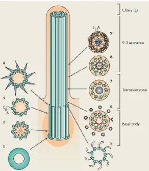
Cilia are generated during interphase, when cells exit the cell cycle from mitosis into a stationary or differentiated state and are reabsorbed when cells entry again into the cell cycle7,8. In this process, called ciliogenesis, cilia emerge from a plasma membrane-associated foundation called basal body that templates the nucleation of the axoneme, the structural core of cilia that consist of nine doublets of microtubules8. The basal body is also made by microtubules, nine triplets of microtubules (Figure 1.1). Between the basal body and ciliary body exists a transition zone, which starts where the triplet microtubules end and where the axoneme of cilia begins (Figure1.1). In combination with the internal structure of basal body, it is thought that the transition zone acts as filter for the cilium, regulating the molecules that can pass into or out of the cilium9. The ciliary tip is another part of the cilium that contains a specialized protein complex1. This structure is an important region to axonemal growth and reabsorption, and is thought to be a main point of regulation and remodeling of intraflagellar transport (IFT)7. This transport is needed to move the organelle´s structure components from cell body to ciliary tip and then to return proteins from the cilium to the cell body (anterograde transport is done by the motor subunit kif3 and retrograde transport by dynein motors) (Cole 1993), once the proteins for cilia assembly and function are synthetized in cell body and not in cilia11.
2
1.2.1. Types of ciliaAccording to the axonemal structure of cilia, its function and its ability to move, they can be classified into two groups: Primary and Motile cilia.
Primary cilia are typically short and immotile and are present on almost vertebrate cell types8 as a single copy per cell12. These types of cilia have a 9+0 axoneme, so they do not have a central pair of microtubules and lack inner (IDAs) and outer (ODAs) dynein arms. The absence of dynein arms and radial spokes makes them immotile.
Primary cilia were long thought to be vestigial, but were recently found to act as a complex signaling center, because a high concentration of signaling molecules were found near this type of cilium. Many of cilia are highly adapted to serve specialized sensory functions13. They participate in numerous biological process ranging from chemo and mechanosensation to the transduction of an expanding list of signaling cascades, that are essential to regulate cellular and tissue homeostasis8. Moreover, cilia are organelles functionally very important, involved in a growing list of diseases caused by defects at its level, such as, PKD (polycystic kidney disease) BBS (Bardet-Biedl syndrome), Joubert Syndrome and others (Goetz & Anderson 2010).
Motile cilia are usually longer and the presence of dynein arms confers them the possibility to beat 14. The axoneme of motile cilia is composed of nine peripheral microtubule doublet and two central microtubules (the central pair),15 so they have a 9+2 axoneme12,16.
The motility of these cilia depends on the presence of IDAS and ODAs, that are attached to the microtubules, the dynein regulator complex (DRC), radial spokes15 and central pair
Figure 1.1 Cilia subcompartments
Scheme of a cross section of the microtubular structure in each subcompartment of cilia: Basal body (1-4) – 9 x 3 microtubular structure; Transition zone (5-8) – converts the triplet microtubular structure of the basal body into the axonemal doublet structure; Axoneme (9) – 9+2 doublet microtubular structure; and Ciliary tip and (Fliegauf et al 2007)
3
projections12. Motile cilia have also nexin links that are responsible for the connection of microtubules doublets7 and the constraints caused by inter-doublet sliding translate into ciliary bending 6.
In mammalians, motile 9+2 cilia normally concentrate in large numbers on the cell surface and beat in an orchestrated wave like fashion being involved in fluid and cell movement13. Motile cilia are responsible for the generation of fluid-flow over epithelia such as mucus clearance in the respiratory tract and circulation of cerebrospinal fluid within the brain and spinal cord14. Thus, traditionally, motile cilia were thought not to be involved in sensory mechanisms.
However, recently, some studies have described the possibility that motile cilia also possess sensory skills, which means that functional differences between these two types of cilia are not as strictly demarcated17. Moreover, it has been described that there are many exceptions on this definition that favor the distinction into four subtypes: motile 9+2 cilia (respiratory and ependymal cilia), motile 9+0 (nodal cilia in node mouse that create a leftward flow of the surrounding fluid and this flow is essential for left-right development18, immotile 9+2 (kinocilium of hair cells) and non-motile 9+0 (renal monocilia and photoreceptor-connecting cilia)7.
1.3. Ciliopathies
As already mentioned, cilia are present in almost all organs of the human body, and there is increasing evidence that dysfunction of cilia are involved in many different human disorders16, called ciliopathies, like polycystic kidney disease (PKD) and primary cilia dyskinesia (PCD)7,19. The ciliary defects can occur at structural level or in protein localization 19
and in motile or immotile cilia. A ciliopathy that affects immotile cilia is PKD or Autosomal dominant PKD (ADPKD) by mutations in two genes: pkd1 and pkd2, and is related with defects is Ca2+ signaling20. This disease is a devastating common genetic disorder, with an estimated prevalence of between 1:400 and 1:1000 individuals21. PKD is characterized by defects in renal epithelial cells, leading to an increase of cell proliferation and cyst formation in kidney, liver and pancreas22,23.
Besides causing cysts formation, PKD is related with ciliary defects and in this disease, many ciliary proteins, localized to the ciliary membrane or basal body, are disrupted 24. The disruption of ciliary proteins influence the Ca2+ signaling mediated by Pkd2, leading to a relation between ciliary signaling and cyst formation 25. This topic will be discussed further ahead.
4
Dysfunctions in motile cilia are more specific, with major manifestations in mammalians as early embryonic death due to failure of embryonic turning, respiratory dysfunction, reproductive sterility and hydrocephalus 26. An example of a disease that affects the motile cilia is the PCD, firstly identified by Afzelius in 1976 27. This disease is characterized by bronchiectasis, infertility and defects in body situs, as situs inversus. This triade of symptoms is called the Kartagener syndrome. Patients with PCD have mutations in dynein arms as DNAH1 and DNAH5, that are necessary for ciliary motility in order to generate nodal flow and to allow normal mucociliary clearance (absent in PCD patients) 16. Moreover, half of individuals with PCD exhibit situs inversus totalis (which is consistent with randomization of left-right (LR) asymmetry). This observation primed Afzelius (1976) to first hypothesize that cilia motility was important in the establishment of LR body asymmetry. But how this regulation is made, will be discussed below 28 .
1.4. Cilia in left-right establishment
The generation of LR asymmetry occurs in two key steps. The first step is characterized by symmetry-breaking in the node. Here, an asymmetric signal that is generated in the node is transferred to the left side of the lateral plate mesoderm (LMP). This transference leads to an asymmetric expression of the gene nodal in the left LPM, which correspond to the second step 29.
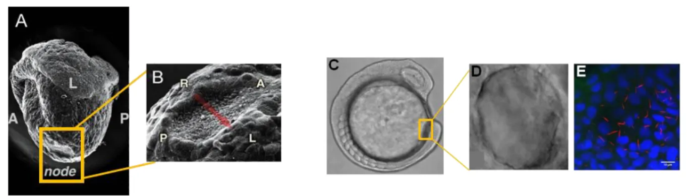
In normal mouse embryos, nodal cilia rotate in a clockwise direction and create a leftward fluid flow (nodal flow) which influences the asymmetric cascade of gene expression, that moves across the node (Okada 2005). The mouse node or the homologue Kupffer’s vesicle (KV) in zebrafish are transient organs present only in the first stages of embryonic development 31 and they are common among vertebrates (Lourenço and Saúde, 2010). The mouse node is a cavity filled with extra embryonic fluid and the zebrafish Kupffer´s vesicle is a elipsoid vesicle also full with fluid at the tailbud region (Figure 1.2)31. These organs are constituted by monociliated cells, and whereas in mouse cilia protrude from cells located on the ventral side of the node into the node cavity 33, in zebrafish cilia are all around the cavity34.
5
The nodal flow generated by nodal cilia leads to a transference of information to the left side of the node and subsequently to the asymmetric expression of nodal on the left side of LPM29,35. In LPM, nodal, exerts a positive feedback on itself and will activate its own negative regulators, the lefty genes32. lefty1 is expressed in the midline and prevents the nodal activation on the right LPM and lefty2 is expressed on the left LPM and restricts the domain of nodal expression into the prospective heart region 29,32. Absence of lefty genes leads to a symmetric nodal expression in LPM, which influences normal internal organ positioning. Nodal expression in LPM also activates the transcription factor Pitx2, which is expressed on the left side of LPM. This transcription factor persists until later stages, maintained by Nkx2, and is responsible for the regulation of asymmetric organogenesis29. Mice that lack Pitx2 exhibit laterally defects in most visceral organs29. Likewise, other mutants have been crucial to the interpretation of the left-right cascade of events.
The iv (Inversus viscerum) mouse embryos have a mutation in motor protein left-right dynein (lrd), only present in nodal cells that leads a loss of motility of motile nodal cilia. The absence of motility results in the absence of nodal flow and consequently leads a randomization in LR asymmetry. This finding indicates that nodal flow is necessary for the establishment of the LR body axis36–38, Furthermore, Nonaka et al (2002) showed that an artificial generated nodal flow to the right side of the node, independent from ciliary motility, is sufficient to reverse the laterality markers on the LPM39.
1.4.1. The two model to explain the LR establishment
In order to explain how LR establishment occurs, there are two theoretical models proposed, based on some experimental data. The “morphogen model” proposes that morphogens become concentrated on the left side of the node in response to flow18,40, while the other
Figure 1.2 Vertebrates left-right organizer
Scanning electron micrographs of the mouse node at 8.0 days post coitum (A). In higher magnification view are the ventral node cells (B). The anteroposterior and left-right axes are indicated by A/P and L/R, respectively (Adapted by Hamada 2008). Light microscopy image of a live zebrafish embryo at 13.5 hpf (9 somites stage) (C). Kupffer´s Vesicle is shown in higher magnification by: high resolution DIC microscopy (D) and confocal microscopy of all z-sections spaced 0.5 μm apart (in red alexa546), cilia labeled with anti-acetylated α-tubulin; and in blue (DAPI) the monociliated cells´ nuclei (E); white bar – 10 μm (Adapted from Lopes 2010)
6
model, the so called “two cilia model”, suggests that there are two types of cilia in the node that perform different functions 41,42
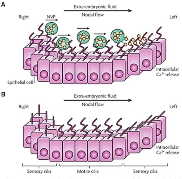
The Morphogen model was proposed by Nonaka and co-workers,18 who saw that in the mouse node there was a leftward fluid flow that they named as nodal flow (Figure1.3). From this observation they proposed that there was a putative secretory factor that was being concentrated on the left-side of the mouse node leading to activation of signaling pathways on this side of the node18,36. In 2005, Tanaka and co-workers identified membrane-sheathed objects, that they termed “nodal vesicular parcels” (NVPs), which carry Sonic Hedgehoc (SHH) and Retinoic acid (RA). They saw that when they abolished the fibroblast growth factor (FGF) pathway, they observed a reduction of Ca2+ signaling on left side of the node that was rescued when SHH and RA were added40. They also showed that FGF signaling triggered the secretion of these NVPs that are transported to the left side by nodal flow and are fragmented on the ciliated surface releasing their contents in proximity to the left wall40. This process was thought to lead to an activation of the downstream signaling pathways starting with an elevation of Ca2+ levels on the left side40, thus starting symmetry breaking 7. The symmetry breaking is declared based on asymmetric expression of mRNAs for signaling molecules in the LPM, such as Nodal, Lefty2 and transcription factor Pitx27. The Physicists Cartwright and co-workers, based on Tanaka´s findings, modeled the movement of the NVPs across the node and demonstrated that the flow should indeed cause them to accumulate on the left side of the node, as necessary for symmetry breaking43. However, they studied also the rupture of NVPs and reported that the rupture cannot be triggered by flow forces and must be induced by an as yet undiscovered biochemical mechanism43.
Figure 1.3 Current models of the LR establishment
The morphogen model (A) argues that nodal vesicular parcels (NVPs) full of retinoic acid and sonic hedgehog are secreted from the right side of the node and transported by nodal flow to the left side. Here NVPs are smashed, releasing their content. By enhancing intracellular Ca2+ levels in node left side cells, an asymmetric gene expression is induced. The Two cilia model (B) defend that there are two types of cilia (motile and immotile) in the node. Motile cilia, in the centre of node, generate a leftward nodal flow that is mechanically sensed by immotile cilia present in node periphery. Bending of immotile cilia leads to intracellular Ca2+ realize in left side of the node that activates an asymmetric gene expression (Fliegauf 2007).
7
The alternative model, the ‘two cilia model’ or mechanosensory model was proposed by McGrath and co-workers in 2003 when they observed two different populations of monocilia in the mouse node. They identified that cells located centrally in the node had motile monocilia that expressed both the motor protein: left-right dynein (Lrd) and the cation channel Pkd2, whereas those cells in periphery of the node had immotile monocilia that contained only Pkd241. This model proposes that the motile cilia present at the center generate the leftward fluid flow, and the immotile cilia located at the periphery of the node might detect this flow by mechanosensory mechanisms and initiate downstream calcium mediated events16,41,42,44 (Figure 1.3).
A stronger support for the mechanosensory model was the finding that LR signaling in humans was disrupted by mutations in human polycystic kidney disease gene pkd245,46 . Mutations in Pkd2 had already been linked to randomization of left-right body asymmetry in mice47. A very important evidence for involving Pkd2 in mechanosensation was given by Nauli et al48 when they reported that the primary cilia of renal epithelia cells without Pkd1 or Pkd2 triggered the appearance of cystic kidneys48 In zebrafish KV, Essner et al 31 observed that KV cells are monociliated and upon injection of fluorescent beads they saw that KV cilia are capable of generating a consistent directional counterclockwise flow inside KV31. Moreover, the knockdown of lrd1 in zebrafish disrupts the fluid flow in KV, resulting in a randomization of nodal-related gene, spaw, expression in LPM and cardiac laterality which means that fluid flow is also required for a correct LR patterning in zebrafish, as in the mouse31,49. However it is not completely understood how (in zebrafish) fluid flow inside a closed vesicle triggers an asymmetric gene expression important for LR determination. However, it is known that this process also results in asymmetric calcium elevation on the left side of the KV44,50. In summary, both models are plausible and still apply to mouse and zebrafish model systems13. However new hypothesis have appeared and claim that all cilia (motile and immotile) have sensory functions51. Bloodgood and co-workers reported that motile cilia of mammalian respiratory epithelium and oviductal epithelium exhibit chemosensory proprieties17. Further studies are necessary to clarify the mechanism that determinates how LR axis patterning information is conveyed by fluid flow and, maybe, sensed by specialized cilia13.
1.5. Objectives
In my project we proposed to study these two models using the zebrafish embryo and studying the role of two different genes. To test the mechanosensation (two cilia model) we
8
studied the pkd1 gene that codes for the potential mechanosensor partner of Pkd2. In order to test the chemosensation (morphogen model) we used tas1r1 gene that codes for a G-protein-coupled receptor (GPCR), and is a taste receptor from the T1R family. These two genes were both present in an unpublished microarray from Lopes lab. The microarray analysed the genes specifically expressed in the KV cells of wT and dld-/- mutant embryos, which have the Notch signalling affected. In the microarray results, pkd1 was significantly down regulated in deltaD-/- embryos, revealing that pkd1 transcription might be under the Notch signalling regulation. On the other hand, tas1r1 was not deferentially regulated by Notch signalling and it had a relatively low expression in wt embryos.
The reasoning that guided us to investigate tas1r1 role in LR development was a recent publication by Shah et al 52 showing that taste receptors surprising have a role in motile cilia, totally unrelated to taste buds52. Vertebrates perceive a variety of substances using the two chemosensory systems, taste and olfaction. For taste reception, vertebrates express the two families of G-protein-coupled receptors (GPCRs), T1Rs and T2Rs, in their taste buds 53–56. Moreover, taste buds also express PLCβ2 and G-protein α-gustducin, which are present in the transduction pathway of taste. Recently, Shah and co-workers discovered that ciliated cells of airway epithelia express multiple T2Rs only in motile cilia and also detected the transcription of PLCβ2 and G-protein α-gustducin in cilia52
. They speculated that the presence of T2R in airway epithelia is useful to hasten elimination of noxious and harmful substances52. In addition, last year, Meyer and colleagues found that tas1r1 and G-protein α-gustducin, were both expressed in mammalian spermatozoa. These data, raised the possibility that taste receptors act as chemosensors in sperm during the passage through the female reproductive tract57. These findings, together with the expression data from our microarray of three other genes that are involved in the taste pathway in taste buds, such as gnat2, trpm5 and plcβ2 gave us a good reason to investigate the chemosensory hypothesis in the ciliated cells of KV using tas1r1 as a starting point.
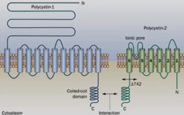
Relatively to pkd1, it is described that mutations in both pkd1 and pkd2 genes lead to PKD. pkd1 and pkd2 codify for two integral membrane proteins, polycystin 1 (Pkd1) and polycistin 2 (Pkd2), respectively48. These proteins are localized in primary cilia of kidney cells acting together to form a channel complex where Pkd1 has a long N-terminal extracellular domain, a short C-terminal intracellular domain and eleven transmembranar domains48. It was proposed that Pkd1 acts as mechanosensor to detect the bending of each cilium induced by flow stimulation48,58 and transmits these extracellular signals to Pkd2 through their connecting C-terminal regions59 (Figure 1.4). It is thought that the signals that Pkd1 senses mechanically, can be transduced into cellular responses that regulate proliferation, adhesion, differentiation and cell morphology48,58,60. On the other hand, Pkd2 is a membrane protein
9
Figure 1.4 Pkd1 - Pkd2 complex at the ciliated kidney cells’ membrane
Pkd1 has a large extramembranar domain, responsible for sense the external stimulus. Pkd2 is a transmembranar protein that functions as non-selective cationic channel. Pkd1 and Pkd2 interact via their C-terminal coiled-coil domains faced to the cell cytoplasm.
with six transmembranar domains and is a non-selective cation channel that mediates Ca2+ influx 48,58, found not only in primary cilia of kidney cells but also in the plasma membrane and in endoplasmic reticulum 59,61,62.
Pkd1 and Pkd2 are unequivocally present in kidney cells primary cilia, but although many authors have also described the presence of Pkd2 in cilia of the mouse node, xenopus node and Kupffer´s vesicle of medaka fish and zebrafish 47,63–67.,the role of the Pkd1-Pkd2 complex in the establishment of the LR axis is still highly debated. Experiments with knockouts and knockdowns of pkd2 in mice, zebrafish and medaka resulted in kidney cysts and laterally defects in all models 47,63–67. From these results we can conclude that Pkd2 involvement in the LR establishment is conserved, but how it occurs and which partner it uses in this developmental process has been controversial 29,68,69.
In mouse nodal cilia and in KV cilia of medaka fish, the presence of Pkd1 has not been reported 67,70. Moreover, in kidney cells, Pkd1 is required for the localization of Pkd2 in cilia 48 but the same does not happen in mouse node and in kupffer´s vesicle of medaka fish 69,71. Recent studies developed by Kamura and Field 67,71 revealed that the partner of Pkd2 in the mouse node and in medaka KV is Pkd1l1. Pkd1l1 is also a member of the PKD family of large membrane proteins, which are also known as the TRPP 67.
In summary, in mouse and medaka it is current thought that Pkd1l1 and Pkd2 act together downstream of nodal flow to mediate LR patterning 67,71. Whether this complex is acting in the cilia membrane or in other cell compartment it has not been formally demonstrated yet. In our microarray we do not have detected expression of pkd1l1, but we do have detected pkd1 transcripts. Our aim was to identify if pkd1 transcripts code for a true Pkd1 or Pkd1l1, and then study its implication in LR development of the zebrafish embryo.
The zebrafish (Danio rerio) is a good vertebrate model in genetic and molecular studies as well as in development. A most important characteristic of this model is the external
10
fertilization, which allows us to see all steps of embryonic development, since first cellular division, once the embryos are transparent 72. This transparency also allows the directly visualization of internal organs, such as the KV73,74. Moreover, this vertebrate has a relatively short generation span and produces large amounts of eggs 75. Zebrafish have many mutants, is easily genetically manipulated by gene knockdown and gene overexpression and, as this model has been widely used, there are many transgenic lines reporting genes of interest in several tissues and organs 76,77.
The main goal of this project was to test the two hypothesis of LR determination in the zebrafish embryo.
A. Test the chemosensation hypothesis by knockdown of tasr1r1 gene. B. Test the mechanosensation hypothesis by pkd1 knockdown.
In order to test these hypotheses we tried to abolish the translation of both genes (separately) with morpholino oligonucleotide technology and evaluated the results in the establishment of LR patterning by observation of internal organ position.
2. M
ATERIAL AND
M
ETHODS
In order to answer our proposed objectives, we performed different experiments that are explained bellow.
2.1. Phylogeny
In order to understand if the pkd1, present in the microarray, is a pkd1 gene or a pkd1l1 gene we did a phylogenetic tree with the protein sequences of these two genes and several other vertebrate organisms, with the help of the IGC Bioinformatics facility.
The protein sequences of Danio rerio (Zebrafish) Pkd1 and Pkd1l1 were obtained in Ensemble Genome Browse, and the homologue sequences of other organisms were retrieved from GenBank and Ensemble Genome Browser after BLAST in NCBI. We obtained 15 species, including mouse, human and medaka fish (the number ID is in Table 1 in annex II). Next, we aligned these sequences according to their primary structure similarity using ClustalW 78 with default gap values. The multiple sequence alignment was manually corrected being the numbers of amino acids of all sequences adjusted to the length of the
11
smallest representative. As outgroup we chose Drosophila melanogaster, because is an arthropod. The phylogenetic analysis was inferred from the genetic distance between pairs of sequences using Kimura two parameters method (PROTDIST, Phylip Package, version 3.5c)79,80 and the tree was constructed by Neighbor Joining (NEIGHBOR, Phylip Package)81. The branching order reliability of the tree was evaluated by bootstrap analysis of 1000 replicates.
2.2. Zebrafish mating for embryo production
To obtain embryos, the males and females are collected from a main tank-system and are placed in a matting box in the evening. Each mating box has one male and female separated by a transparent partition to maintain the visual contact. In the following morning the partition is removed the water level is lowered and that triggers a mating ritual between the couple, after which females release the eggs and by external fertilization, the male fertilizes the eggs. As in each mating box there is a grate, the eggs stay protected from the parents, and sink to the bottom of the mating box. After that, the fish go back to the main tank-system, eggs are collected and placed in a Petri dish with embryonic medium (5 mM NaCl, 0.2 mM KCl, 0.3 mM CaCl2, 0.3 mM MgSO4,ddH2O—pH 6.5). The Petri dish with eggs is incubated at 25oC or 28oC until reaching the desired stage for experiments.
2.3. Quantitative Real-Time Polymerase Chain Reaction (qPCR)
The qPCR provides measurement of gene expression and is suitable when a small number of cells are available. In small embryos during in early stages of development qPCR can be very useful.
With tas1r1, which had very low expression in the microarray dataset, we wanted to confirm its expression at 10 hpf and 13 hpf. The data obtained at these stages and 4 dpf were normalized using the elf1a. After that we decided to compare the tas1r1 expression at 10 hpf and 13 hpf with its expression at 4dpf. We used this latter stage to compare the levels of transcription because it was described that at 4dpf this gene is highly expressed in the zebrafish mouth, and we wanted to know how much less tas1r1 expression we had relatively to 4dpf 82.
Relatively to pkd1, we compared the levels of gene expression in wt and deltaD-/- embryos at bud stage, in order to confirm the microarray data, where this gene was down regulated in deltaD mutants. Here, we also normalized the gene expression with reference genes, elf1a and sox17 at 10 hpf in wt and deltaD-/- mutants.
12
To evaluate the transcription levels and validate our microarray results of tas1r1 and pkd1, we utilized the technique of qPCR in wt and deltaD embryos. For tas1r1 we used wt embryos at 10 hpf (bud stage) and 13 hpf (8 somites stage), while for pkd1 we only used wt and deltaD-/- mutant embryos at 10 hpf because at 13 hpf this gene is also expressed in the zebrafish developing kidney, which would influence the data. For tas1r1 we performed a positive control of gene expression with cDNA from 4days post fertilization (dpf) when the taste buds are reported to have this receptor expressed.
To perform the qPCR experiments we extracted RNA from 100 embryos of wt and deltaD -/-mutants with RNeasy® micro Kit (Qiagen Inc., Valencia, CA) according to the instructions of the manufacture. Next we quantified the RNA obtained in NanoDrop 2000 Spectrophotomer (Thermo Scientific), a very important step to evaluate the concentration and purity of the RNA. Then we performed the reverse transcription of total RNA samples into cDNA. We used 4 μg of RNA from wt and deltaD-/- mutant embryos in a reverse transcription reaction with RevertAidTM First Strand cDNA Synthesis Kit (Fermentas, LIFE SCIENCES), according to the manufactures instructions.
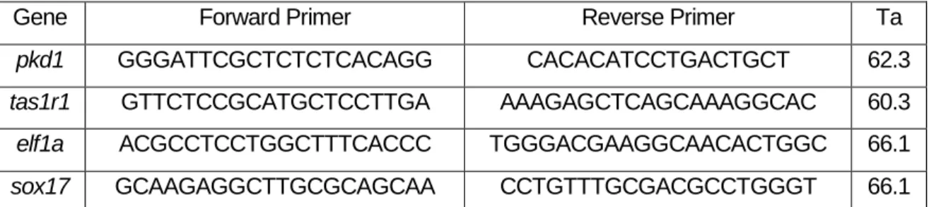
Next, we designed primers for amplification of the cDNAs. We designed primers with the bioinformatic tool Primer-BLAST (NCBI) and primer sequences are described in Table 2.1. The primer sequence of elf1a, a house keeping gene used as control, was according to Silva et al 83.
The qPCR reactions were performed on the CFX96™ Real-Time PCR Detection System (BIO-RAD PAIS), using Evagreen Supermix (BIO-RAD).
In order to avoid nonspecific annealing and primer-dimmer formation, we tested different annealing temperatures that ranged from 72ºC to 56ºC, as way to determine the optimal annealing temperature for each primer pair. The optimal temperature is chosen based on the lowest Ct value obtained for each primer pair. In the end of this reaction, the program
Gene Forward Primer Reverse Primer Ta
pkd1 GGGATTCGCTCTCTCACAGG CACACATCCTGACTGCT 62.3
tas1r1 GTTCTCCGCATGCTCCTTGA AAAGAGCTCAGCAAAGGCAC 60.3
elf1a ACGCCTCCTGGCTTTCACCC TGGGACGAAGGCAACACTGGC 66.1
sox17 GCAAGAGGCTTGCGCAGCAA CCTGTTTGCGACGCCTGGGT 66.1
Tabela 2.1 Nucleotide sequence of the primers (forward and reverse) used in qPCR assays for each gene in study and respective annealing temperatures (Ta).
13
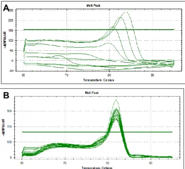
provides as a melting curve, which shows the specificity of primer annealing. This specificity is represented by a single sharp peak which represents one specific amplicon. If we obtained two sharp peaks in the same melting curve that would mean that we had more than one amplicon being transcribed in the same reaction for the same primer pair. If we obtained two unique peaks in different reaction wells containing the same reaction mix that indicates two different amplicons being obtained with same prime pair.
In each qPCR reaction for each primer pair we performed a calibration curve with four different concentration of cDNA (1:1, 1:10, 1:100 and 1:1000) with 2 replicates for each concentration. No template control (NTC) reaction was included for each primer pair to test buffers, solutions and DNA contamination and to assess for primer-dimers84. Cycling parameters used were as follows: 95oC for 2 min and 95oC for 10s, 62.3oC (for pkd1) or 60.3oC (for tas1R1) or 66.1oC (for elf1a and sox17) for 20s in 49 cycles, 60oC for 5s and 95oC for 5s.
We obtained a standard curve which represents the reaction efficiency. This curve is constructed by plotting the log of starting quantity of the template against the Cq values obtained. All data obtained were normalized with the reference gene. Here we utilized the elf1a which was described as having an expression very high and maintained along all developmental stages in zebrafish85. We also used sox17 as reference gene for pkd1 because this gene has a constant expression in bud stage in wt and deltaD-/- mutants. Reference genes are important to normalize the data and to correct the differences in amplification by stochastic variation during qPCR.
A
B
Figure 2.1 qPCR melting curves.
Panel A shows an unspecific primer annealing (with more than one sharp peak) indicating the presence of more than one amplicon. In B, is represented an ideal melting curve with only one sharp peak and, thus, only one amplicon.
14
The data obtained were treated using delta-delta-Cq method available in BIO-RAD software, “BIO-RAD software Manager”.
2.4. Immunofluorescence
We performed an immunofluorescence assay to test if Pkd2 localizes to the cilia in absence of Notch signalling (and presumably in the absence of Pkd1). In order to do that, we used deltaD-/- mutant embryos and wt embryos as controls.
The embryos were collected and then were dechorinated at 13 hpf (8 somites stage). After that the embryos were fixed in methanol: DMSO (80:20) during 1 minute. Next, embryos were placed in PBS: Methanol gradually: 75%, 50%, 25% and finally stored in PBS 1x at 4oC and ready to use. Embryos were washed in PBS (250 μl Triton X-100, 10% in 50 ml of PBS and blocked with Foetal Bovine Serum to diminished non-specific binding of primary antibody. Posteriorly, primary antibodies, mouse anti-acetylated α-tubulin (1:400) and rabbit anti-pkd2 (1:400) (a gift from Ian Drummonds’ lab) were incubated in blocking solution (1% DMSO, 2% Foetal Bovine Serum, 0, 1% Tween-20 and PBS) overnight at 4oC.
In the next day embryos were washed in blocking solution and incubated with secondary antibody, anti-mouse alexa 546 (1:500) and anti-rabbit alexa 488 (1:500) overnight at 4oC. These antibodies have fluorophores which emit green and red light when excited with a wave-length of 488nm and 546 nm respectively.
In the last day, embryos were submitted to several washes and cleared in 50% Glycerol: PBS. Next, embryos were transferred to PBS and stored at 4oC until mounted to confocal microscopy.
Using this antibody we also did an immunofluorescence assay with the transgenic line foxj1a: GFP which has the advantage of having the cells of Kupffer´s vesicle labelled with GFP. The objective of this assay was to test in which type of cilia, Pkd2 co-localizes, immotile or motile. This was done in these transgenic embryos, because foxj1a is used as a marker for cells with motile cilia. As fixation in methanol leads to a loss of fluorescence, the embryos were fixed in PFA instead and the block solution was PBDX (50 ml PBS 1x, 0.5g BSA and 0.250 ml 10% Triton X-100). The antibodies used in this assay were the same that I referred above, however we changed the secondary antibody in order to obtain other colour for the visualisation of acetylated tubulin. So we used anti-mouse alexa 647 (1:500) and anti-rabbit alexa 546 (1:500). The light emitted by these antibodies is magenta and red, respectively.
15
2.5. Preparation of whole embryos for confocal microscopy
The embryos used in the immunofluorescence assay were flat mounted to visualise the organ of interest – Kupffer´s vesicle with cilia. In a glass slide it was made a circle with silicone and within this circle the embryo was placed in PBS. Finally when the embryo was well orientated, the sample was covered with a lamella and a little pressure was applied to seal the preparation and remove air bubbles. The samples were observed in the confocal microscope - LSM710 (ZEISS) – with an objective 40X water and 1.2 of numerical aperture and each stack produced slices with 0,4μm -1μm of thickness.
The stacks of images obtained were analysed and treated with the free software “image J” 86.
2.6. Morpholino Injection
Morpholinos (MOs) are synthetic molecules constituted by oligonucleotides that are similar to the structure of natural nucleic acids. Structurally, the difference between morpholinos and DNA is that while morpholinos have standard nucleic acid bases, those bases are bound to morpholino rings instead of deoxyribose rings and linked through phosphorodiamidate groups instead of phosphates. MOs are used to knockdown the expression of proteins by interaction with a complementary mRNA to block its translation to protein or create an alternative splicing site in pre-mRNA. This interaction does not lead to degradation of mRNA and has very few toxic effects.
2.6.1. Morpholino designing
In this experiment we used two different MOs, one of them was to block the translation of tas1r1 and the other to block pkd1 translation. However, pkd1 sequence was not very well annotated, and did not have the initiation codon. Thus, we could not design a MO for the initiation codon to impair the translation of pkd1 and had designed a MO that binds to an intron-exon boundary to create an alternative splicing site, which according to GeneTools’ online advice should lead to an out of frame mutation. As this MO was designed to put exon 3 out of frame, we expected that the protein produced was very small and could be degraded by the proteosome.
We also injected a MO against pkd2, as a positive control for our injections, because this morpholino was already described by Schottenfeld et al. and causes LR defects in zebrafish embryos 65.
The morpholino for knockdown of tas1r1 (tas1r1 augMO) was designed by GeneTools® in order to bind to the mRNA near of its start codon (ATG). Its sequence was:
16
5´TGCTCAACATCGTTCCCATAATA 3´. As this morpholino binds to an UTR region, the translation is abolished and the protein should not be expressed.
The Morpholino for pkd1 (pkd1 splicingMO) was designed to bind to an intron and lead to an alternative splicing site. This interaction should lead to the removal of exon 3 and cause the sequence to go out of frame. The sequence of pkd1 morpholino was: 5´GTACTGTTGGTGACTCACCACAAGT 3´.
The morpholino for knockdown of pkd2 (pkd2 augMO) was designed according to Schottenfeld et al 65, beginning at the start AUG and extending into the first exon. This morpholino prevents de translation of mRNA to protein, and its sequence was: 5´AGGACGAACGCGACTGGAGCTCATC 3´. Morpholinos arrived lyophilized and then were re-suspend in Milli-Q water, according to the manufacturer's instructions, in order to obtain a final concentration of 1mM stock.
2.6.2. Microinjection of morpholinos
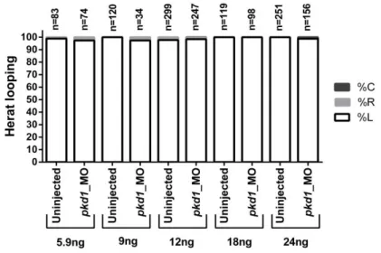
The technique of microinjection is made with precision micro-injectors (Narishigi). These micro-injectors allow injection of very small solution quantities. In these two experiments we injected a range of MO amounts to optimize mortality rates versus phenotypes. We injected: 5.9ng, 9ng, 12ng, 18ng and 24ng for pkd1 splicingMO at 0.5mM, 0.75mM and 1mM; 1.8ng, 3.5ng, 7ng, 11,9ng and 23ng for tas1r1 augMO at 0.5mM and 1mM. For pkd2 augMO we only used 1.8ng at 0.5mM, because this morpholino was already optimized in lab.
The embryos were obtained in the same way, as explained previously, however in this experiment we injected morpholinos at one cell stage. So, when we removed the partition of the matting box, we waited a few minutes for the fish to lay and fertilize the eggs and collected them immediately. Then we put the embryos on the lid of a petri dish that contained a glass-slide and aligned them against the glass-slide to form a string of eggs. Next, we pierced the choreon and the yolk with the sharp needle and injected 1 discharge of MO with a foot pedal device. In the end the embryos were put at 28oC for the next days to evaluate the position of the internal organs, such as heart at 30 hpf, and liver and pancreas at 50 hpf as will be described below.
2.7. Validation for Pkd1 splicingMO
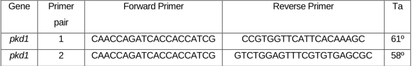
As pkd1 splicingMO was a splicing MO, we were able to perform a RT-PCR with cDNA from uninjected and injected embryos, using primers designed before and after the splicing site. This assay should have allowed us to obtain a band in wt embryos of 1080 bp or 1600 bp, for
17
first and second primer pairs. In contrast in injected embryos we expected to amplify only 917 bp or 1357 bp, respectively.
We did the PCR with My TaqTM Red Mix (BIOLINE) with the following protocol: 95ºC 2min, 95ºC 1min, 61ºC/59ºC (primer pair 1 and 2) 1min, 72º, 2min and 72ºC, 7min.
Gene Primer
pair
Forward Primer Reverse Primer Ta
pkd1 1 CAACCAGATCACCACCATCG CCGTGGTTCATTCACAAAGC 61º
pkd1 2 CAACCAGATCACCACCATCG GTCTGGAGTTTCGTGTGAGCGC 58º
2.8. Evaluation of gut and heart position
After MO injection we wanted to see if there were defects in left-right asymmetry. To access these defects we observed the position of the heart and gut (pancreas and liver), first visualizing under the stereoscope (Nikon, SMZ745) and then by performing an in situ hybridization with riboprobes that label these organs unequivocally.
The first organ that appears and allows an evaluation of the effects of the MO is the heart. The heart can be clearly observed at 30 hpf and is positioned on the left side below the left eye in normal embryos. In embryos with laterally defects the heart could be below the right eye or in the middle of the two eyes. At 50 hpf it is already possible to see de gut (liver on the left side and pancreas on the right side). When we want to evaluate gut position we have do previously add PTU (f 1-phenyl 2-thiourea), to the embryo medium, in order to eliminate the pigment (it inhibits melanogenesis by blocking all tyrosinase-dependent steps in the melanin pathway) to facilitate the visualization of internal organs. The evaluation of the gut loop is also possible to do in live embryos when we inject MOs in the transgenic line, sox17:GFP, which has the gut clearly labelled with GFP. These injected transgenic sox17:GFP embryos were scored in the fluorescence stereoscope (SteREO Discovery.V8 , ZEISS). When the injections were made in wt embryos we performed an in situ hybridization to report the position of both organs.
2.9. Whole mount in situ hybridization (WISH)
The in situ hybridization allows detection of specific acid nucleic sequences in morphologically preserved cells or embryos. The whole mount in situ hybridization (WISH) normally is used when we want to describe the expression pattern of developmentally Tabela 2.2 Nucleotide sequence of the primers (forward and reverse) used in RT-PCR assays and respective annealing temperatures (Ta).
18
regulated genes87 . In this methodology we used digoxigenin-labeled antisense RNA probes, which bind to the mRNA of interest in a specific tissue of the embryo.
The probes used to mark the gene expression in the heart and gut were: cmlc2 (cardiac myosin light chain2) as a marker of heart position, and foxA3 (forkhead box A3) as a marker of gut position. The in situ probes used here bound to mRNA specific for these genes in these two organs and allowed us to observe the position and integrity of these organs inside
the embryos. These two probes were already available in the lab. In this experiment we followed the Thisse´s protocol 87. The embryos injected with MOs were
fixed in PFA overnight until the pretended stage (30 hours for the heart and 50 hours for the gut). In the next day, embryos were placed in methanol 100% and stored at -20oC until the first day of the protocol.
In first day the embryos were put through a series of 75%, 50%, 25% MeOH-PBS for 5 min each at RT and then twice for 5 min in PBT (PBS/ Tween20 – 0.1%). Next the embryos were dechorionate and after that were permeabilized with Proteinase K (5mg/ml in PBT) at RT during 30 min or 50 min (time recommend for latter stages). After this step, embryos were fixed in 4% PFA for 20 min, followed by a series of washes in PBT.
The pre-hybridization and hybridization were did at 70ºC for the two ribo-probes in HYB-mix solution (Formamide, 20x SSC, Tween20 10%, 1M citrix acid to pH6 and heparine (0.63mg/ml). After two hours of pre-hybridization, the probes were added to the embryos and incubated overnight.
In the second day the probes were recovered and embryos were submited to 100%, 75%, 50% and 25% of HYB-mix/2x SSC (NaCl 175.3g, Citric acid trisodium salt 88.2g dissolved in 1l of water) washes for 15 min each at 70ºC and then once for 15 min in 2x SSC at 70ºC. After, embryos were washed twice in 0.2 SSC for 30 min and then through 75%, 50% and 25% 0.2 SSC/PBT for 10 min each at RT, finishing with a RT wash in PBT for 10 min. Embryos were blocked for 2 hours at RT with BSA, GS (goat serum) and PBT (50ml PBT with 100mg BSA and 1ml GS). In situ hybridization signals were detected at a 1:5000 dilution in blocking solution, overnight at 4ºC.
In the last they, antibody solution was removed and embryos were washed in PBT during 6 series of 15min washes. After that, the embryos were equilibrated in staining buffer (1M TRIS pH9.5, 1M MgCl, 5M NaCl and Tween20 10%), 3 times for 5 min each RT. The color staining was carried out with NBT/BCIP (Roche Mannheim, Germany), alkaline phosphatase subtracts, until probes reached the desired level of purple staining. When the revelation was concluded we stopped the reaction with PFA 4% for 20 min at RT and several washes with
19
PBT. The embryos were cleared with 50% Glycerol: PBS and photographed in SteREO Lumar.V12 (ZEISS) with the High Resolution Microscopy Camera AxioCam MRc Rev. 3 FireWire (ZEISS).
2.10. Mounting Zebrafish for Live Imaging
In order to understand if Fox1a is a good marker for motile cilia, we did live imaging using the transgenic line Foxj1a: GFP, which has the KV cells labelled with GFP. To observe all cilia of the KV we injected a construct of mRNA: Arl13b-GFP 400pg. To visualize the KV cilia of the zebrafish embryo we used embryos with 13 hpf and used a mould made with agarose 2% that enables each embryo to stay oriented with KV towards the inverted objective). As live imaging was done in an inverted microscope with the camera recording from the bottom, the embryos, dechorionated, were mounted with the dorsal roof of the KV facing the objective lens – named as “dorsal view”. The embryos were obtained as explained initially and are visualized at 13 hpf (8 somites stage). The images were obtained in confocal microscope- LSM710 (ZEISS) – with an objective 40X water and each stack produced slices with 0,4μm of thickness.
2.11. Statistic analysis
The data analysis was done using the software GraphPad – Prism. The statistical analysis for qPCr and cilia counting cilia in wt and deltaD-/- was done with Student’s t-test, two tails for two samples assuming unequal variances. We considered values below p<0.05 as statistically significant (p<0.05 is represented by * and p< 0.01 by **)
3. R
ESULTS ANDD
ISCUSSIONTo facilitate the description of results I divided the results according to the two different objectives proposed. First I will present the results from objective A: Test the chemosensory pathway using tas1r1 and then I will discuss the mechanosensory hypothesis with pkd1.
A: Test the chemosensory pathway using tas1r1 gene
3.1. Expression of tas1r1 in cilia of KV at 10 hpf and 13 hpf
The tas1r1 had a relatively low expression in the microarray screen performed in our lab. However, as the reported expression of this gene in cilia of airway epithelia and sperm was
20
very surprising and interesting so we decide to test if this gene had some role in LR patterning.
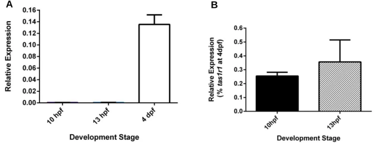
To do that, we evaluated the transcription levels of tas1r1 in wt embryos at 10 hpf and 13 hpf to confirm its’ expression at these stages. The values obtained by q-PCR were normalized relatively to the reference gene, elf1a (Figure 3.1).
In Figure 3.1A we observed that expression of tas1r1 at 10 hpf and 13 hpf was very low compared with expression of tas1r1 at 4 dpf. From this first analysis, we decided to normalize the values with levels of expression of tas1r1 at 4dpf (Figure 3.1B.) At 4 dpf it was reported that this gene was unequivocally expressed in the mouth of the zebrafish larvae. With this new normalization we can observe that the expression was higher at 13 hpf than at 10 hpf, however this difference was not significant (p > 0.05). Moreover, the expression in both stages continued to be very low, between 0.3% and 0.4% (Figure 3.1B).
3.2. Knockdown of tas1r1 by morpholino injection
We injected different concentrations and amounts of morpholino, to test their toxicity and their effect in LR establishment. After that we evaluated the position of internal organs, such as the heart (at 30 hpf) and gut (50 hpf) to see if tas1r1 atgMO caused any effects in LR patterning.
For the different MO concentrations and amounts used in this experiment, we did not obtain any laterally defects. The majority (97%-100%) of embryos injected with this morpholino had the heart on the left side of the body as the uninjected control embryos (Figure 3.2)
A B
Figure 3.1 Expression analysis of tas1r1 by qPCR at 10 hpf, 13 hpf and 4 dpf in wt embryos
Gene expression results were normalized with eif1a as reference gene (A and B) and with gene expression at 4dpf (B).