Universidade de Aveiro 2014
Departamento de Biologia
Ana Rita Guimarães
Rodrigues da Silva
Codon ambiguities as a mechanism to alter
the genetic code in Saccharomyces cerevisiae
Ambiguidade de codões como mecanismo de
alteração do código genético em
Universidade de Aveiro 2014
Departamento de Biologia
Ana Rita Guimarães
Rodrigues da Silva
Codon ambiguities as a mechanism to alter
the genetic code in Saccharomyces cerevisiae
Ambiguidade de codões como mecanismo de
alteração do código genético em
Saccharomyces cerevisiae
Tese apresentada à Universidade de Aveiro para cumprimento dos requisitos necessários à obtenção do grau de Mestre em Biologia Molecular e Celular, realizada sob a orientação científica da Doutora Ana Rita Macedo Bezerra, investigadora Pós-Doutoramento do RNA Biology Lab, Departamento de Biologia da Universidade de Aveiro
I would like to dedicate my thesis to my beloved grandfather. You gave me my tender memories and always had proud and seen the best of me. You will never be forgotten…
O júri
Presidente Doutora Maria Adelaide de Pinho Almeida
Professora auxiliar do Departamento de Biologia da Universidade do Aveiro
Arguente Doutora Ana Catarina Gomes
Diretora científica da Unidade de Genómica do Biocant
Orientadora científica Doutora Ana Rita Macedo Bezerra
Investigadora Pós-Doutoramento do RNA Biology Lab, Departamento de Biologia da Universidade de Aveiro
Agradecimentos First, I would like to thank Professor Manuel Santos for giving me the opportunity to work in his laboratory and in this project. Also for receiving me so well and for the guidance and confidence in my work. A special thank you for broaden the horizons of my mind.
I am deeply grateful to my supervisor, Drª. Ana Rita Bezerra, for all the knowledge, advice, readiness, patience and specially, for the friendship built between us. Thank you for all the guidance and support during the course of this work. You are the best.
I would like to thank to all the extraordinary team from the RNA Biology Laboratory for welcoming me so well in the laboratory and for good working atmosphere. Thank you for all the support, fruitful discussions and for the good friendship. Also, I would like to thank to the past lab-member João Simões for the advice, helpful insights and support in the beginning of my project.
I would like to thank also to Dr. Yitzhak Pilpel for kindly providing the
Saccharomyces cerevisiae deletion library used in this work. As well to
University of Aveiro, specially the Biology Department, for providing me the conditions for the development of my Master Thesis.
Finally, but not least, a special thank you to my friends and my family, specially my mother, grandfathers and brother, for all the support and encouragement. Thanks to my friends from Aveiro for the love, support and joyful moments. A special thank you to my second family for their support, friendship and kind words.
Palavras-chave Saccharomyces cerevisiae, código genético, tRNA, evolução, erros de tradução, ambiguidade do codão
Resumo O código genético é geralmente visto como imutável, no entanto várias alterações à sua forma padrão são conhecidas. Uma das mais notáveis acontece em várias espécies do género Candida, onde o codão Leu-CUG é descodificado como serina por um novo RNA transferência (Ser-tRNACAG). O
laboratório de acolhimento fez um grande progresso ao reverter a alteração atípica do código genético do fungo patogénico humano C. albicans, usando uma combinação de tRNAs mutantes, recombinação genética e evolução forçada. Estes resultados levantaram a hipótese que as ambiguidades sintéticas do codão, combinadas com evolução experimental, poderem libertar os codões do seu estado fixo.
Nesta tese testamos esta hipótese usando S. cerevisiae como modelo biológico. Geramos ambiguidade em codões específicos, de forma bifásica, envolvendo a deleção de genes de tRNA, seguida pela expressão de tRNAs não-cognatos capazes de compensar o tRNA eliminado. Tendo como base a ideia que os codões raros são mais suscetíveis a alterações que aqueles usados frequentemente, usamos duas estirpes knock-out, nas quais não existem os tRNAs cognatos capazes de descodificar os codões raros CUC-Leu e AGG-Arg.
Exploramos então a vulnerabilidade destes codões pela construção de tRNAs mutantes que incorporam erradamente Ser nestes locais. Estas estirpes recombinantes foram evoluídas ao longo do tempo, usando evolução experimental. Apesar de ter havido um forte impacto negativo na taxa de crescimento de estirpes que expressam o tRNA mutante a altos níveis, esta expressão a baixos níveis teve pouco impacto no fitness celular. Descobrimos que não só a ambiguidade do codão, mas também destabilizações da pool de tRNAs endógenos têm um impacto negativo na taxa de crescimento. Após a evolução, as estirpes com elevada expressão do tRNA mutante recuperaram significativamente em vários parâmetros de crescimento, o que mostra que estas adaptam-se e exibem maior tolerância à ambiguidade do codão. Através do sistema repórter fluorescente desenvolvido monitorizamos a incorporação errónea de Ser, o que nos indica que a Ser está de facto a ser incorporada e que, possivelmente, a alteração da identidade do codão foi atingida.
Apesar das consequências negativas gerais da ambiguidade do codão, demonstramos que os codões capazes de tolerar a perda do seu tRNA cognato, conseguem também tolerar a incorporação elevada de Ser. Isto levanta a hipótese que estes codões podem ser recodificados para outros aminoácidos naturais e/ou artificiais para a produção de proteínas com novas propriedades, contribuindo assim para o campo da Biologia Sintética e Biotecnologia.
Keywords Saccharomyces cerevisiae, genetic code, tRNA, evolution, mistranslation, codon ambiguity
Abstract Although the genetic code is generally viewed as immutable, alterations to its standard form occur in the three domains of life. A remarkable alteration to the standard genetic code occurs in many fungi of the Saccharomycotina CTG clade where the Leucine CUG codon has been reassigned to Serine by a novel transfer RNA (Ser-tRNACAG). The host laboratory made a major breakthrough by reversing
this atypical genetic code alteration in the human pathogen Candida albicans using a combination of tRNA engineering, gene recombination and forced evolution. These results raised the hypothesis that synthetic codon ambiguities combined with experimental evolution may release codons from their frozen state.
In this thesis we tested this hypothesis using S. cerevisiae as a model system. We generated ambiguity at specific codons in a two-step approach, involving deletion of tRNA genes followed by expression of non-cognate tRNAs that are able to compensate the deleted tRNA. Driven by the notion that rare codons are more susceptible to reassignment than those that are frequently used, we used two deletion strains where there is no cognate tRNA to decode the rare CUC-Leu codon and AGG-Arg codon.
We exploited the vulnerability of the latter by engineering mutant tRNAs that misincorporate Ser at these sites. These recombinant strains were evolved over time using experimental evolution. Although there was a strong negative impact on the growth rate of strains expressing mutant tRNAs at high level, such expression at low level had little effect on cell fitness. We found that not only codon ambiguity, but also destabilization of the endogenous tRNA pool has a strong negative impact in growth rate. After evolution, strains expressing the mutant tRNA at high level recovered significantly in several growth parameters, showing that these strains adapt and exhibit higher tolerance to codon ambiguity. A fluorescent reporter system allowing the monitoring of Ser misincorporation showed that serine was indeed incorporated and possibly codon reassignment was achieved.
Beside the overall negative consequences of codon ambiguity, we demonstrated that codons that tolerate the loss of their cognate tRNA can also tolerate high Ser misincorporation. This raises the hypothesis that these codons can be reassigned to standard and eventually to new amino acids for the production of proteins with novel properties, contributing to the field of synthetic biology and biotechnology.
15 15 15 15 16 17 17 17 19 20 20 20 20 21 21 21 1 1 2 2 3 4 5 7 9 10 12 13
Contents
Chapter 1 1. Introduction ……….…………... 1.1 Genetic code ……… 1.1.1 Reading the code ..……… 1.1.1.1 Transfer RNAs ….………..…. 1.1.1.2 Aminoacyl-tRNA synthetases ………... 1.1.2 Protein biosynthesis ..………... 1.2 Alternative genetic codes ..………..…. 1.2.1 Evolution of the genetic code ..……….….... 1.2.2 Mechanisms of reassignment ..……….……... 1.2.3 Codon ambiguity and mistranslation ..……….…. 1.3 Saccharomyces cerevisiae as a biological model ..……….… 1.4 Working hypothesis and objectives..………..Chapter 2
2. Materials and methods ………... 2.1 Strains and growth conditions ………... 2.1.1 Strains ………... 2.1.2 Growth conditions ………... 2.2 Oligonucleotides ………... 2.3 Plasmids ………... 2.3.1 Original plasmids ………... 2.3.2 Constructed plasmids ………... 2.3.3 Site directed mutagenesis ………... 2.4 Preparation of E. coli competent cells ………... 2.5 Plasmidic DNA purification from E. coli ………... 2.5.1 Miniprep kit ………... 2.5.2 “Homemade” minipreps ………... 2.6 Plasmidic DNA purification from S. cerevisiae ………... 2.7 Transformation procedures ………... 2.7.1 Escherichia coli ………...
21 22 22 22 23 23 23 24 25 27 27 29 29 31 32 34 37 41 41 42 42 45 47 48 50 54 2.7.2 Saccharomyces cerevisiae ………...
2.7.3 Saccharomyces cerevisiae (alternative) ………...
2.8 Colony PCR ………... 2.9 Growth curves and rates………... 2.10 Evolution experiments ………... 2.11 Northern Blot Analysis ………... 2.11.1 Total RNA extraction ………... 2.11.2 Northern blot ………... 2.12 Epifluorescence microscopy …...
Chapter 3
3. Characterization of codon ambiguities ……….…...…… 3.1 Overview ………..………..….……… 3.2 Results ………..……….…….…… 3.2.1 Expression of mutant Ser-tRNAs in S. cerevisiae ………..…….… 3.2.2 Leucine set ……….…… 3.2.3 Arginine set ……….………..……….. 3.2.4 Impact of CUC and AGG ambiguity ………... 3.3 Discussion ……….………..….………..
Chapter 4
4. Evolution of codon ambiguities ………..…..…… 4.1 Overview ……….………..……… 4.2 Results ……….……….………. 4.2.1 Evolution of the Leucine set ……….……….……… 4.2.2 Evolution of the Arginine set ..……….……….…….. 4.2.3 Northern blot analysis of mutant tRNA expression ....………..…… 4.2.4 Mutant tRNA sequencing ……….……….…… 4.2.5 Monitoring of Ser misincorporation ……….………….. 4.2.6 Discussion ……….……….……….…..
57 57 59 Chapter 5
5. Discussion ………...……….. 5.1 General discussion and future conclusions ………. 5.2 Future work ……….………..
List of abbreviations
5-FOA – 5-Fluoro-orotic Acid aa-AMP – Aminoacyl-adenylate aaRS – Aminoacyl-tRNA synthetase aa-tRNA – Aminoacylated tRNA AMP – Adenosine monophosphate ATP – Adenosine triphosphate BiP – Immunnglobulin-binding protein eIF – Eukaryotic translation initiation factors ER – Endoplasmatic reticulum
eRF – Eukaryotic release factors f5C – 5-formylcytidine
GFP – Green fluorescent protein m7G
34 – 7-methylguanosine
MC groups – Strains constructed with multi-copy vectors mRNA – Messenger RNA
OMP – Orotidine-5'-phosphate PKA – cAMP-protein kinase A Pyl – Pirrolysine
SC groups – Strains constructed with single-copy vectors SCS – Stop codon suppression method
Sec – Selenocisteine
SPI – Supplementation based incorporation method t6A
37 – N6-threonylcarbamoyladenosine
tRNA – Transfer RNA WT – Wild-type
yEGFP – Yeast-enhanced green fluorescent protein ΔtL – Abbreviation of the knock-out strain ΔtL(GAG)G ΔtR – Abbreviation of the knock-out strain ΔtR(CCU)J ψ – Pseudouridine
List of figures
Figure 1.1 Standard genetic code
Figure 1.2 tRNA structure and translation overview Figure 1.3 mRNA translation
Figure 1.4 Genetic code diversity
Figure 1.5 Mechanisms of codon reassignments
Figure 1.6 Delivery of aminoacylated tRNAs translational machinery
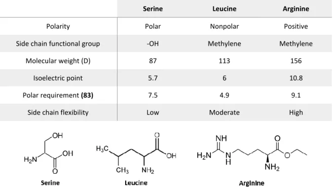
Figure 3.1 Aminoacylation with canonical (a) and non-canonical amino acids for protein translation Figure 3.2 Structure of serine, leucine and arginine
Figure 3.3 Representation of mutant tRNAs in the strain background of this work Figure 3.4 Impact of mutant tRNAs on cell fitness of ΔtL(GAG) mutants
Figure 3.5 Impact of mutant tRNAs on cell fitness of ΔtR(CCU) mutants
Figure 3.6 Leu-CUC (A) and Arg-AGG (B) codon distribution over S. cerevisiae genome
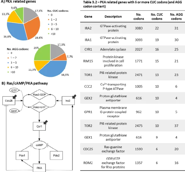
Figure 3.7 CUC and AGG codon distribution over the PKA related genes group (A) and a schematic of the Ras/cAMP/PKA pathway (B)
Figure 4.1 Different types of experimental evolution
Figure 4.2 Comparison between the growth rate of non-evolved (grey) and evolved (black) Leucine mutants Figure 4.3 Comparison between the lag phase duration, in hours, of non-evolved (dark grey) and evolved Leucine mutants (light grey)
Figure 4.4 Comparison between the growth rate of non-evolved ΔtL strains highly expressing the endogenous Ser-tRNAUGA (blue bar), and evolved strains with 100 (orange bar) and 200 generations (grey bar)
Figure 4.5 Comparison between the growth curve profiles of non-evolved (ΔtL, light blue) and evolved (EΔtL) Leu mutants
Figure 4.6 Comparison between the growth rate of non-evolved (grey) and evolved (black) Arginine (ΔtR) mutants
Figure 4.7 Comparison between the lag phase duration, in hours, of non-evolved (dark grey) and evolved (light grey) Arginine mutants
Figure 4.8 Northern blot analysis of the mutant tRNAs
Figure 4.9 Sequencing data from strains highly expressing Ser-tRNAGAG and the theoretical structure of the
mutant tRNA
Figure 4.10 Scheme of the reporter system built to monitor serine incorporation at AGG-Arg codons Figure 4.11 Scheme of the reporter system built to monitor serine incorporation at CUC-Leu codons Figure 4.12 Quantification of the mean fluorescence of evolved Leu mutants expressing Ser-tRNAGAG Figure 4.13 Area measurements from non-evolved and evolved Leu mutants
List of tables
Table 2.1 Yeast strains used in this study
Table 2.2 List of oligonucleotides used in this thesis Table 2.3 Original plasmids used in this study Table 2.4 List of the engineered plasmids
Table 2.5 List of oligonucleotides used for northern blot analysis Table 3.1 Principal characteristics of amino acids involved in this study Table 3.2 PKA related genes with 6 or more CUC codons
Chapter 1
1. Introduction
1.1 Genetic code
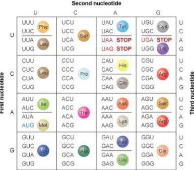
The genetic code was established 3.5 billion years ago (1) and is one of the oldest and most conserved characteristics of life. It is universally used across the three domains of life and can be defined as a series of biochemical reactions that establish the system of rules by which the information encoded by a nucleotide sequence is translated into a protein. Each three-nucleotide combination represents a codon which encodes a single amino acid. Since there are 4 nucleotides, there is a total of 64 combinations where 3 of them represent the termination codons UGA, UAA and UAG, and the remaining 61 encode for the 20 canonical amino acids (figure 1.1). This means that most amino acids are specified by more than one codon, showing the redundancy of the code. Codons that encode the same amino acid are named synonymous codons and are important to minimize the harmful effects of potential incorrectly placed nucleotides (2). However these synonymous codons are not equivalent and are used with different frequencies in high or low expressing genes, and in different organisms (3). This codon usage biases are linked with gene expression levels, translation efficiency and protein folding (4). (5)
Figure 1.1 – Standard genetic code. The genetic code is composed of 64 codons and each one is composed of 3 nucleotides. The initiation codon is highlighted as blue (AUG) and termination codons are highlighted as red (UAA, UAG, UGA), and the rest of the codons show their specified amino acid. Adapted from Clancy and Brown, 2008.
1.1.1 Reading the code
Codon assignments are established by two major components: transfer RNAs (tRNA) and aminoacyl tRNA synthetases (aaRS). Codons are recognized by the complementary anticodon of its cognate tRNA, which in turn is aminoacylated by aaRS that are highly selective for the correspondent amino acid (6). This relationship is central to the genetic code and pivotal to protein biosynthesis (figure 1.2).
1.1.1.1 Transfer RNAs
Transfer RNAs are the interface between the genetic information encoded in messenger RNA (mRNA) and proteins. tRNAs are small molecules composed of a single polynucleotide chain with 73-90 nucleotides which are arranged in a tridimensional L-shaped structure. Its secondary structure is often represented as a cloverleaf structure (figure 1.2a) composed by the acceptor stem, D arm, TψC arm, variable arm and the anticodon arm (7). The acceptor stem is composed of 7 base pairs followed by an unpaired nucleotide at position 73 and the 3’-CCA terminal where the amino acid is attached, whereas the TψC and anticodon arms have a 5 base pair stem and a 7 nucleotide loop. Stems are stabilized by Watson-Crick interactions and an occasional wobble G-U base pair is often observed. The major deviations seen in tRNA size are due to variations in nucleotide number in the variable and D loops. (8). One important structural motif is the anticodon Figure 1.2 – tRNA cloverleaf structure (a) and translation overview (b) highlighting the role of tRNAs in translation, and aminoacyl-tRNA synthetases (aaRS) in their aminoacylation. Adapted from Knight et al., 2001
U-turn that involves the ubiquitous U33. This is responsible for an abrupt reversion in direction
(~180°) of the tRNA chain which exposes the anticodon nucleotides to anticodon-codon pairing during posterior mRNA decoding (9). In turn, so that the tRNA is aminoacylated, aaRS must recognize the correct tRNA which involves a series of identity elements.
Transfer RNAs have many post-transcriptional modifications and are by far the most and more diversely modified biological molecule. While some modifications simply involve the addition of a methyl group, others are rather complex and involve multi-step reactions catalyzed by a series of specialized enzymes. These ensure tRNA proper folding and function, and can be divided in two major groups: those that affect structure, and those that tackle critical positions to mRNA decoding and aaRS recognition (10). Although modified nucleotides can be present all over the tRNA molecule, the two most frequent modifications are found in positions 34 and 37 present in the anticodon loop. For example, modifications in position 37 aid in the maintenance of the U-turn and influence frameshifting, and therefore have an impact in translational accuracy (11). Modifications in the wobble position 34 can account for some of the degeneracy of the genetic code; for example an U34 enables stacking with A and G nucleotides (the latter by wobble interaction), while a modified
I34 (inosine-34) enables recognition of C, A and U nucleotides, thus altering tRNA decoding capacity
(12). Generally, modifications in these two positions contribute to efficient anticodon-codon pairing and to stability of the tRNA-mRNA interaction during mRNA translation (11). Also, some modified nucleotides in the anticodon domain are crucial to aaRS’s ability to accurately aminoacylate tRNAs (13).
Noteworthy, tRNAs have other parts on the biological theatre beyond its role as adaptor in protein synthesis, not only in its aminoacylated state but also in its uncharged form, and even in fragmented form (7). Uncharged tRNAs have been shown to act as molecular sensors of external stresses such as amino acid deprivation, which regulates global gene expression to counteract nutritional stress (14, 15). Beside the canonical function of aminoacyl-tRNAs, they have also been implicated in other non-ribosomal functions. In bacterial cell wall formation, they act as substrates for building peptidoglycan bridges (16); and also mediate aminoacylation of phospholipids in the bacterial cell membrane (17). They also have a role in antibiotic biogenesis and in protein labeling for degradation (7).
1.1.1.2 Aminoacyl-tRNA synthetases
Aminoacyl-tRNA synthetases catalyze the aminoacylation reaction, which corresponds to the attachment of the correct amino acid to its cognate tRNA. This occurs through a two-step mechanism by first activating the amino acid and then transferring it to the tRNA. The aaRS binds and activates the amino acid with ATP, forming aminoacyl-adenylate (aa-AMP), and then this complex recognizes the cognate tRNA and transfers the amino acid to its 3’ end, releasing the AMP. This is a rather complex task for aaRS, as they have to recognize the correct tRNA (between a large family of structurally similar tRNAs), as well as discriminate amino acids (18). Despite this, aaRS have an overall error rate of 10-4 which is achieved by a series of quality control mechanisms (19).
There is one aaRS for each one of the 20 amino acids, and their recognition is usually done in two phases: the first activating step is inaccurate and is followed by a second step where non-cognate amino acids are hydrolyzed by an intrinsic proofreading activity, namely editing. While some amino acids have quite distinct physical and chemical properties which facilitates their discrimination, some amino acids, such as valine and isoleucine, only differ by a methyl group and their discrimination cannot be accomplished without the aaRS editing activity (20).On the other hand, tRNA is a large molecule that allows a series of intricate contacts with aaRS during recognition, mainly with the discriminator base (N73), the acceptor stem, and the anticodon. These so called identity elements can be determinants or antideterminants whether they promote or prevent, respectively, the interaction between the aaRS and the tRNA. By so, aminoacyl tRNA synthetases establish the genetic code by accurately pairing cognate tRNAs with their corresponding amino acids (19).
1.1.2 Protein biosynthesis
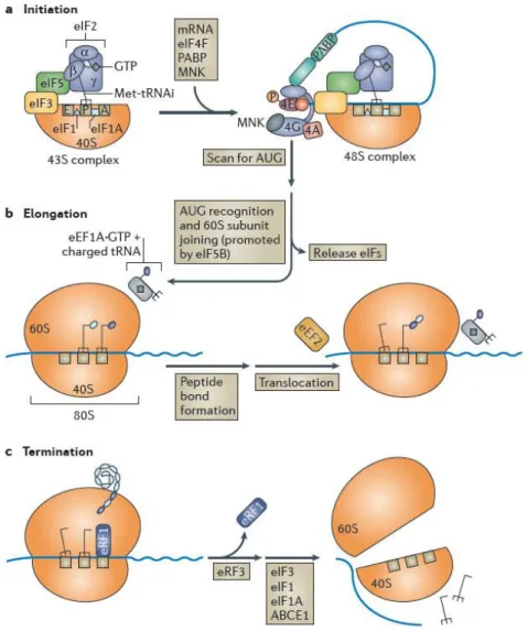
After the transcription of a particular gene, the synthesis of its protein product can begin (figure 1.3). Translation can generally be divided in three major steps: initiation, elongation and termination. This process is orchestrated by the ribosome, together with several auxiliary factors that bind to mRNA.
Initiation begins with the loading of the initiator methionyl-tRNA (Met-tRNAi) in the ribosomal
subunit 40S P site, forming a 43S pre-initiation complex. This is accomplished by eIF2·GTP complex, together with the eukaryotic translation initiation factors 1, 3 and 5 bound to the ribosomal subunit. With the help of eIF4F, the complex is positioned onto the 5′ end of a capped and polyadenylated mRNA and scans it until an initiator codon AUG is found, which in turn, is recognized by the
Met-tRNAi. Assemblage of the ribosomal subunit 60S to the later complex is facilitated by eIF3, eIF1 and
1A, forming the ribosome and enabling the elongation step. While Met-tRNAi is bounded to peptidyl
(P) site, another aminoacylated tRNA is delivered by eEF1A·GTP to the acceptor (A) site of the ribosome. If the latter is the cognate tRNA, a peptide bond between the carried amino acid and the previous amino acid is formed. The deacylated tRNA is released and the complex is translocated, exposing the next codon to the A site and the process is repeated, codon by codon. When a termination codon is encountered, the eukaryotic release factor 1 (eRF1) binds to the A site, which triggers ribosome arrest and the finished peptide is released. With the aid of eRF3, the ribosomal subunits, along with deacylated tRNAs and auxiliary factors dissociate from mRNA, and are free to initiate another round of translation (21, 22).
Figure 1.3 - mRNA translation. This process has three main phases: initiation (a), elongation (b) and termination (c), which requires a series of specific translation factors. Adapted from Walsh and Mohr, 2011
1.2 Alternative genetic codes
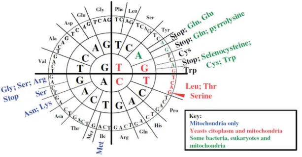
The genetic code is universally used by all forms of life and was initially postulated by Crick as a “frozen accident” unable to further evolve, since any alteration to the code would produce aberrant proteins, leading to proteome mayhem that would be lethal to the cell (2). Nevertheless, several natural deviations from its standard form are known in various microorganisms and mitochondria (figure 1.4). Together with the expansion of the amino acid repertoire by incorporation of two natural non-canonical amino acids (23), it shows that the code is not as frozen as initially thought.
The first natural alterations were discovered in human mitochondria and involved the decoding of the UGA stop codon as Trp, and the AUA-Ile codon as Met (24). Subsequent discoveries have shown that mitochondria are rather prone to codon identity reassignments and significant diversity of nonsense-to-sense and sense-to-sense reassignments is often found in Euglenozoans, Haptophytes, Stramenophiles, alveolates, green plants, red algae, fungi and metazoans (23). There are 16 known alterations in mitochondria and the rationalization is that they are particularly tolerant to reassignments due to their reduced genome size and complexity, when compared to nuclear genomes. Still, 10 alterations in nuclear codes were also found and termination codons are usually the only ones that are reassigned by the eukaryotic cytoplasmic translational machineries (24). For example, the UGA stop codon has been reassigned to Cys in Euplotes spp. (25) and to Trp in the Colpoda sp. and in various heterotrichs (26). The UUA and UAG stop codons have also been
Figure 1.4 – Genetic code diversity found in mitochondria and in eubacteria, archaea and eukaryotic cytoplasm. Unchanged codons are in black. Adapted from Miranda et al., 2006
reassigned to Gln in diplomonads (27), ciliates (26) and green algae (28). Termination codons appear to be particularly flexible, as they are also the target for the incorporation of the non-canonical amino acids selenocysteine, in a wide range of prokaryotes and eukaryotes (29), and pyrrolysine in archaeal Methanosarcina species (30), producing novel classes of proteins with unique catalytic properties. The only known sense-to-sense reassignment in nuclear genomes is found in many fungi of the Saccharomycotina CTG with the decoding of the Leu-CUG codon as Ser (31, 32).Another interesting observation in nuclear genomes is that, in a small number of species, some codons
like the Arg-AGA and Ile-AUA codons in Micrococcus spp., and the Arg-CGG
codon is Mycoplasma capricolum are either extremely rare or absent, and appear
unassigned (33). All together, t
hese observations invalidate the hypothesis of a non-mutable code and shows that it is flexible and evolvable.1.2.1 Evolution of the genetic code
Codon reassignments are an intriguing biological brainteaser since they show that the genetic code is still evolving, although these events are strongly selected against due to their intrinsic negative impact in proteome stability. Two evolutionary theories have arisen to explain how these alterations could emerge without leading to species extinction – the codon capture theory and the ambiguous intermediate theory (figure 1.5) (34).
The codon capture theory (figure 1.5.a) was initially proposed by Osawa and Jukes as a neutral mechanism in which genetic code changes arise from fluctuations in the GC/AT balance in the evolving genome. It postulates that evolution of codon reassignment happens through a stage in which a particular codon disappears from the genome, possibly leading to the disappearance of the cognate tRNA. At a later time in evolution, the codon may reappear by genetic drift and be decoded through a non-cognate tRNA, misreading the codon and thus allowing its identity alteration (24, 35). The disappearance of CGG-Arg codon from the AT-rich genome (75%) of Mycoplasma
capricolum, rendered the hypothesis that an AT-rich genome has a genomic bias against GC
content, which can drive GC-rich codons to disappear. This is further supported by the similar disappearance of AGA-Arg and AUA-Ile codons from the GC-rich genome (74%) of Micrococcus
luteus (34). However, there are reassignments in genomes with no obvious GC or AT bias, and even
Alternatively, Schultz and Yarus proposed the ambiguous intermediate theory (figure 1.5.b) as a non-neutral mechanism driven through tRNA mutations (37). It postulates that codon reassignments can appear through an intermediate stage of codon ambiguity when a single codon is both decoded by its cognate tRNA and a mutant tRNA. This translational ambiguity is the initial step for a gradual codon reassignment, and if selected, will lead to a gradual decrease of the cognate tRNA usage, and eventually to its loss. (34, 37). This theory applies both to sense and nonsense codon reassignments, but in the latter case the termination codon is being disputed by the release factor (RF) and a mutant tRNA capable of decoding it (37). Since codon ambiguity leads to proteome wide amino acid misincorporation and consequently to the synthesis of aberrant proteins, it is expected to be strongly selected against, as it would be highly detrimental to cell survival. However, this theory is credited by the ambiguous decoding of the CUG-Leu codon as Ser in several extant Candida species (32)and by the dual meaning of the Bacillus subtilis UGA codon as Trp or stop (38). Contrary to codon capture theory, this does not impose codon disappearance but it states that rarely used codons are more prone to reassignment than frequently used ones, so the potential negative impact of codon ambiguities in the proteome is minimized (37).
Figure 1.5 – Mechanisms of codon reassignments, where (a) illustrates the codon capture theory and (b) the ambiguous intermediate theory. Adapted from Santos et al., 2004
1.2.2 Mechanisms of reassignment
The molecular mechanisms of codon reassignment are poorly understood, however several studies provide interesting insight on how genetic code alterations may occur. They link them to alterations in components of the translational machinery accountable for interpreting the genetic code, namely tRNAs, aminoacyl-tRNA synthetases, and the release factors that recognize stop codons (39). As presented above (figure 1.4), some codons appear reassigned more frequently than others, particularly termination codons. Stop codon alterations are associated with critical structural changes in the codon recognition domain of release factors and in the anticodon of the misreading tRNAs. Such is the case of UAA and UAG decoding as Gln and UGA decoding as Trp or Cys, which occurs in several species of ciliates (39). It is proposed that this kind of reassignment happens through a first stage of codon ambiguity due to the appearance of a mutant tRNA capable of reading stop codon(s), and so competing with the RF. As ambiguity increases, the RF would fail in its ability to recognize the respective codon, enabling full reassignment (34). Another interesting way of exploiting the code was found by yeast mitochondria, where the four leucine CUN codons are decoded as threonine. This came as result of the loss of the Leu-tRNAUAG capable of decoding
the CUN codons and the appearance of a mutant Thr-tRNAUAG with an atypically large anticodon
loop. Surprisingly, this mutant Thr-tRNA has evolved from a His-tRNA, in which a single-nucleotide change converted it to a substrate for the yeast mitochondrial threonyl-tRNA synthetase (40). A distinct mechanism involves the reassignment of the nuclear Leu-CUG codon to Ser in various
Candida species, where a mutation in the Ser-tRNACAG produced a novel tRNA that is recognized
both by SerRS and LeuRS. This renders the CUG codon ambiguous, as it is decoded both as Ser and Leu in vivo (41).
An important question raised by these studies is if, during the course of codon reassignment, tRNA selection can affect codon usage in a way that might lead to codon final reassignment. In this way, we have to consider two key aspects: tRNA abundance which is correlated with codon usage, and the strength of codon-anticodon interaction. Since codon reassignments tend to happen in less abundant codons, they are also translated by less abundant tRNAs (34) and such rarity renders them more vulnerable to competition during translation if a novel non-cognate tRNA would emerge (42). Strength of codon-anticodon interactions is modulated by tRNA structure and modified nucleosides, particularly those in the anticodon loop at positions N34, N37, and in some cases N35. Modified nucleosides in these positions have impact in codon recognition, and some have the potential to restrict the decoding capacity of tRNAs, while others expand it (39). For example, in squid mitochondria, Ser-tRNAGCU contains m7G34 which expands its capacity to read Arg-AGA and
AGG codons, inserting serine at these sites (43). The mitochondrial Asn-tRNA of echinoderms has a 5’-Gψ35U-3’ anticodon which enables it to recognized AAA-Lys codons. Also, the decoding of
mitochondrial AUA-Ile codons as methionine by Met-tRNACAU is enabled by t6A37 which stabilizes
codon-anticodon interaction (34).
1.2.3 Codon ambiguity and mistranslation
Maintaining the integrity of the proteome is essential for cell viability as erroneous translation can induce protein misfolding and aggregation, and eventually cell death (figure 1.6). So codon ambiguities pose a striking problem to cellular homeostasis as they produce statistical proteins.
High error rate in protein synthesis has been shown to cause disease phenotypes in mouse models where global mistranslation produce tissue-specific neurodegeneration (44, 45). For example, the sticky mutation was identified by Lee and coworkers in mice that exhibited progressive neurodegeneration and cerebellar Purkinje cell loss in the cerebellum. Due to a mutation in the editing domain of alanyl-tRNA synthetase, its proofreading activity was compromised which increased mischarching of non-cognate amino acids into Ala-tRNAs,
Figure 1.6 – Delivery of aminoacylated tRNAs translational machinery. (A) Correctly aminoacylated tRNAs will lead to production of correctly folded WT proteins. (B) Misacylated tRNAs will produce errors that induce misfolding, which cause initiation of the unfolded protein response. Adapted from Lee et al., 2006.
consequently leading to global mistranslation of codons decoded by these tRNAs, which led to accumulation of misfolded/unfolded proteins (44). Misfolded proteins are associated with multiple neurodegenerative diseases such as Alzheimer’s, Parkinson’s and Huntington’s diseases, and amino acid misincorporation may be a key trigger in the pathology of multiple sclerosis and amyotrophic lateral sclerosis (46, 47). Nevertheless, in natural conditions, the protein quality control machinery maintains a basal threshold of translational errors at a frequency of around 10-4 (46). Molecular
chaperones are crucial to these mechanisms as they recognize misfolded proteins, which can either be refolded, degraded via the ubiquitin-proteasome pathway, or delivered to specialized compartments that sequester potentially toxic species (47). In the same way as the sticky mice, the woozy mutation was described with a similar phenotype of Purkinje cell loss (with exception of the cells in the most distal caudal lobule) as result of the accumulation of misfolded/unfolded proteins, due to a disruption in one of the chaperones of BiP, which is essential in misfolded protein translocation and folding, and ER stress sensing (45).
Despite the inherent negative impact on proteome, ambiguous decoding of mRNA can create selective advantages in some circumstances. Reassignment of stop codons to selenocysteine (Sec) and pyrrolysine (Pyl) clearly proves this point. Selenium is an essential dietary micronutrient with antioxidant properties and its biological effects are delivered by selenium-containing proteins, the selenoproteins. These are present in all three domains of life but not in all species. Selenocysteine is the biological form of selenium in proteins, which is inserted at specific UGA codons by a complex selenosome machinery (39, 48). In eukaryotes, a unique Sec-tRNA is serylated by SerRS which is then converted to Sec. Selenocysteine is inserted in response to UGA stop codons in a very complex mechanism coordinated by an extraordinary number of factors (49). Also, in Euplotes crassus, specific UGA codons are decoded both as Sec and Cys and such ambiguity is necessary to produce fully functional proteins (50). On the other hand, in the methanogen family Methanosarcinaceae, UAG stop codons in specific genes are translated as pyrrolysine. This amino acid is incorporated in methylamine methyltransferases, rendering these methanogenic archaea their unique capability of methane synthesis using methylamines as precursors (51). This demonstrates that codon ambiguity is tolerable and indeed functional.
It has been shown recently in HeLa cells grown under optimal conditions, that methionine misacylation corresponds to ~1.5% of all methionylated tRNAs, unveiling an unexpected basal level of mismethionylation in mammalian cells. Interestingly, upon viral infection the level of tRNA mismethionylation increased ~13%, and further increases were observed upon exposure to ROS-inducing agents, and upon induction of the unfolded protein response. The authors found that the
trigger to this induction was oxidative stress (52), which is in concordance with the protective role conferred by Met residues in proteins against damage mediated by ROS, seen in E. coli (53). Noteworthy, mismethionylation has been proved to occur in mammalian (52), yeast (54) and bacterial cells (55). On the other hand, mistranslation induced by severe oxidative stress has been proved to be toxic in E. coli. Hydrogen peroxide is capable of oxidizing a critical cysteine residue in the editing domain of ThrRS, leading to serine misacylation of Thr-tRNAs, which induced protein mistranslation and reduced growth rate (56).
In yeast, expression of the mutant Candida albicans Ser-tRNACAG induced highly detrimental
ambiguity in the Leu-CUG codon and major decrease in fitness due to high level synthesis of aberrant proteins. These triggered the expression of a stress response that provided important selective advantages under stress conditions and allowed ambiguous cells to survive in otherwise lethal environments (e.g. toxic doses of heavy metals, salts and cycloheximide) (57). Recently, the first microorganism with an altered genetic code was engineered by reversion of the CUG identity in C. albicans from serine back to leucine. Surprisingly, these strains adapted to increasing Leu misincorporation, recovered growth rate to wild-type levels and displayed unexpected phenotypic variability, with highly variable colony and cell morphologies, and increased tolerance to antifungals (58). Altogether this indicates that genetic code alterations are not mere abnormalities and can, in fact, represent a potential to adaptation, allowing species to colonize new ecological niches.
1.3 Saccharomyces cerevisiae as a biological model
Yeast is a good biological model to study cellular processes conserved in Eukaryotes and its genome can be easily manipulated. It has a rapid growth and generation time, and is particularly easy to manipulate, replicate and maintain. S. cerevisiae cells are round and stable in haploid and diploid form. Their size depend on their state, strain and growth phase, and normally ranges from 4 to 10 µm (59).
Yeasts genes are organized in 16 chromosomes and approximately 6,600 open reading frames (ORFs) have been annotated, with more than 80% functionally characterized (59, 60). In 1996, S.
cerevisiae genome was the first sequenced eukaryotic genome (61). Due to conservation of
homologous genes to human, yeasts are used as model systems for the study of human diseases. In fact, 60% of yeast genes have human homologues or at least one conserved domain with human genes (60). In addition, up to 30% of genes implicated in human diseases have a close yeast homologue (62).
There are a vast repertoire of well-established and widely used yeast cellular and molecular techniques, together with modern high-throughput tools (60). Particularly, DNA transformation techniques in yeast proved a powerful useful tool for genomics and proteomics studies as plasmids can be easily introduced in yeast cells either as replicating molecules or by genome integration (59, 63). This proves yeast as a practical and resourceful model organism.
Gene deletion techniques are also easily applicable. One example is the knock-out collection from Bloom-Ackermman and colleagues, used in this thesis, consisting of 204 nuclear encoded tRNA genes deletion, and even some double deletions. Their work exposed the highly complex architecture of the RNA pool, which revealed an extensive network of backup-compensation between and within tRNA families and, interestingly, that genes encoding identical tRNAs within the same family contribute differently to cell fitness. Also, deletions in single-copy and multi-copy tRNA families elicited different transcriptional responses. Of particular interest to this work, they identified two single-copy tRNA families, tL(GAG)G and tR(CCU)J, as non-essential upon deletion. This genetic perturbation resulted in a strong growth defect of the cells due to the inexistence of a cognate tRNA to decode the CUC-leucine codon and AGG-arginine codon (64), rendering them “orphan codons”.
1.4 Working hypothesis and objectives
The host laboratory has made a major breakthrough by engineering the first complete codon reassignment in the human pathogen Candida albicans, using a combination of tRNA engineering, gene recombination and forced evolution (58). This raised the hypothesis that synthetic codon ambiguities combined with experimental evolution have the power to re-code rare sense codons.
In this project, we used the described above yeast tRNA gene deletion library, particularly the two strains with viable single-copy tRNA gene deletions to test the hypothesis that rare codons can be reassigned using experimental evolution. For this, we engineered a mutant serine tRNA (58) to misincorporate Ser at CUC-leucine and AGG-arginine sites on a proteome wide scale, and evolved the ambiguous strains using experimental evolution. The specific objectives of this Masters Thesis are the following:
1 – Construction of Saccharomyces cerevisiae strains with ambiguity in rare Leu and Arg codons; 2 – Experimental evolution of ambiguous strains to reassign sense codons;
3 – Development of a reporter system to monitor Ser misincorporation at CUC-leucine and AGG-arginine sites.
Chapter 2
2. Materials and Methods
2.1 Strains and growth conditions 2.1.1 Strains
Escherichia coli strain JM109 (recA1 SupE44 endA1 hsdR17 gyrA96 relA1 thi Δ[Lac-proAB]
F’[traD36 proAB-lacI lacZΔM15) was used as a host for all DNA manipulations.
Saccharomyces cerevisiae strains used in this thesis are haploid and based on the genetic
background of Y5565 (table 2.1). ΔtL(GAG)G and ΔtR(CCU)J are knock-out strains that have a deletion of the single-copy genes tL(GAG)G and tR(CCU)J that encode tRNALeu
GAG and tRNAArgCCU,
respectively. These strains belong to a tRNA gene deletion library that was kindly provided by Dr. Yitzhak Pilpel (64).
Strain Genotype
Y5565 MATα, can1Δ::MFA1pr-HIS3, mfα1Δ::MFα1pr-LEU2, lyp1Δ, ura3Δ0,
leu2Δ0, his3Δ1, met15Δ0
ΔtL(GAG)G ΔtL(GAG)G::Hyg, MATα, can1Δ::MFA1pr-HIS3, mfα1Δ::MFα1pr-LEU2,
lyp1Δ, ura3Δ0, leu2Δ0, his3Δ1, met15Δ0
ΔtR(CCU)J ΔtR(CCU)J::Hyg, MATα, can1Δ::MFA1pr-HIS3, mfα1Δ::MFα1pr-LEU2,
lyp1Δ, ura3Δ0, leu2Δ0, his3Δ1, met15Δ0
2.1.2 Growth conditions
E. coli cells were grown at 37 ºC in LB broth medium or LB with 2% agar (Formedium)
supplemented with ampicillin (75 µg/ml; Sigma-Aldrich).
Saccharomyces cerevisiae cells were grown at 30ºC in YPD (2% glucose). Transformed S. cerevisiae cells were grown in minimal medium lacking uracil (MM-Ura; 0,67% yeast nitrogen base
without amino acids, 2% glucose, 2% agar and 100 µg/ml required amino acids – drop-out mixture in annexes).
After evolution experiments, yeast cells were grown in 5-fluoro-orotic acid (5-FOA) plates (description in annexes). The purpose of this assay was to select cells that lost the plasmid containing the URA3 marker, so that transformation of the same cells with another plasmid (with
the same marker) was possible. For this, 100 µl of an evolved clone from each strain were plated in 5-FOA plates and left at 30ºC for 2 days, until colonies were visible.
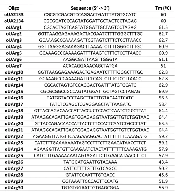
2.2 Oligonucleotides
Oligonucleotides (table 2.2) were purchased from IDT – Integrated DNA Technologies and ressuspended in miliQ water to a final concentration of 100 mM.
Oligo Sequence (5’ -> 3’) Tm (ºC) oUA2133 CGCGTCGACGTCCAGGACTGATTTATGTGCATC 60 oUA2134 CGCGGATCCCAGTATGGATTGCTAGTCCTAGAG 60 oUArg1 CGCACTAGTCAGTATGGATTGCTAGTCCTAGAG 61.5 oUArg2 GGTTAAGGAGAAAGACTACGAATCTTTTGGGCTTTGC 62.7 oUArg3 GCAAAGCCCAAAAGATTCGTAGTCTTTCTCCTTAACC 62.7 oUArg4 GGTTAAGGAGAAAGACTTAAAATCTTTTGGGCTTTGC 60.9 oUArg5 GCAAAGCCCAAAAGATTTTAAGTCTTTCTCCTTAACC 60.9 oUArg6 AAGGCGATTAAGTTGGGTA 51.1 oUArg7 ACACAGGAAACAGCTATGA 51 oUArg10 GGTTAAGGAGAAAGACTGAGAATCTTTTGGGCTTTGC 62.8 oUArg11 GCAAAGCCCAAAAGATTCTCAGTCTTTCTCCTTAACC 62.8 oUArg14 CGCACTAGTGTCCAGGACTGATTTATGTGCATC 62.9 oUArg15 CGCGCGGCCGCCAGTATGGATTGCTAGTCCTAGAG 69.9 oUArg16 TATGGTACCCTAGCTTATTTGTACAATTCATC 56.5 oUArg17 TATCTCGAGCTCGAGGAGCTATTAAGATC 58.4 oUArg18 GTTACCAGACAACCATTACCUCTCCACTCAATCTGCCTTAT 64.4 oUArg19 ATAAGGCAGATTGAGTGGAGAGGTAATGGTTGTCTGGTAAC 64.4 oUArg20 GTTACCAGACAACCATTACTCTTCCACTCAATCTGCCTTAT 63.5 oUArg21 ATAAGGCAGATTGAGTGGAGAGGTAATGGTTGTCTGGTAAC 64.4 oUArg22 AGAAGGTTATGTTCAAGAAAGGACTATTTTTTTCAAAGATG 59.2 oUArg23 CATCTTTGAAAAAAATAGTCCTTTCTTGAACATAACCTTCT 59.2 oUArg24 AGAAGGTTATGTTCAAGAATCTACTATTTTTTTCAAAGATG 57.9 oUArg25 CATCTTTGAAAAAAATAGTAGATTCTTGAACATAACCTTCT 57.9 oUArg26 TATGGATGAATTGTACAAA 43.4 oUArg27 CATTCTTTTGTTTGTCAGCC 50.2 oUArg28 GTATTCCAATTTGTGACC 45.6 oUArg29 GGTAAATTGCCAGTTCCATG 51.9 oUArg30 TGTGTGGAATTGTGAGCGGA 56.9
2.3 Plasmids
2.3.1 Original plasmids
In this work we used the following plasmids (table 2.3) as backbone for the new constructions (section 2.3.2):
Plasmid Description
pUA261 Plasmid based on pRS315 with a single copy of the C. albicans tRNASerUGA gene inserted between SalI e BamHI cloning sites
pRS426
Multi-copy plasmid with 5726 bp containing the AmpR and URA3 gene, allowing for selection of transformed cells in LB media with ampicillin or minimal media lacking uracil, respectively
pRS316 Single-copy plasmid with 4887 bp belonging to a series of pBluescript-based
centromere vectors. Contains the AmpR and URA3 gene
pACT1-GFP Plasmid based on CIP10 containing the codon-optimized yeast enhanced GFP (yEGFP) gene (65)
2.3.2 Constructed plasmids
For expression of the chimeric C. albicans tRNASer
UGA gene in S. cerevisiae, we used the plasmid
pUA261 to amplify the tRNASer
UGA gene with the primers oUA2133 and oUA2134. The gene was then
cloned into SalI e BamHI restriction sites of pRS426 and pRS316, originating the plasmids pUA715 and pUA716, respectively (figure 2.1 in annexs).
The anticodon of the tRNASer
UGA gene, inserted in pUA715 and pUA716, was altered by
site-directed mutagenesis to CCU and GAG in order to generate mutant misreading tRNA genes. Expression of tRNASer
CCU from multi and single-copy plasmids (pUA717 and pUA718) promotes the
insertion of serine at AGG arginine sites in strains lacking the tRNA that recognizes this codon (ΔtR(CCU)J). Expression of tRNASer
GAG from multi and single-copy plasmids (pUA719 and pUA720)
promotes the insertion of serine at CUC leucine sites in strains lacking the tRNA that recognizes this codon (ΔtL(GAG)G).
In order to monitor Ser misincorporation, we developed a loss-of-function reporter system based on the codon-optimized yeast enhanced GFP (yEGFP) gene and assembled it on the multi-copy plasmids pRS426, pUA715, pUA717 and pUA719, in different versions. For that, first we amplified the yEGFP gene plus the promoter ACT1 from pACT1-GFP, using the primers oUArg16 and oUArg17, and then inserted it between the KpnI e XhoI cloning sites of the referred plasmids. This
originated the plasmids pUA721 (figure 2.2 in annexes), pUA728, pUA733 and pUA736, respectively.
To monitor misincorporation of serine at leucine sites, we used a strategy previously optimized in the host laboratory for C. albicans (58) on the plasmids pUA721, pUA728 and pUA733. We changed the leucine codon UUA in position 201 (L201) of the yEGFP by site-directed mutagenesis to leucine codon CUC, yielding plasmids pUA725, pUA730 and pUA735. In this way, when Ser is incorporated at residue 201, it causes a destabilization in GFP rendering it inactive, permitting the monitorization of Ser misincorporation by tRNASer
GAG. Next, in order to have a negative control, we
altered this same codon to the serine UCU codon, originating plasmids pUA723, pUA729 and pUA734.
Lastly, to monitor misincorporation of serine at AGG arginine sites, we used an analogous strategy on the plasmids pUA721, pUA728 and pUA736. For that, we changed the codon AGA in position 96 (correspondent to arginine, R96) by site-directed mutagenesis to codon AGG, yielding the plasmids pUA727, pUA732 and pUA738. Like in the last case, when Ser is incorporated at this residue renders GFP inactive, allowing the monotorization of Ser misincorporation by tRNASer
CCU.
Next we produced the negative control altering the codon of this residue to UCU, originating plasmids pUA726, pUA731 and pUA737.
Plasmids Description
pUA715 Multi-copy plasmid with insertion of Ser-tRNAUGA gene between SalI e BamHI
pUA716 Single-copy plasmid with insertion of Ser-tRNAUGA gene between SalI e BamHI
pUA717 Plasmid based on pUA715, constructed by site directed mutagenesis of the anticodon of the Ser-tRNAUGA gene to CCU
pUA718 Plasmid based on pUA716, constructed by site directed mutagenesis of the anticodon of the Ser-tRNAUGA gene to CCU
pUA719 Plasmid based on pUA715, constructed by site directed mutagenesis of the anticodon of the Ser-tRNAUGA gene to GAG
pUA720 Plasmid based on pUA716, constructed by site directed mutagenesis of the anticodon of the Ser-tRNAUGA gene to GAG
pUA721 Plasmid pRS426 with insertion of ACT1-GFP between the restriction sites of
KpnI and XhoI
pUA723 Plasmid based on pUA721, constructed by site directed mutagenesis of the codon correspondent to L201 to TCT
pUA725 Plasmid based on pUA721, constructed by site directed mutagenesis of the codon correspondent to L201 to CUC
pUA726 Plasmid based on pUA721, constructed by site directed mutagenesis of the codon correspondent to R96 to TCT
pUA727 Plasmid based on pUA721, constructed by site directed mutagenesis of the codon correspondent to R96 to AGG
pUA728 Plasmid based on pUA715 with insertion of ACT1-GFP between the restriction sites of KpnI and XhoI
pUA729 Plasmid based on pUA729, constructed by site directed mutagenesis of the codon correspondent to L201 to TCT
pUA730 Plasmid based on pUA729, constructed by site directed mutagenesis of the codon correspondent to L201 to CUC
pUA731 Plasmid based on pUA729, constructed by site directed mutagenesis of the codon correspondent to R96 to TCT
pUA732 Plasmid based on pUA729, constructed by site directed mutagenesis of the codon correspondent to R96 to AGG
pUA733 Plasmid based on pUA719 with insertion of ACT1-GFP between the restriction sites of KpnI and XhoI
pUA734 Plasmid based on pUA733, constructed by site directed mutagenesis of the codon correspondent to L201 to TCT
pUA735 Plasmid based on pUA733, constructed by site directed mutagenesis of the codon correspondent to L201 to CUC
pUA736 Plasmid based on pUA717 with insertion of ACT1-GFP between the restriction sites of KpnI and XhoI
pUA737 Plasmid based on pUA736, constructed by site directed mutagenesis of the codon correspondent to R96 to TCT
pUA738 Plasmid based on pUA736, constructed by site directed mutagenesis of the codon correspondent to R96 to AGG
2.3.3 Site directed mutagenesis
This procedure was used with the goal of mutating the anticodon of C. albicans tRNASer
UGA gene
and the codons of leucine and arginine residues (L201 and R96, respectively) of yEGFP gene. For that, polymerase chain reactions (PCR) were prepared with 1x Pfu Buffer with MgSO4, 0.2 mM dNTPs, 0.2 mM of each respective primer, 1.25 U of Pfu polymerase, 5-25 ng of plasmidic DNA, and miliQ water to a final volume of 25 µl. The PCR program used consisted of a cycle of 30 seconds at 95 ºC, 1 minute at 55 ºC and 7 minutes at 68ºC. Finally, the resulting products were treated with 0.5 µl of DpnI for 2 hours, at 37 ºC. The goal was to digest the parental DNA template and select for the mutation-containing DNA. After digestion, plasmids were transformed in E. coli competent cells and plasmid DNA was extracted for DNA sequencing (StabVida sequencing services).
2.4 Preparation of E. coli competent cells
E. coli competent cells were prepared from strain JM109 using the TFB method (66) . First, 200
µl of an overnight culture was inoculated in 5 ml of LB medium and left in the incubator at 37 ºC, 180 rpm, until they reached an OD600 of 0.3. Then, 4 ml of the late culture were inoculated in 100
ml of LB medium and left to grow in the same conditions until they reached an OD600 of 0.3. After
this, cells were collected in two falcons, cooled on ice for 5 minutes and centrifuged at 2500 rpm for 5 minutes, at 4 ºC. The supernatant was discarded and the pellet ressuspended in 20 ml of cold TFBI (0.03 mM potassium acetate, 0.08 mM RbCl2, 0.013 mM CaCl2, 0.08 mM MnCl2, 15.4% glycerol,
pH 5.8). Cell suspension was centrifuged at 2500 rpm for 5 minutes, at 4 ºC and after rejection of the supernatant, the pellet was ressuspended in 2.5 ml of cold TFBII (0.01 mM MOPS Ca, 0.01 mM CaCl2, 0.008 mM RbCl2, 13.4% glycerol, pH 6.5). Cells were then incubated on ice for 5 minutes and
distributed on aliquots of 200 µl each and stored at -80 ºC.
2.5 Plasmidic DNA purification from E. coli 2.5.1 Miniprep Kit
For the extraction and purification of plasmidic DNA, we used GeneJETTM Plasmid Miniprep kit
(Fermentas). Recombinant E. coli cells were inoculated in 5 ml or 10 ml of LB medium with 75 µg/ml ampicillin in the case of multi-copy or single-copy plasmids, respectively, and grown overnight at 37 ºC. Cells were harvested at 8000 rpm for 2 minutes, the supernatant was removed and the procedure was followed according to the manufacturer instructions. After the purification, DNA concentration was quantified using the NanoDrop system and plasmids were stored at -20 ºC.
2.5.2 “Homemade minipreps”
For screening procedures, a cheap miniprep protocol that avoided the use of the kit columns was established. Briefly, 1.5 ml of E. coli cultures grown overnight were collected by two rounds of centrifugation at 13000 rpm, ressuspended in 1 ml of cold solution I (0.5 mM glucose, 250 mM Tris, 10 mM EDTA) and incubated on ice for 5 minutes. Then, it was added 200 µl of solution II (0.2 M NaOH, 1% SDS), at room temperature, and incubated again on ice for 5 minutes. This was followed by the addition of 150 µl of cold solution III (1 mM KOAc saturated with KOH) and by other incubation on ice. The suspension was then centrifuged at 13000 rpm for 10 min and supernatant
was collected. After a series of precipitations with isopropanol and ethanol 70%, the pellet was dried, ressuspended in water and quantified using NanoDrop.
2.6 Plasmidic DNA purification from S. cerevisiae
For the extraction and purification of plasmidic DNA from yeast, we used Wizard® Genomic DNA Purification Kit from Promega, and followed the instructions from the manufacture.
2.7 Transformation procedures
2.7.1 Escherichia coli
Transformation of E. coli cells was performed using the chemical SOC method (66). For this, 10 ng of plasmidic DNA was added to thawed aliquots of competent cells (JM109), mixed and incubated on ice for 30 minutes. This was followed by a heat-shock at 42 ºC for 90 seconds and then cooled on ice for 2 minutes. Next, 800 µl of SOC (for preparation of 100 ml at pH 7, 2 g of tryptone, 0.5 g of yeast extract and 0.05 g of NaCl were weighted, and 1 ml of KCl 250 mM and 20 ml of glucose 1M were added) were added to the transformation mix and then incubated at 37 ºC, 180 rpm for 1 hour. After this, cell suspension was centrifuged at 2500 rpm for 1 minute and the supernatant was removed, leaving approximately 50 µl that were homogenized and plated in solid LB supplemented with 75 µg/ml of ampicillin. Plates were incubated at 37 ºC, overnight.
2.7.2 Saccharomyces cerevisiae
Yeast transformation was accomplished using the lithium acetate (LiAc) method described by Gietz and Woods (63). Briefly, 1.5 ml of overnight cultures were harvested in log phase by centrifugation for 1 minutes at 4000 rpm. The supernatant was discarded and the following reagents were added to the pellet in sequence: 240 µL of PEG 50% (w/v), 36 µL of LiAc 1 M, 25 µL of previously denatured single-stranded salmon sperm carrier DNA (2 mg/mL) and 36 µl dH2O plus
0.1-1 µg of plasmid DNA. The mixture was ressuspended and submitted to a heat-shock treatment at 42 ºC for 1 hour. Cells were harvested by centrifugation for 1 minute at 13000 rpm, the supernatant was removed and the pellet resuspended in minimal medium lacking uracil. The cell suspension was then plated in solid MM-Ura medium and incubated for 3 to 5 days at 30 ºC, until colonies were visible.
2.7.3 Saccharomyces cerevisiae (alternative)
When the LiAc method proved unfruitful, we used an adapted and modified transformation protocol for Candida albicans (67). In this version, 5 ml of overnight cultures were harvested in log phase by centrifugation for 5 minutes at 4000 rpm and the pellet was ressuspended in 150 µl LiAc-sol (10% LiAc 1M, pH 7.5; 10% TE 10x, pH 7.5). 5 µg of plasmid DNA was added to the suspension, followed by 100 µg of single-stranded DNA carrier and 600 µl of PEG/LiAc-sol. (50% PEG 50% (w/v), 50% LiAc-sol), and mixed. The transformation mix was incubated overnight at 30ºC, 180 rpm and submitted to a heat-shock at 44 ºC during 15 minutes and then cooled on ice about 2 minutes. The cell suspension was centrifuged in a bench top centrifuge at maximum speed for 1 minute and the supernatant was discarded. The pellet was ressuspended in 150 µl of MM-Ura and platted in solid MM-Ura medium and incubated for 3 to 5 days at 30 ºC, until colonies were visible.
2.8 Colony PCR
Colony PCR was used to check for the correct transformation of E. coli and yeast cells. For this, individual colonies from selective plates were picked using a P10 tip and were homogenized in 5 µl of miliQ water. Cell suspension was submitted to a heat shock at 95 ºC for 5 min. Next, PCR reagents were added to each tube: 1x DreamTaq Buffer, 0.2 mM dNTP mix, 0.15 mM of each primer, 1.25 U of DreamTaq polymerase and miliQ water to a final volume of 15 µl. General PCR programs consisted an initial denaturation at 95 ºC for 3 minutes, followed by 30 cycles of a denaturation step at 95 ºC for 30 seconds, a annealing step for 30 seconds at variable temperature and an extension step at 72 ºC for 1 minute. Reactions were performed in a MyCyclerTM thermal cycler (BIORAD) and ended with a final extension step at 72 ºC for 5 minutes. The PCR products were subjected to a gel electrophoresis for quality check.
2.9 Growth curves and rates
Growth curves were established for all strains in minimal medium lacking uracil or YPD in the case of non-transformed cells. First, cultures were pre-incubated for 3 days in the appropriate medium at 30ºC with constant shaking at 180 rpm. The optical density (OD) of the pre-inoculum was measured at 600 nm and then the cultures were inoculated in fresh media to an initial OD600
of 0.01. After approximately 7 hours, the OD was monitored each hour until they reached stationary phase. Data obtained was plotted in a graph as log(OD600) as a function of time (h). A trend line was
a fitted to the exponential phase of growth and its slope represented the growth rate of the clones. This procedure was carried out with five different clones from each strain, in the non-evolved and evolved state.
2.10 Evolution experiments
Experimental evolution assays were carried out using 5 clones of each newly constructed strain, as well as wild-type and knock-out control strains. Control cells were grown in 1 ml of YPD, whereas transformed cells were grown in 1 ml of minimal medium lacking uracil at 30ºC, until they reached stationary phase. Then cells were diluted by a factor of 1:200 into fresh medium (number of generations). This procedure was repeated until the strains reached approximately 200 generations. Number of generations was calculated as shown in the equation below.
2.11 Northern Blot Analysis 2.11.1 Total RNA extraction
For total RNA extractions, 50 ml cultures of S. cerevisiae cells were grown overnight in minimal medium and harvested at an OD600 of 1-1.5. The pellets were then frozen overnight at -80ºC.
Frozen cells were resuspended in hot acid-phenol:chloroform 5:1 (pH 4.7) and TES-buffer (10 mM Tris pH 7.5, 10 mM EDTA, 0.5% SDS) and incubated for 1 hour, at 65ºC, with repeated shaking every 10 minutes. Cell suspension was centrifuged at 13000 rpm for 20 minutes, at 4ºC. Then the RNA containing aqueous phase was transferred to new tubes and the same volume of acid-phenol:chloroform 5:1 (pH 4.7) was added. The suspension was mixed using a vortex and centrifuged at 13000 rpm for 10 minutes, at 4ºC and this step was repeated. The aqueous phase was transferred to a new tube with the same volume of chloroform:isoamil-alchool 24:1, vortexed hard and centrifuged at 13000 rpm for 10 minutes, at 4ºC. Then 350 µl of the aqueous phase was transferred to a new tube containing 35 µl of sodium acetate (3 mM NaAc, pH 5.2) and 800 µl of ethanol 100% (-20ºC). The aqueous phase was precipitated overnight at -20ºC. Tubes were then centrifuged at 13000 rpm for 5 minutes, at 4ºC. The fluid was carefully removed without touching
the RNA-pellet and the later was washed with 500 µl of ethanol 80% (-20ºC) and briefly spinned down at 13000 rpm. The ethanol was removed and the tubes were air-dried so that all traces of ethanol were removed. The pellet was dissolved in 100 µl of sterile miliQ water and the concentration was quantified by Nanodrop.
2.11.2 Northern blot
Fractionation of total tRNAs was carried out on 12-15% polyacrylamide (40% Acril:Bis) gels containing 8 M urea (0.8 mm thick, 30 cm long). In each gel slot, 50 µg of total RNA sample was loaded and gels were electrophoresed at 500 V for 16 hours. Fractioned tRNAs were localized by UV shadowing, the portion of the gel containing tRNAs was cut and transferred onto a nitrocellulose membrane (Hybond N, Amersham) using a Semy-dry Trans Blot (Bio-Rad). For hybridization, probes were prepared using 10 pmol of dephosphorylated oligonucleotide and 4 µl of ɤ-32P-ATP
(5000Ci/mmol) (Perkin Elmer) in 1x T4 kinase buffer, 10 mM spermidine and 16 units of T4 kinase (Takara). Labelling reaction was incubated at 37ºC for 1 hour and then the probe was extracted using phenol:chloroform:isoamyl alcohol (PCIA). The hybridization protocol was performed as described by Jacques Heitzler (68). Membranes were pre-hybridized for 30 minutes at 53 ºC in a hybridization solution [5x Denhardt’s solution (1% Ficol, 1% Polyvinylpyrrolidone and 1% Bovine serum albumin), 6x SSPE (3 M NaCl, 173 mM NaH2PO4, 25 mM EDTA) and 0.05% SDS]. Membrane hybridization was performed overnight in the above buffer using probes oUA2199 for detection of WT and mutant Ser-tRNA and oUA2195 for detection of the WT control Gly-tRNACCC (table 2.5).
Membranes were then washed 4 times (3 minutes each time) in 2x SSPE, 0.5% SDS at 53 ºC. The membranes were exposed overnight with intensifying screens and developed using a Molecular Imager FX (Biorad).
Probe Detection Tm (ºC) Sequence (5’ -> 3’)
oUA2199 WT and mutant Ser-tRNAUGA 51,5 TTAACCGCTCGGACAAGTT
oUA2195 WT Gly-tRNACCC 52-55 GCGGAAGCCGGGAATCGAAC
2.12 Epifluorescence microscopy
To monitor Ser misincorporation, yEGFP expression was visualized in S. cerevisiae cells by epifluorescence microscopy. Strains were grown overnight in liquid media at mid-exponential phase and aliquots were spotted onto microscope slides. Fluorescence was detected using a Zeiss MC80 Axioplan 2 light microscope, equipped for epifuorescence microscopy with filter set HE38. Photographs were taken using an AxioCam HRc camera and images were analysed using ImageJ software. Mean fluorescence intensities (± standard deviation) were quantified in individual Leucine mutant cells expressing the reporter yEGFP Leu-UUA201 (positive control), Ser-UCU201
(negative control) and Ser/Leu-GAG201 (reporter). In Arginine mutants, intensities were quantified
in individual cells expressing the reporter yEGFP Arg-AGA96 (positive control), Ser-UCU96 (negative
control) and Ser/Leu-AGG96 (reporter). yEGFP fluorescence (intensity/pixel) was determined for at