ORIGINAL ARTICLE
Defining Long-Term Survivors Following Resection
of Intrahepatic Cholangiocarcinoma
Fabio Bagante1&Gaya Spolverato1&Matthew Weiss2&Sorin Alexandrescu3&
Hugo P. Marques4&Luca Aldrighetti5&Shishir K. Maithel6&Carlo Pulitano7&
Todd W. Bauer8&Feng Shen9&George A. Poultsides10&Oliver Soubrane11&
Guillaume Martel12&B. Groot Koerkamp13&Alfredo Guglielmi1&Endo Itaru14&
Timothy M. Pawlik15
Received: 3 July 2017 / Accepted: 16 August 2017 / Published online: 24 August 2017 # 2017 The Society for Surgery of the Alimentary Tract
Abstract
Background Intrahepatic cholangiocarcinoma (ICC) is an aggressive primary tumor of the liver. While surgery remains the cornerstone of therapy, long-term survival following curative-intent resection is generally poor. The aim of the current study was to define the incidence of actual long-term survivors, as well as identify clinicopathological factors associated with long-term survival.
Methods Patients who underwent a curative-intent liver resection for ICC between 1990 and 2015 were identified using a multi-institutional database. Overall, 679 patients were alive with≥ 5 years of follow-up or had died during follow-up. Prognostic factors among patients who were long-term survivors (LT) (overall survival (OS)≥ 5) were compared with patients who were not non-long-term survivors (non-LT) (OS < 5).
Results Among the 1154 patients who underwent liver resection for ICC, 5- and 10-year OS were 39.6 and 20.3% while the actual LT survival rate was 13.3%. After excluding 475 patients who survived < 5 years, as well as patients were alive yet had < 5 years of follow-up, 153 patients (22.5%) who survived≥ 5 years were included in the LT group, while 526 patients (77.5%) who died < 5 years from the date of surgery were included in the non-LT group. Factors associated with not surviving to 5 years Fabio Bagante and Gaya Spolverato both authors equally contributed to
the manuscript
Presented as an Oral Presentation at the Annual Americas Hepato-Pancreato-Biliary Association Meeting, Miami, FL 2017.
* Timothy M. Pawlik Tim.Pawlik@osumc.edu
1
Department of Surgery, University of Verona, Verona, Italy
2
Department of Surgery, Johns Hopkins Hospital, Baltimore, MD, USA
3 Department of Surgery, Fundeni Clinical Institute,
Bucharest, Romania
4
Department of Surgery, Curry Cabral Hospital, Lisbon, Portugal
5
Department of Surgery, Ospedale San Raffaele, Milan, Italy
6
Department of Surgery, Emory University, Atlanta, GA, USA
7
Department of Surgery, Royal Prince Alfred Hospital, University of Sydney, Sydney, Australia
8 Department of Surgery, University of Virginia, Charlottesville, VA,
USA
9 Department of Surgery, Eastern Hepatobiliary Surgery Hospital,
Shanghai, China
10
Department of Surgery, Stanford University, Stanford, CA, USA
11
Department of Hepatobiliopancreatic Surgery and Liver Transplantation, AP-HP, Beaujon Hospital, Clichy, France
12 Division of General Surgery, Department of Surgery, University of
Ottawa, Ottawa, ON, Canada
13
Department of Surgery, Erasmus University Medical Centre, Rotterdam, Netherlands
14
Gastroenterological Surgery Division, Yokohama City University School of Medicine, Yokohama, Japan
15 Department of Surgery, The Urban Meyer III and Shelley Meyer
Chair for Cancer Research, The Ohio State University Wexner Medical Center, 395 W. 12th Ave., Suite 670, Columbus, OH, USA
included perineural invasion (OR 4.78, 95% CI, 1.92–11.8; p = 0.001), intrahepatic metastasis (OR 3.75, 95% CI, 0.85–16.6, p = 0.082), satellite lesions (OR 2.12, 95% CI, 1.15–3.90, p = 0.016), N1 status (OR 4.64, 95% CI, 1.77–12.2; p = 0.002), ICC > 5 cm (OR 2.40, 95% CI, 1.54–3.74, p < 0.001), and direct invasion of an adjacent organ (OR 3.98, 95% CI, 1.18–13.4, p = 0.026). However, a subset of patients (< 10%) who had these pathological characteristics were LT.
Conclusion While ICC is generally associated with a poor prognosis, some patients will be LT. In fact, even a subset of patients with traditional adverse prognostic factors survived long term.
Keywords Intrahepatic . Cholangiocarcinoma . Long-term . Survival
Introduction
Intrahepatic cholangiocarcinoma (ICC) is the second most common primary liver cancer.1–6Long-term survival of pa-tients with unresectable ICC is dismal, with only 5–10% of patients alive 5 years from the time of diagnosis.7While sur-gery remains the only hope for long-term cure for patients with resectable disease, 5-year overall survival (OS) remains poor even after hepatic resection (5-year OS, 25–30%). While several clinicopathological variables have been associated with prognosis including Cancer Antigen (CA) 19-9, tumor number and size, lymph node status, margin status, and vas-cular invasion, the incidence of long-term survivors after curative-intent surgery remains poorly defined. Traditionally, the American Joint Committee on Cancer (AJCC) staging manual has been the main means of stratifying patients with regard to prognosis8; however, new prognostic tools have also been accepted and utilized.9In particular, several studies have reported on nomograms based on patient and tumor-specific factors, as well as the impact of perioperative complications on long-term prognosis for patients with ICC undergoing surgery.10–18
Following resection of abdominal malignancies, most stud-ies have examined short-term survival, while fewer studstud-ies have reported data specifically defining long-term survivors.19 In one study of 618 patients who underwent resection for pancreatic adenocarcinoma, the authors reported an actual 5-year survival of only 12% following resection.20AJCC stage and negative surgical margins were predictive of long-term survival, while patient age, gender, and tumor location were not associated with actual 5-year survival. In a separate study, Zheng et al. reported on a large Western series of actual 10-year survivors following resection of hepatocellular carcino-ma (HCC) and noted that surgical carcino-margin status was the carcino-main factor associated with long-term survival.21Despite these re-ports, no previous study has focused on actual long-term sur-vivors following resection of ICC. As such, the objective of the current study was to define the incidence of actual long-term survivors following curative-intent resection of ICC using a large, international, multi-center cohort of patients. In addition, we sought to characterize the impact of
clinicopathological factors on the likelihood of patients to sur-vive long-term after surgical resection of ICC.
Materials and Methods
Patient Demographic and Clinical Data
Patients who underwent a liver resection for histologically confirmed ICC between 1990 and 2015 were identified from a multi-institutional database including 14 major hepatobiliary centers in the USA, Europe, Australia, and Asia (Johns Hopkins Hospital, Baltimore, MD, n = 89, 7.7%; Stanford University, Stanford, CA, n = 45, 3.9%; University of Virginia, Charlottesville, VA, n = 22, 1.9%; Emory University, Atlanta, GA, n = 72, 6.2%; Fundeni Clinical Institute of Digestive Disease, Bucharest, Romania, n = 103, 8.9%; Curry Cabral Hospital, Lisbon, Portugal, n = 48, 4.2%; Ospedale San Raffaele, Milan, Italy, n = 88, 7.6%; Royal Prince Alfred Hospital, University of Sydney, Sydney, Australia, n = 38, 3.3%; Eastern Hepatobiliary Surgery Hospital, Shanghai, China, n = 312, 27.0%; Beaujon Hospital, Clichy, France, n = 76, 6.6%; University of Ottawa, Ottawa, Ontario, Canada, n = 26, 2.3%; Erasmus University Medical Centre, Rotterdam, Netherlands, n = 51, 4.4%; Yokohama City University School of Medicine, Yokohama, Japan, n = 79, 6.9%; University of Verona, School of Medicine, Verona, Italy, n = 105, 9.1%). Only pa-tients who underwent curative intent surgery for non-metastatic ICC were included, while patients who underwent a palliative operation were excluded. In addition, patients who underwent only ablation or intra-arterial therapy (IAT) were excluded. The Institutional Review Board of each institution approved the study.
Standard patient demographic, clinicopathologic, tumor-specific, treatment-related data were collected, as previously reported.18Demographic and clinicopathologic data on age, gender, American Society of Anesthesiologists (ASA) classi-fication, presence of cirrhosis, HBVor HCV infections, serum level of carcinoembryonic antigen (CEA), and CA 19-9 were collected. Treatment-related data included receipt of neoadju-vant chemotherapy, type of surgery, and receipt of adjuneoadju-vant treatments. Of note, resection margin status was classified as microscopically negative (R0) or microscopically positive (R1). Patients who underwent non-radical resection
(macroscopically residual disease (R2)), as well as patients who underwent palliative surgery or received only non- sur-gical treatments (ablation or intra-arterial therapies) were ex-cluded. Tumor-specific characteristics included tumor size and number, presence of intrahepatic metastasis (multiple he-patic lesions in separate segments) or satellite lesions (domi-nant mass with nodules in same segment),22 liver capsule involvement, vascular/perineural/biliary invasion, and direct invasion of contiguous organs. Data on histological grade, morphological type, number of achieved, and metastatic lymph nodes were also collected. Tumor and lymph node stages were categorized according to the 8th edition of the AJCC.8
For the purpose of the study, patients who were alive yet whose follow-up time was less than 5 years were excluded. The analytic cohort was categorized into patients who sur-vived at least 5 years after surgery (i.e., long-term survival group (LT)) vs. patients who died within 5 years from the date of surgery (i.e., non-long term survival group (non-LT)). Statistical Analysis
Continuous variables were summarized as medians with inter-quartile ranges (IQR) while categorical variables were report-ed as whole numbers and percentages. Overall survival (OS) was defined as the time interval between the date of surgery and the date of death. Time was censored at the date of last follow-up for living patients. OS estimates were calculated using the Kaplan-Meier method. Logistic regression models were used to evaluate associations between clinicopathologi-cal variables and long-term survival. The coefficients from the logistic regression models were subsequently reported as odds ratios (OR) with corresponding 95% confidence intervals (CI). All analyses were carried out with STATA version 12.0 (StataCorp, College Station, TX). All tests were two sided, and a p value < 0.05 was considered statistically significant.
Results
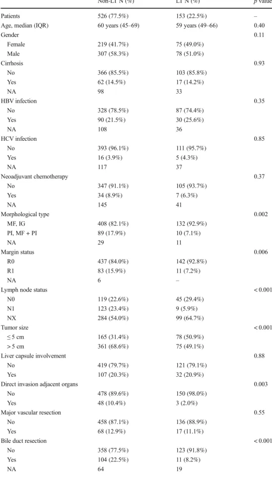
Among 1154 patients who underwent liver resection for ICC, overall actuarial survival at 5 and 10 years was 39.6 and 20.3%, respectively (Fig.1). In the entire cohort, the incidence of actual survivors was 13.3% (n = 153) at 5 years, and among these patients, the incidence of actual survivors at 10 years was 10.4% (n = 16). Excluding 475 patients who were alive with a follow-up < 5 years, the analytic cohort consisted of 153 patients (22.5%) in the long-term survival group (LT) vs. 526 patients (77.5%) who died within 5 years of surgery in the non-long-term survival group (non-LT).
Many clinical and demographic factors among LT and non-LT patients were comparable (Table1). For example, the pro-portion of men (non-LT, n = 788, 51.0% vs. LT, n = 306,
58.3%), median age (non-LT, 60 years, IQR, 45–69 vs. LT, 59 years, IQR, 49–66), as well as the incidence of cirrhosis (non-LT, n = 62, 14.5% vs. LT, n = 17, 14.2%), HBV (non-LT, n = 90, 21.5% vs. LT, n = 30, 25.6%), and HCV (non-LT, n = 16, 3.9% vs. LT, n = 5, 4.3%) were similar (all p > 0.05). In addition, utilization of preoperative chemothera-py was the same among non-LT (n = 7, 6.3%) and LT (n = 34, 8.9%) patients (p = 0.37). The use of adjuvant chemotherapy and radiotherapy also did not differ among non-LT and LT patients (adjuvant chemotherapy: non-LT group, n = 165, 33.3% vs. LT group, n = 52, 34.7%, p = 0.75) (adjuvant radiotherapy: non-LT: n = 30, 7.4% vs. LT, n = 7, 5.5%, p = 0.46).
A number of prognostic factors were, however, different among non-LT vs. LT patients. In particular, the incidence of periductal infiltrating/mass forming + periductal infiltrating (PI/MF + PI) tumor morphology and R1 surgical margin sta-tus was lower among LT patients compared with non-LT pa-tients (PI/MF + PI: non-LT: n = 89, 17.9% vs. LT, n = 11, 7.1%, p = 0.002) (R1: non-LT, n = 83, 15.9% vs. LT, n = 11, 7.2%, p = 0.006). Moreover, the incidence of N1 disease and tumor size > 5 cm was also lower in the LT vs. non-LT group (N1: non-LT group, n = 123, 23.4% vs. LT group, n = 9, 5.9%, p < 0.001) (tumor size > 5 cm: non-LT group, n = 361, 68.6% vs. LT group, n = 75, 49.1%, p < 0.001). ICC involvement of adjacent organs was present in 48 (12.9%) non-LT patients compared with 3 (2.2%) LT patients (p = 0.003). In addition, the incidence of microvascular (microvascular invasion: non-LT group, n = 171, 33.3% vs. non-LT group, n = 34, 22.2%, p = 0.009) and perineural invasion (perineural invasion: non-LT group, n = 111, 23.2% vs. LT group, n = 12, 8.8%, p < 0.001) (Fig. 2a) were lower in the LT group. Median values of CA 19-9 and CEA were also lower in the LT group than in the non-LT group (CA 19-9: non-LT group, 84.2, IQR, Fig. 1 Kaplan-Meier estimates of overall survival among the 1154 patients undergoing liver resection with curative intent for intrahepatic cholangiocarcinoma
Table 1 Clinical and pathologic
features of patients (n = 679) Non-LT N (%) LT N (%) p value
Patients 526 (77.5%) 153 (22.5%) –
Age, median (IQR) 60 years (45–69) 59 years (49–66) 0.40
Gender 0.11 Female 219 (41.7%) 75 (49.0%) Male 307 (58.3%) 78 (51.0%) Cirrhosis 0.93 No 366 (85.5%) 103 (85.8%) Yes 62 (14.5%) 17 (14.2%) NA 98 33 HBV infection 0.35 No 328 (78.5%) 87 (74.4%) Yes 90 (21.5%) 30 (25.6%) NA 108 36 HCV infection 0.85 No 393 (96.1%) 111 (95.7%) Yes 16 (3.9%) 5 (4.3%) NA 117 37 Neoadjuvant chemotherapy 0.37 No 347 (91.1%) 105 (93.7%) Yes 34 (8.9%) 7 (6.3%) NA 145 41 Morphological type 0.002 MF, IG 408 (82.1%) 132 (92.9%) PI, MF + PI 89 (17.9%) 10 (7.1%) NA 29 11 Margin status 0.006 R0 437 (84.0%) 142 (92.8%) R1 83 (15.9%) 11 (7.2%) NA 6 –
Lymph node status < 0.001
N0 119 (22.6%) 45 (29.4%) N1 123 (23.4%) 9 (5.9%) NX 284 (54.0%) 99 (64.7%) Tumor size < 0.001 ≤ 5 cm 165 (31.4%) 78 (50.9%) > 5 cm 361 (68.6%) 75 (49.1%)
Liver capsule involvement 0.88
No 419 (79.7%) 121 (79.1%)
Yes 107 (20.3%) 32 (20.9%)
Direct invasion adjacent organs 0.003
No 478 (89.6%) 150 (98.0%)
Yes 48 (10.4%) 3 (2.0%)
Major vascular resection 0.55
No 458 (87.1%) 136 (88.9%)
Yes 68 (12.9%) 17 (11.1%)
Bile duct resection < 0.001
No 358 (77.5%) 123 (91.8%)
Yes 104 (22.5%) 11 (8.2%)
23.7–400.0 vs. LT group, 25.5, IQR, 11.0–107.8, p < 0.001; CEA: non-LT group, 2.8, IQR, 1.6–5.4 vs. LT group, 1.8, IQR, 1.0–3.2, p < 0.001). More patients in the non-LT group had a poorly/undifferentiated tumor (n = 114, 23.3%) com-pared with LT patients (n = 18, 12.8%, p = 0.007; Table1).
After controlling for competing risk factors on multivari-able analysis, several factors remained strongly associated with LT (Table2). For example, patients with direct invasion of adjacent organs had an almost fourfold decreased odds of surviving to 5 years compared with patients without direct invasion of adjacent organs (OR 3.98, 95% CI, 1.18–13.4, p = 0.026) (Fig. 2b). Patients with intrahepatic metastasis (OR 3.75, 95% CI, 0.85–16.6, p = 0.082) (Fig.2c) and satel-lite lesions (OR 2.12, 95% CI, 1.15–3.90, p = 0.016) (Fig.2d) were also less likely to be LT survivors. Patients with an ICC > 5 cm had an increased risk to be in the non-LT group than in the LT group (OR 2.40, 95% CI, 1.54–3.74, p < 0.001)
(Fig.2e). Accordingly, in the Kaplan-Meier analysis, 5- and 10-year OS was 32 and 16%, respectively, for patients with ICC≥ 5 cm vs. 52 and 27% for patients with an ICC < 5 cm. Of note, N1 status (OR 4.64, 95% CI, 1.77–12.2; p = 0.002) and perineural invasion (OR 4.78, 95% CI, 1.92–11.8; p = 0.001) were the strongest independent predictors of poor prognosis and decreased likelihood of LT. In the LT group, while only three (2.0%) patients had intrahepatic metastasis and direct invasion of adjacent organs, 5% of patients in the LT group had satellite lesion (n = 18, 11.8%), perineural inva-sion (n = 12, 8.8%), N1 status (n = 9, 5.9%), and ICC≥ 5 cm (n = 75, 49.1%).
In comparing LT vs. non-LT patients, the AJCC 8th edition T1b, T2, and T4 patients were under-represented in LT group vs. non-LT group (T1b: non-LT group, n = 129, 80.1% vs. LT group, n = 32, 19.9%; T2: non-LT group, n = 205, 84.4% vs. LT group, n = 38, 15.6%; T4: non-LT group, n = 48, 94.1% vs. Table 1 (continued) Non-LT N (%) LT N (%) p value Grade 0.007 Well/moderate 377 (76.8%) 123 (87.2%) Poorly/undifferentiated 114 (23.2%) 18 (12.8%) NA 35 12 Microvascular invasion 0.009 No 342 (66.7%) 119 (77.8%) Yes 171 (33.3%) 34 (22.2%) NA 13 – Perineural invasion < 0.001 No 368 (76.8%) 125 (91.2%) Yes 111 (23.2%) 12 (8.8%) NA 47 16 Satellite lesion < 0.001 No 370 (70.9%) 135 (88.2%) Yes 152 (29.1%) 18 (11.8%) NA 4 – Intrahepatic metastasis 0.002 No 474 (90.6%) 150 (98.0%) Yes 49 (9.4%) 3 (2.0%) NA 3 – Ca 19–9, median (IQR) 84.2 (23.7–400.0) 25.5 (11.0–107.8) < 0.001
CEA, median (IQR) 2.8 (1.6–5.4) 1.8 (1.0–3.2) < 0.001
Adjuvant chemotherapy 0.75 No 331 (66.7%) 98 (65.3%) Yes 165 (33.3%) 52 (34.7%) NA 30 3 Adjuvant radiotherapy 0.46 No 378 (92.6%) 121 (94.5%) Yes 30 (7.4%) 7 (5.5%) NA 118 25 NA not available
LT group, n = 3, 5.9%) (all p < 0.05). In contrast, more LT patients were noted to be AJCC T1a and T3 than in T1b
and T2 (T1a: non-LT group, n = 71, 58.2% vs. LT group, n = 51, 41.8%; T3: non-LT group, n = 73, 71.6% vs. LT group, n = 29, 28.4%, p < 0.001) (Table3). When nodal status was restricted to patients who had at least six LNs harvested as recommended by the AJCC, the proportion of LT survivors who were N0 or NX was 31% (n = 16) and 25.8% (n = 128), respectively, whereas only a mi-nority of LT patients were N1 (n = 9, 6.8%) (Table 3). Only 158 (30.0% of 526 patients in the non-LT group) and 25 (16.3% of 153 patients in the LT group) patients in non-LT and LT groups, respectively, had an adequate nodal staging and were staged according to the AJCC 8th edition TNM staging system. The incidence of LT was 80% in stage Ia, 25% in stage Ib, 35% in stage II, 12% in stage III, and 7% in stage IV. In contrast, the incidence of non-LT was 93% in stage IIIb and decreased to only 20% in stage Ia (Table 3).
Fig. 2 Histogram showing the incidence of patients stratified by a perineural invasion, b invasion of adjacent organs, c intrahepatic metastasis, d satellite lesions, and e tumor size in the long-term vs. non-long-term groups
Table 2 Multivariable analysis
OR 95% CI p value
Intrahepatic metastasis 3.75 0.85–16.6 0.082
Direct invasion adjacent organs 3.98 1.18–13.4 0.026
Perineural invasion 4.78 1.92–11.8 0.001 Satellite lesion 2.12 1.15–3.90 0.016 Lymphnode status N0 – – – N1 4.64 1.77–12.2 0.002 NX 1.28 0.76–2.15 0.36 Size > 5 cm 2.40 1.54–3.74 < 0.001
Discussion
Although complete surgical resection remains the treatment of choice for patients with ICC, the prognosis of ICC re-mains unfavorable with 5-year survival ranging from 20 to 40%.23 Several studies have identified clinicopathological factors associated with long-term outcomes of patients un-dergoing liver surgery for ICC, such as preoperative CA 19-9 levels, tumor number and size, lymph node status, margin status, as well as vascular invasion.2, 13, 16, 24–30 Several predictive models have been applied to patients with ICC in order to better define prognosis.9, 26, 31 The most com-monly used staging system for ICC is the TNM classifica-tion system. In the recently released new 8th ediclassifica-tion of the AJCC TNM manual, several new revisions were introduced into the staging for ICC.8Specifically, in the 8th edition, T1 disease has been revised to include tumor size (≤ 5 cm vs. > 5 cm); T2 now reflects an equivalent prognostic value of vascular invasion and multifocal disease; while T4 disease is defined as involving local extrahepatic structures by direct invasion. Hyder et al. reported a prognostic nomogram for resectable ICC based on the clinicopathologic data of 367 patients with ICC that included six factors, such as age, tumor size, number of lesions, nodal status, vascular inva-sion, and presence of cirrhosis.9Most of these past studies focused, however, on short-term prognosis within 5 years of surgery. To our knowledge, no past studies has specifically focused on the actual long-term survivors following curative-intent resection of ICC to identify clinicopathologi-cal factors associated with long-term survival. The current study is important because it is one of the first studies to examine the incidence of actual long-term survivors in a
large, multi-center cohort of over 1000 patients undergoing surgery for ICC at 1 of 14 major hepatobiliary centers in the USA, Europe, Australia, and Asia, as well as measure the impact of clinicopathological factors on long-term survivors. When analyzing the data, it was interesting to note that analysis of actual survivors provided additional information compared with simple, standard Kaplan-Meier survival esti-mates. In particular, while the calculated actuarial OS at 5 years was roughly 40%, there were only 153 (13%) actual long-term survivors among the 1154 patients following curative-intent surgery. In turn, data from the current study provide more accurateBactual^ data on the long-term prog-nosis of patients undergoing curative intent surgery of ICC. While Kaplan-Meier analyses are helpful in estimating prog-nosis, this type of survival analysis may underestimate the effect of patients lost to follow-up and who are therefore censored in the analysis. Lost to follow-up may be particu-larly important among patients with ICC given the high risk of recurrence and death of disease in the first years follow-ing surgery for ICC.32 As such, patients lost to follow-up are likely to adversely impact any estimation of prognosis and the Kaplan-Meier method may overestimate true survival.14, 33 In turn, some groups, like our own, have proposed using nonmixture cure models as a means to bet-ter estimate the chance of statistical cure following surgical resection.15, 34, 35In fact, using a noncore statistical model, we had previously estimated that the overall probability of cure was approximately 10% for Ball comer^ patients un-dergoing hepatic resection for ICC. Interestingly, data from our previous statistical model (10%) very closely approxi-mated the 13% incidence of actual long-term survivors re-ported in the current study.
Table 3 Long-term survivors and AJCC staging system 8th edition
Non-LT N (%) LT N (%) p value OR 95% IC p value
AJCC T stages < 0.001 T1a 71 (58.2%) 51 (41.8%) – – – T1b 129 (80.1%) 32 (19.9%) 2.89 1.71–4.91 < 0.001 T2 205 (84.4%) 38 (15.6%) 3.87 2.35–6.38 < 0.001 T3 73 (71.6%) 29 (28.4%) 1.81 1.03–3.17 0.038 T4 48 (94.1%) 3 (5.9%) 11.5 3.39–38.9 < 0.001 AJCC N stages < 0.001 N0 35 (68.6%) 16 (31.4%) – – – N1 123 (93.2%) 9 (6.8%) 6.25 2.54–15.3 < 0.001 NX 368 (74.2%) 128 (25.8%) 1.31 0.70–2.45 0.39 AJCC TNM stages < 0.001 Ia 1 (16.7%) 5 (83.3%) – – – Ib 8 (72.7%) 3 (27.3%) 12.0 0.79–180.9 0.07 II 17 (70.8%) 7 (29.2%) 7.42 0.69–79.9 0.09 IIIa 9 (90.0%) 1 (10.0%) 28.0 1.35–580.6 0.031 IIIb 123 (93.2%) 9 (6.8%) 54.7 5.51–541.7 0.001
Perhaps not surprisingly, LT patients had many more favor-able prognostic factors compared with non-LT patients. In particular, patients who were in the LT group had a lower incidence of PI/MF + PI ICC vs. the non-LT group (p = 0.002). As our group recently reported, PI/MF + PI tu-mors were associated with more aggressive features than MF/ IG ICC.36Specifically, patients with a PI/MF + PI tumor had a 5-year OS of 25.5% vs. 41.8% for patients with a MF/IG ICC (p < 0.001). Also, the incidence of tumors≥ 5 cm was lower in the LT group, with over a twofold increased odds that patients with an ICC ≥ 5 cm were in the non-LT group (OR 2.40; p < 0.001). The correlation between tumor size and long-term outcome has been previously evaluated.24 Hyder et al. reported that tumor size was a prognostic factor for survival after surgical resection for ICC and noted that the association of tumor size on survival plateaued at 7 cm.9The effect of tumor size on outcome has been correlated to the underlying presence of microscopic vascular invasion and higher tumor grade in larger ICC.13In a study by Spolverato et al., one third and one half of patients with tumors measuring 7 to 15 and≥ 15 cm, respectively, had microscopic vascular invasion and one in three patients with tumors≥ 15 cm had evidence of major vascular invasion.13 Consistent with these previous findings, patients in the LT group were more likely to have well/moderately differentiated ICC and to have tumors with microvascular invasion. The importance of N status has also been reported in previous studies.7, 10, 11, 37–44In a recent meta-analysis, LN metastasis was associated with increased risk of death in pool data (HR 2.09),7and Kim et al. confirmed these findings reporting a similar relative risk of death associ-ated with LN metastasis (HR 2.42; p < 0.001).12In the current study, lymph node metastasis (OR 4.64; p = 0.002) was one of the strongest independent predictors of poor prognosis and decreased chance of long-term survival. In fact, when the AJCC recommended cut-off of six LNs harvested was applied to identify N0 patients, the incidence of LTs in N0 stage in-creased to 31%, which was higher than the incidence of LTs in either the N1 and NX categories. Collectively, these data serve to emphasize both the prognostic value of N status and the importance of adequate nodal staging.
LT patients were more likely to have T1a-T1b-T3 tumors (i.e., solitary tumor measuring≤ 5 cm (T1a); solitary tumor > 5 cm (T1b); tumor perforating the visceral peritoneum (T3)), rather than T2 tumors (i.e., solitary tumor with intrahepatic vascular invasion or multiple tumors, with or without vascular invasion). These data suggest that perforation of the visceral peritoneum may not carry as poor a prognostic impact as vascular invasion. In fact, Spolverato et al. previously reported that, while T1b patients had a better 5-year OS (37.3%) than T2 patients (21.3%), T3 patients paradoxically had a better 5-year OS than either of these lower T categories (45.8%).18As such, an advanced T stage tumor may not preclude long-term survival. Moreover, while stage T2 includes both patients with
intrahepatic metastases (multiple lesions in different seg-ments) and satellite lesions (dominant mass with nodules in same segment), in the current analyses, these two distinct pat-terns of multifocal disease were associated with different chances of LT survival (intrahepatic metastasis: OR 3.75; sat-ellite lesions: OR 2.12). Based on these results, stages T2 and T3 might need to be redefined to better stratify patient prognosis.
Another interesting finding of the current study was that traditional adverse prognostic factors did not categorically preclude LT survival. To this point, a subset of patients in the LT survival group were characterized by a number of tra-ditionally poor prognostic factors including R1 disease (7%), T3 tumors (19%), moderate/poor tumor differentiation (13%), and N1 disease (6%). These data emphasize how prognostic factors cannot be utilized to rule out the possibility of LT survival even in patients with predicted poor outcomes. To this end, several groups have reported on using conditional survival estimates to provide quantitative information about the changing probability of survival over time among patients with cancer.34, 35, 45, 46 Spolverato and colleagues reported that, while actuarial OS decreased over time from 39% at 3 years to 16% at 8 years, 3-year conditional increased over time among those patients who survived.15In fact, the 3-year conditional survival at 5 years—the probability of surviving to postoperative year 8 after having already survived to postop-erative year 5—was 65% compared with an 8-year actuarial OS estimate of 16%. Taken together, while certain factors may be strongly associated with LT survival, data in the current study, as well as previous data, demonstrate that LT can occur even in a subset of patients with traditional adverse prognostic factors.
The current study had several limitations. The multi-center nature of the study also did not allow for standardization of operative or perioperative approach, especially in terms of performance and extent of lymphadenectomy, neoadjuvant and adjuvant chemotherapy, and follow-up. Although this is a possible limitation, it is also a strength of the study as it contributes to the generalizability of the data. Due to the ret-rospective nature of the study, selection bias should be taken into account when interpreting the results; however, such con-founding was unlikely to impact the evaluation of the prog-nostic features of long-term survivors.
In conclusion, while ICC is generally associated with a poor prognosis, some patients can survive more than 5 years after surgery. The incidence of LT survivors following curative-intent surgery was low, as only one in ten patients actually survived past 5 years. Several pathological factors were associated with the likelihood of LT survivorship, yet LT survival did occur among a small subset of patients who had poor prognostic features. ICC is an aggressive disease with few LT survivors even after curative-intent surgery. Efforts should be aimed to better understand the pathogenesis
and molecular underpinnings of ICC in order to identify more effective systemic therapeutic agents. Only through the dis-covery and implementation of novel therapeutic approaches will we be able to improve the LT outcomes of patients with ICC.
References
1. El-Serag HB, Engels EA, Landgren O, Chiao E, Henderson L,
Amaratunge HC et al. Risk of hepatobiliary and pancreatic cancers after hepatitis C virus infection: a population-based study of U.S.
veterans. Hepatology. 2009;49(1):116–23.https://doi.org/10.1002/
hep.22606.
2. Endo I, Gonen M, Yopp AC, Dalal KM, Zhou Q, Klimstra D et al.
Intrahepatic cholangiocarcinoma: rising frequency, improved sur-vival, and determinants of outcome after resection. Annals of
Surgery. 2008; 248(1):84–96. https://doi.org/10.1097/SLA.
0b013e318176c4d3.
3. Malhi H, Gores GJ. Cholangiocarcinoma: modern advances in
un-derstanding a deadly old disease. Journal of Hepatology. 2006;
45(6):856–67.https://doi.org/10.1016/j.jhep.2006.09.001.
4. Shaib YH, Davila JA, McGlynn K, El-Serag HB. Rising incidence
of intrahepatic cholangiocarcinoma in the United States: a true
in-crease? Journal of Hepatology. 2004; 40(3):472–7.https://doi.org/
10.1016/j.jhep.2003.11.030.
5. Welzel TM, McGlynn KA, Hsing AW, O’Brien TR, Pfeiffer RM.
Impact of classification of hilar cholangiocarcinomas (Klatskin tu-mors) on the incidence of intra- and extrahepatic cholangiocarcino-ma in the United States. Journal of the National Cancer Institute.
2006; 98(12):873–5.https://doi.org/10.1093/jnci/djj234.
6. Khan SA, Toledano MB, Taylor-Robinson SD. Epidemiology, risk
factors, and pathogenesis of cholangiocarcinoma. HPB: the Official Journal of the International Hepato Pancreato Biliary Association.
2008; 10(2):77–82.https://doi.org/10.1080/13651820801992641.
7. Mavros MN, Economopoulos KP, Alexiou VG, Pawlik TM.
Treatment and prognosis for patients with intrahepatic cholangio-carcinoma: systematic review and meta-analysis. JAMA Surgery.
2014.https://doi.org/10.1001/jamasurg.2013.5137.
8. Amin MB EiC, American Joint Committee on Cancer. Springer.
2017.
9. Hyder O, Marques H, Pulitano C, Marsh JW, Alexandrescu S,
Bauer TW et al. A nomogram to predict long-term survival after resection for intrahepatic cholangiocarcinoma: an Eastern and
Western experience. JAMA Surgery. 2014.https://doi.org/10.
1001/jamasurg.2013.5168.
10. Amini N, Ejaz A, Spolverato G, Maithel SK, Kim Y, Pawlik TM.
Management of lymph nodes during resection of hepatocellular carcinoma and intrahepatic cholangiocarcinoma: a systematic re-view. Journal of Gastrointestinal Surgery: Official Journal of the
Society for Surgery of the Alimentary Tract. 2014; 18(12):2136–
48.https://doi.org/10.1007/s11605-014-2667-1.
11. Bagante F, Gani F, Spolverato G, Xu L, Alexandrescu S, Marques
HP et al. Intrahepatic cholangiocarcinoma: prognosis of patients who did not undergo lymphadenectomy. Journal of the American
College of Surgeons. 2015; 221(6):1031–40 e4.https://doi.org/10.
1016/j.jamcollsurg.2015.09.012.
12. Kim Y, Spolverato G, Amini N, Margonis GA, Gupta R, Ejaz A
et al. Surgical Management of intrahepatic cholangiocarcinoma: defining an optimal prognostic lymph node stratification schema.
Annals of Surgical Oncology. 2015; 22(8):2772–8.https://doi.org/
10.1245/s10434-015-4419-1.
13. Spolverato G, Ejaz A, Kim Y, Sotiropoulos GC, Pau A,
Alexandrescu S et al. Tumor size predicts vascular invasion and histologic grade among patients undergoing resection of intrahepatic cholangiocarcinoma. Journal of Gastrointestinal Surgery: Official Journal of the Society for Surgery of the
Alimentary Tract. 2014; 18(7):1284–91.https://doi.org/10.1007/
s11605-014-2533-1.
14. Spolverato G, Kim Y, Ejaz A, Alexandrescu S, Marques H,
Aldrighetti L et al. Conditional probability of long-term survival after liver resection for intrahepatic cholangiocarcinoma: a multi-institutional analysis of 535 patients. JAMA Surgery. 2015; 150(6): 538–45.https://doi.org/10.1001/jamasurg.2015.0219.
15. Spolverato G, Vitale A, Cucchetti A, Popescu I, Marques HP,
Aldrighetti L et al. Can hepatic resection provide a long-term cure for patients with intrahepatic cholangiocarcinoma? Cancer. 2015;
121(22):3998–4006.https://doi.org/10.1002/cncr.29619.
16. Spolverato G, Yakoob MY, Kim Y, Alexandrescu S, Marques HP,
Lamelas J et al. The impact of surgical margin status on long-term outcome after resection for intrahepatic cholangiocarcinoma.
Annals of Surgical Oncology. 2015.https://doi.org/10.1245/
s10434-015-4472-9.
17. Spolverato G, Yakoob MY, Kim Y, Alexandrescu S, Marques HP,
Lamelas J et al. Impact of complications on long-term survival after resection of intrahepatic cholangiocarcinoma. Cancer. 2015;
121(16):2730–9.https://doi.org/10.1002/cncr.29419.
18. Spolverato G BF, Weiss, M, Alexandrescu S, Marques HP,
Aldrighetti L, Maithel SK, Pulitano C, Bauer TW, Shen F, Poultsides GA, Soubrane O, Martel G, Koerkamp BG, Guglielmi A, Itaru E, Pawlik TM. Comparative performances of the 7th and the 8th editions of the American Joint Committee on Cancer Staging Systems for intrahepatic cholangiocarcinoma. Journal of Surgical Oncology.
19. Ronnekleiv-Kelly SM, Pawlik TM. Staging of intrahepatic
cholan-giocarcinoma. Hepatobiliary Surg Nutr. 2017; 6(1):35–43.https://
doi.org/10.21037/hbsn.2016.10.02.
20. Ferrone CR, Brennan MF, Gonen M, Coit DG, Fong Y, Chung S
et al. Pancreatic adenocarcinoma: the actual 5-year survivors. Journal of Gastrointestinal Surgery: Official Journal of the Society
for Surgery of the Alimentary Tract. 2008; 12(4):701–6.https://doi.
org/10.1007/s11605-007-0384-8.
21. Zheng J, Kuk D, Gonen M, Balachandran VP, Kingham TP, Allen
PJ et al. Actual 10-year survivors after resection of hepatocellular
carcinoma. Annals of Surgical Oncology. 2017; 24(5):1358–66.
https://doi.org/10.1245/s10434-016-5713-2.
22. Baheti AD, Tirumani SH, Shinagare AB, Rosenthal MH, Hornick
JL, Ramaiya NH et al. Correlation of CT patterns of primary intrahepatic cholangiocarcinoma at the time of presentation with the metastatic spread and clinical outcomes: retrospective study of
92 patients. Abdominal Imaging. 2014; 39(6):1193–201.https://
doi.org/10.1007/s00261-014-0167-0.
23. De Jong MC, Nathan H, Sotiropoulos GC, Paul A, Alexandrescu S,
Marques H et al. Intrahepatic cholangiocarcinoma: an international multi-institutional analysis of prognostic factors and lymph node assessment. Journal of Clinical Oncology: Official Journal of the
American Society of Clinical Oncology. 2011; 29(23):3140–5.
https://doi.org/10.1200/jco.2011.35.6519.
24. Dodson RM, Weiss MJ, Cosgrove D, Herman JM, Kamel I, Anders
R et al. Intrahepatic cholangiocarcinoma: management options and emerging therapies. Journal of the American College of Surgeons.
2013; 217(4):736–50e4.https://doi.org/10.1016/j.jamcollsurg.
2013.05.021.
25. de Jong MC, Pulitano C, Ribero D, Strub J, Mentha G, Schulick RD
et al. Rates and patterns of recurrence following curative intent surgery for colorectal liver metastasis: an international multi-institutional analysis of 1669 patients. Annals of Surgery. 2009;
26. Nathan H, Aloia TA, Vauthey JN, Abdalla EK, Zhu AX, Schulick RD et al. A proposed staging system for intrahepatic
cholangiocar-cinoma. Annals of Surgical Oncology. 2009; 16(1):14–22.https://
doi.org/10.1245/s10434-008-0180-z.
27. Spolverato G, Kim Y, Alexandrescu S, Popescu I, Marques HP,
Aldrighetti L et al. Is hepatic resection for large or multifocal intrahepatic cholangiocarcinoma justified? Results from a multi-institutional collaboration. Annals of Surgical Oncology. 2014.
https://doi.org/10.1245/s10434-014-4223-3.
28. DeOliveira ML, Cunningham SC, Cameron JL, Kamangar F,
Winter JM, Lillemoe KD et al. Cholangiocarcinoma: thirty-one-year experience with 564 patients at a single institution. Annals of
Surgery. 2007; 245(5):755–62.https://doi.org/10.1097/01.sla.
0000251366.62632.d3.
29. Nakagohri T, Kinoshita T, Konishi M, Takahashi S, Gotohda N.
Surgical outcome and prognostic factors in intrahepatic cholangio-carcinoma. World Journal of Surgery. 2008; 32(12):2675–80.
https://doi.org/10.1007/s00268-008-9778-3.
30. Hatzaras I, Schmidt C, Muscarella P, Melvin WS, Ellison EC,
Bloomston M. Elevated CA 19-9 portends poor prognosis in pa-tients undergoing resection of biliary malignancies. HPB: the Official Journal of the International Hepato Pancreato Biliary
Association. 2010; 12(2):134–8.
https://doi.org/10.1111/j.1477-2574.2009.00149.x.
31. Nathan H, Pawlik TM. Staging of intrahepatic cholangiocarcinoma.
Current Opinion in Gastroenterology. 2010; 26(3):269–73.https://
doi.org/10.1097/MOG.0b013e328337c899.
32. Hyder O, Hatzaras I, Sotiropoulos GC, Paul A, Alexandrescu S,
Marques H et al. Recurrence after operative management of intrahepatic cholangiocarcinoma. Surgery. 2013; 153(6):811–8.
https://doi.org/10.1016/j.surg.2012.12.005.
33. Bollschweiler E. Benefits and limitations of Kaplan-Meier
calcula-tions of survival chance in cancer surgery. Langenbeck’s Archives
of Surgery/Deutsche Gesellschaft fur Chirurgie. 2003; 388(4):239– 44.https://doi.org/10.1007/s00423-003-0410-6.
34. Bagante F, Spolverato G, Merath K, Postlewait LM, Poultsides GA,
Mullen MG et al. Neuroendocrine liver metastasis: the chance to be
cured after liver surgery. J Surg Oncol. 2017.https://doi.org/10.
1002/jso.24563.
35. Spolverato G, Bagante F, Ethun CG, Poultsides G, Tran T, Idrees K
et al. Defining the chance of statistical cure among patients with
extrahepatic biliary tract cancer. World J Surg. 2017; 41(1):224–31.
https://doi.org/10.1007/s00268-016-3691-y.
36. Bagante F, Spolverato G, Weiss M, Alexandrescu S, Marques HP,
Aldrighetti L, Maithel SK, Pulitano C, Bauer TW, Shen F, Poultsides GA, Soubrane O, Martel G, Koerkamp BG, Guglielmi A, Itaru E, Pawlik TM. Impact of morphological status on
long-term outcome among patients undergoing liver surgery for intrahepatic cholangiocarcinoma. Ann Surg Oncol. 2017; 24:
2491–2501.
37. Adachi T, Eguchi S. Lymph node dissection for intrahepatic
chol-angiocarcinoma: a critical review of the literature to date. Journal of
Hepato-Biliary-Pancreatic Sciences. 2014; 21(3):162–8.https://doi.
org/10.1002/jhbp.30.
38. Ribero D, Pinna AD, Guglielmi A, Ponti A, Nuzzo G, Giulini SM
et al. Surgical approach for long-term survival of patients with intrahepatic cholangiocarcinoma: a multi-institutional analysis of 434 patients. Archives of Surgery. 2012; 147(12):1107–13.
39. Ercolani G, Vetrone G, Grazi GL, Aramaki O, Cescon M, Ravaioli
M et al. Intrahepatic cholangiocarcinoma: primary liver resection and aggressive multimodal treatment of recurrence significantly prolong survival. Annals of Surgery. 2010; 252(1):107–14.
https://doi.org/10.1097/SLA.0b013e3181e462e6.
40. Saxena A, Chua T, Sarkar A, Chu F, Morris D. Clinicopathologic
and treatment-related factors influencing recurrence and survival after hepatic resection of intrahepatic cholangiocarcinoma: a 19-year experience from an established Australian Hepatobiliary Unit. Journal of Gastrointestinal Surgery. 2010; 14(7):1128–38.
https://doi.org/10.1007/s11605-010-1203-1.
41. Guglielmi A, Ruzzenente A, Campagnaro T, Pachera S,
Valdegamberi A, Nicoli P et al. Intrahepatic cholangiocarcinoma: prognostic factors after surgical resection. World Journal of
Surgery. 2009; 33(6):1247–54.
https://doi.org/10.1007/s00268-009-9970-0.
42. Yedibela S, Demir R, Zhang W, Meyer T, Hohenberger W,
Schonleben F. Surgical treatment of mass-forming intrahepatic cholangiocarcinoma: an 11-year western single-center experience in 107 patients. Annals of Surgical Oncology. 2009; 16(2):404–12.
43. Uenishi T, Kubo S, Yamazaki O, Yamada T, Sasaki Y, Nagano H
et al. Indications for surgical treatment of intrahepatic cholangiocar-cinoma with lymph node metastases. Journal of
Hepato-Biliary-Pancreatic Surgery. 2008; 15(4):417–22.https://doi.org/10.1007/
s00534-007-1315-5.
44. Yamashita Y, Taketomi A, Morita K, Fukuhara T, Ueda S, Sanefuji
K et al. The impact of surgical treatment and poor prognostic factors for patients with intrahepatic cholangiocarcinoma: retrospective
analysis of 60 patients. Anticancer Research. 2008; 28(4C):2353–9.
45. Rama R, Swaminathan R, Venkatesan P. Cure models for
estimat-ing hospital-based breast cancer survival. Asian Pac J Cancer Prev.
2010; 11(2):387–91.
46. Rasouli M, Ghadimi MR, Mahmoodi M, Mohammad K, Zeraati H,
Hosseini M. Survival analysis of patients with esophageal cancer using parametric cure model. Asian Pac J Cancer Prev. 2011; 12(9):