2013/2014
Pedro Fernando Magalhães Valente
Epithelial dysplasia of the stomach
with gastric immunophenotype shows
features of biological aggressiveness
Mestrado Integrado em Medicina
Área: Anatomia Patológica
Trabalho efetuado sob a Orientação de: Doutora Maria de Fátima Machado Henriques Carneiro
Trabalho organizado de acordo com as normas da revista: Gastric Cancer
Pedro Fernando Magalhães Valente
Epithelial dysplasia of the stomach
with gastric immunophenotype shows
features of biological aggressiveness
Agradecimentos
Esta dissertação que marca o fim de um ciclo da minha formação académica não teria sido possível sem a colaboração e apoio de muitas pessoas às quais não poderia deixar de exprimir a minha mais sincera gratidão:
À Professora Doutora Fátima Carneiro, que me abriu a porta do Serviço de Anatomia Patológica e amavelmente aceitou orientar este projecto de investigação, tornando-o uma experiência pedagógica e pessoal verdadeiramente enriquecedora.
Ao Serviço de Anatomia Patológica, em especial aos técnicos Bárbara Gomes, Helena Pereira, Armando Castro, Armindo Pereira e Manuel Moutinho.
Ao Dr. Franclim Ribeiro por toda a formação e disponibilidade paciente.
A todos que, de alguma forma, contribuíram para esta dissertação, nomeadamente a Dr.ª Irene Gullo, Dr.ª Helena Baldaia, Dr. Francisco Baldaque-Silva e Dr.ª Joanne Lopes.
À Mónica, que me motivou para a realização conjunta deste projecto e pela construção partilhada de um futuro em comum.
A todos os meus,
Original article
Epithelial dysplasia of the stomach with gastric immunophenotype shows features of biological aggressiveness
Pedro Valente1*, Mónica Garrido1*, Irene Gullo1,2, Helena Baldaia3, Margarida Marques3, Francisco Baldaque-Silva3,4, Joanne Lopes3, Fátima Carneiro1,3,5
1
Faculty of Medicine of the University of Porto, Porto, Portugal
2
Faculty of Medicine and Surgery, Genova, Italy
3
Centro Hospitalar de São João, Porto, Portugal
4
Department of Gastroenterology and Hepatology, Karolinska University Hospital, Stockholm, Sweden
5
Institute of Molecular Pathology and Immunology of the University of Porto (IPATIMUP), Porto, Portugal
*contributed equally
Figures: 2 Tables: 5
Short title: Aggressiveness of gastric-type dysplasia Word Count: 2870
Potential competing interest: The authors have no competing interest to declare.
Correspondence to:
Fátima Carneiro, M.D., Ph.D.
Institute of Molecular Pathology and Immunology, University of Porto (IPATIMUP)
Rua Dr. Roberto Frias S/N, 4200-465 Porto, Portugal Tel.: +351 225570700
Fax: +351 225570799
ABSTRACT
Background: Gastric dysplasia is classified as adenomatous/type I (intestinal
phenotype) and foveolar or pyloric/type II (gastric phenotype) according to
morphological (architectural and cytological) features. The immunophenotypic
classification of dysplasia, based on the expression of mucins, CD10 and
CDX2, recognizes the following immunophenotypes: intestinal (MUC2, CD10
and CDX2); gastric (MUC5AC and/or MUC6, absent of CD10 and absent or low
expression of CDX2); hybrid (gastric and intestinal markers) and null.
Methods: Sixty-six cases of non-polypoid epithelial dysplasia of the stomach
were classified according to morphological features (histotype and grade) and
immunophenotype. Immunohistochemical staining was performed with
antibodies against MUC2, MUC5AC, MUC6, CD10, CDX2, chromogranin,
synaptophysin, Ki-67 and TP53. HER2 alterations were analysed by
immunohistochemistry and silver-enhanced in situ hybridization (SISH).
Results: By conventional histology, dysplasia was classified as
adenomatous/intestinal (n=42; 64%) and foveolar or pyloric/gastric (n=24; 36%)
and graded as low-grade (n=37; 56%) and high-grade (n=29; 44%).
Immunophenotypic classification showed intestinal (n=22; 33.3%), gastric
(n=25; 37.9%), hybrid (n=17; 25.8%) or null (n=2; 3.0%) phenotypes. In 20
cases a coexistent intramucosal carcinoma was identified.
The intestinal immunophenotype was shown to be significantly associated with
low-grade dysplasia (p=0.001), high expression of CDX2 (p=0.015), TP53
(p=0.034), synaptophysin (p=0.003) and chromogranin (p<0.0001); the gastric
immunophenotype was significantly associated with high-grade dysplasia
intramucosal carcinoma (p=0.013). HER2 amplification was observed in 3
cases, typed as gastric or hybrid.
Conclusions: Epithelial non-polypoid dysplasia of the stomach with gastric
immunophenotype shows features of biological aggressiveness and may
represent the putative precursor lesion in a pathway of gastric carcinogenesis
originated de novo from the native gastric mucosa, leading to gastric type
adenocarcinoma.
MINI ABSTRACT
Epithelial dysplasia of the stomach encompasses two major
immunophenotypes, intestinal and gastric, the latter significantly associated with
features of biological aggressiveness: high-grade, high proliferative index and
coexistent carcinoma.
Key words: Gastric carcinogenesis; dysplasia; HER2; immunophenotype; mucins.
INTRODUCTION
At present, gastric carcinoma (GC) has a significant morbi-mortality impact,
being the fourth most incident cancer worldwide and the second deadliest one
(1).
According to Laurén's classification (2), there are two main subtypes of
GC - intestinal and diffuse - that differ in epidemiology, pathogenesis,
morphology and molecular features (2, 3). According to the Correa model,
gastric cancer develops along a cascade of lesions encompassing Helicobacter
pylori induced chronic superficial gastritis, chronic atrophic gastritis, intestinal metaplasia, dysplasia, and ultimately invasive adenocarcinoma (4). Gastric
dysplasia is neoplastic in nature and is a direct precursor of gastric carcinoma,
as well as a risk factor of carcinoma in other locations of the stomach (5, 6).
Dysplasia is graded as low- and high-grade on the basis of architectural
and cell features. Further, according to the histomorphological profile, dysplasia
may be classified as adenomatous/type I (intestinal phenotype) and foveolar or
pyloric/type II (gastric phenotype). The two types may be distinguished by the
immunoexpression of mucins, CD10 and CDX2 (intestinal/adenomatous: MUC2, CD10, and CDX2; gastric/foveolar: MUC5AC and/or MUC6, absence of CD10 and low or absent expression of CDX2) (7-9). Cases with hybrid differentiation may also occur as well as null cases in which there is no expression of the aforementioned markers (8).
A relationship has been reported between the histological grade and the
immunohistochemical profiles of dysplasia: in one study, 81.8% of low-grade
dysplasia expressed intestinal markers, and 72.2% of high-grade dysplasia
markers (10). In another study, foveolar and hybrid subtypes were also
significantly associated with high-grade dysplasia (8).
In this study we aimed at analysing the relationship between different
types of gastric dysplasia (based on histotypes and grading) and the
immunohistochemical profile according to the expression of markers of cell
differentiation (MUC5AC, MUC6, MUC2, CD10). The expression of CDX2,
Ki-67, TP53, HER2 and neuroendocrine markers (chromogranin and
synaptophysin) was also evaluated.
MATERIALS AND METHODS
A series of 66 cases of non-polypoid epithelial dysplasia of the stomach
identified in Endoscopic Submucosal Dissection (ESD) specimens were
retrieved retrospectively from the files of the Department of Pathology, Centro
Hospitalar São João, between June/2010 and June/2013. In 20 cases a
coexistent intramucosal carcinoma was identified. The study was approved by
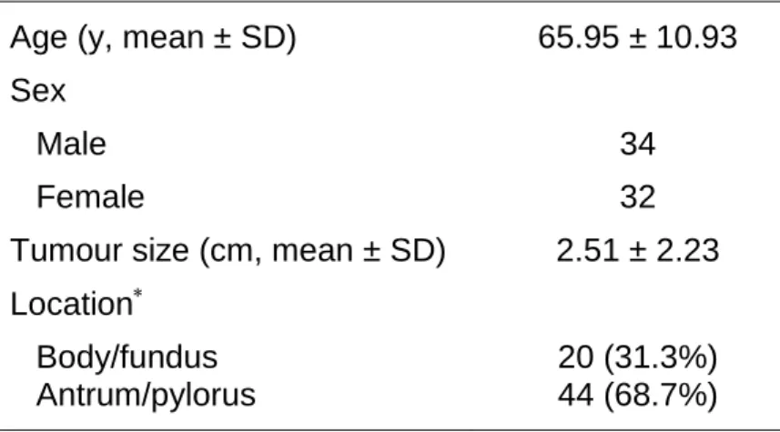
the Ethical Committee of the Hospital. The clinicopathological features of the
cases are summarized in Table 1.
Tissues were fixed in neutral buffered 10% formalin, embedded in
paraffin and cut into 3 µm-thick sections.
The lesions were classified in H&E stained slides as: adenomatous/type I
(intestinal phenotype) and foveolar or pyloric/type II (gastric phenotype),
according to the WHO classification (3). The adenomatous/intestinal subtype
resembles colonic adenomas, with crowded, tubular glands lined by atypical columnar cells with overlapping, pencillate, hyperchromatic and/or pleomorphic nuclei, with pseudostratification and mucin. The foveolar or pyloric/gastric
phenotype is characterized by cuboid/low columnar cells, with round to oval
nuclei and clear or eosinophilic cytoplasm.
The grade of the dysplasia was evaluated according to WHO 2010
criteria (3): low-grade dysplasia shows minimal architectural disarray and only
mild to moderate cytological atypia; the nuclei are elongated/oval, polarized and
basally-located and the mitotic activity is mild to moderate; high-grade dysplasia
presents pronounced architectural disarray, such as complex branching or
fusion of glands; the neoplastic cells are usually cuboidal, rather than columnar,
with a high nuclear/cytoplasm ratio, high number of mitoses, occasionally
atypical, and nuclei within the luminal zone of the epithelium with loss of
polarity. The diagnosis of invasive carcinoma was performed when invasion of
the lamina propria or deeper occurred.
Immunohistochemical staining was performed with antibodies against
MUC2, MUC5AC, MUC6, CD10, CDX2, chromogranin, synaptophysin, Ki-67,
HER2 and TP53 (Table 2). Samples were processed in the automatic
equipment Benchmark ULTRA using the Ultraview Universal DAB kit (Ventana
Medical Systems, Roche group). Each sample was heated and deparaffinized,
followed by antigen recovery through heat and high-pH buffer solution. Each
primary antibody was incubated in an individually optimized time and
temperature, followed by application of the detection system and contrast with
hematoxilin and bluing reagent from the same manufacturer.
Immunoreactivity was scored as follows: the immunoexpression of
MUC2, MUC5AC, MUC6, CD10, synaptophysin and chromogranin was scored as positive when ≥5% of the dysplastic cells displayed immunoreactivity; HER2
immunoreactivity; 1+ – tumour cell cluster with faint or barely perceptible
membrane reactivity irrespective of percentage of immunoreactive cells; 2+ –
tumour cell cluster with weak to moderate (complete, lateral or basolateral)
reactivity irrespective of the percentage of immunoreactive cells; 3+ – tumour
cell cluster with moderate to strong (complete, lateral or basolateral) reactivity
irrespective of the percentage of immunoreactive cells; for scoring purposes any
nuclear or cytoplasmatic backgroung staining was disregarded. The
immunoexpression of CDX2 was considered positive when ≥25% of the
dysplatic cells displayed immunoreactivity (9); immunoexpression of Ki-67 and
TP53 was classified as absent/low when immunoreactivity was displayed in
<50% of the dysplastic cells, and high in the presence of ≥50% positive cells
(12).
The detection of the number of copies of the HER2 gene was performed
in the cases scored as 2+ and 3+ by immunohistochemistry with SISH automatized technique using the BenchMark XT equipment and the INFORM™
HER2 SISH probe, manufactured by Ventana Medical Systems. The
HER2/Chr17 ratio of each case was calculated using a minimum of 40 cells in two independent areas of dysplasia. Cases were assigned a score based on the
ASCO/CAP guideline recommendations for HER2 testing in breast cancer as
follows (13): negative - ratio HER2:Cr17<2.0 with <4 copies of HER2 gene; borderline - ratio HER2:Cr17<2.0 with ≥4 and <6 copies of HER2 gene; positive
Statistical Analysis
Appropriate statistical methods were used regarding the type of sample and its
distribution. The data was analysed with SPSS software v. 19.0 (SPSS Software, Chicago, IL, USA), using chi-square or Fischer’s test. P-value <0.05
was considered statistically significant.
RESULTS
The study group was composed of 66 cases (Table 1), classified by
conventional histology in H&E stained slides as foveolar or pyloric/gastric
(n=24; 36%) or adenomatous/intestinal (n=42; 64%) (Fig.1) and graded as
low-grade (n=37; 56%) or high-low-grade (n=29; 44%). According to the
immunophenotype, the cases were classified as gastric type (n=25; 37.9%) (Fig. 2 – a, c, e, g, i), intestinal (n=22; 33.3%) (Fig. 2 – b, d, f, h, j), hybrid (n=17;
25.8%) or null (n=2, 3.0%). The latter were not considered for subsequent
analysis.
Table 3 summarizes the expression of the different markers in the three
immunophenotypes of gastric dysplasia. Statistically significant differences were
observed between the immunophenotypes regarding the expression of MUC2
(p=0.002), CD10 (p<0.0001), MUC5AC (p<0.0001) and MUC6 (p<0.0001).
Cases with low/absent expression of CDX2 were observed only in the gastric
immunophenotype (p=0.015).
The frequency of cases with high expression of Ki-67 was significantly
higher in the gastric and hybrid (84.0% and 94.1%, respectively) than in the
The frequency of cases with high expression of TP53 was significantly
higher in the intestinal and hybrid (52.9% and 52.9%, respectively) than in the
gastric (16.0%) immunophenotypes (p=0.034).
The expression of HER2 (2+ and 3+) was observed in 11 cases with
gastric or hybrid immunophenotypes (28.0% and 23.5%, respectively) and not
detected in the intestinal immunophenotype (p=0.029).
Amplification of HER2 gene was observed only in three cases,
immunophenotyped as gastric (n=1) and hybrid (n=2).
Regarding the neuroendocrine markers, the frequency of the expression
of synaptophysin was significantly higher in intestinal (81.8%) than in hybrid and
gastric (58.8% and 32.0%, respectively) immunophenotypes (p=0.003). Similar
observations were made for the expression of chromogranin, displayed
predominantly in intestinal (95.2%) in comparison with hybrid and gastric
(70.6% and 28.0%, respectively) immunophenotypes (p<0.0001). In some
cases, immunophenotyped as intestinal, small nests of neuroendocrine cells
were observed.
Table 4 shows the relationship between the immunophenotypes and the
histotypes of gastric dysplasia (adenomatous/intestinal and foveolar or
pyloric/gastric), and grade (low- and high-grade). The frequency of high-grade
dysplasia was significantly higher in the gastric immunopenotype (68.0%) than
in the other immunophenotypes (47.1% and 13.6%, in hybrid and intestinal,
respectively). Within cases with intestinal immunophenotype, dysplasia was
graded as low in most cases (86.4%) (p=0.001). Gastric immunophenotype
encompassed cases classified by conventional histology as gastric (72.0%) and
histology as gastric (35.3%) and intestinal (64.7%) and all cases of the intestinal
immunophenotype displayed features of the adenomatous/intestinal histotype
(p<0.0001).
Table 5 shows the relationship between the presence of the coexistent
intramucosal adenocarcinoma and the features of dysplasia (grade, histo and
immunophenotypes). In 20 of 66 cases (30.3%), there was a coexistent
carcinoma at the periphery of the dysplastic lesions, the latter displaying the
following features: high-grade (75.0%; p=0.001); gastric histotype (60.0%;
p=0.024); gastric immunophenotype (65.0%, 20.0% and 15.0% for gastric,
hybrid and intestinal immunophenotypes, respectively; p=0.013). Gastric
dysplasia at the periphery of invasive carcinoma, when compared with gastric
dysplasia in the absence of invasive carcinoma, displayed significantly lower
frequency of expression of synaptophysin and chromogranin (30.0% and
40.0%; p=0.006 and p=0.025, respectively).
DISCUSSION
Gastric carcinogenesis is a complex process, still requiring the elucidation of
putative distinct pathways. According to the so-called Correa model (4), gastric
carcinogenesis is a multistep and multifactorial process that, in many cases,
appears to involve a progression from normal mucosa, through chronic atrophic
gastritis and intestinal metaplasia, to dysplasia and invasive carcinoma.
However, evidence from literature points to the possibility of the existence of
alternative pathways in which intestinal metaplasia may not play a role.
Evidence stems mainly from the study of tiny early gastric carcinomas arising in
the studies of the expression of markers of gastric differentiation in dysplasia
and gastric adenocarcinoma (7, 10, 16, 17). The latter demonstrate that both
types of lesions may express, predominantly or exclusively, markers of gastric
differentiation, raising the possibility of an origin in native gastric mucosa, rather
than in intestinal metaplastic lesions. It remains to be elucidated the role of
spasmolytic polypeptide-expressing metaplasia (SPEM) in the pathogenesis of
the lesions with gastric immunophenotype. Other evidences stem from
hereditary gastric cancer models (Hereditary Diffuse Gastric Cancer and Gastric Adenocarcinoma and Proximal Polyposis of the Stomach – HDGC and GAPPS)
in which gastric carcinoma, diffuse/poorly cohesive type and intestinal/tubular
types, respectively, originate in non-metaplastic gastric epithelium (fundic gland
polyps in the case of GAPPS) (18, 19).
Our study provides additional evidence in favour of de novo neoplastic
transformation from native gastric mucosa (37.9% of the dysplastic lesions displayed “pure” gastric immunophenotype).
Another relevant issue is the risk of malignant transformation of the
different types of gastric dysplasia. Our results show that within the group of
cases immunophenotyped as gastric, the majority were classified as high-grade
dysplasia (68.0%; p=0.001). At variance, within cases immunophenotyped as
intestinal, low-grade dysplasia was the most frequent (86.4%; p=0.001). These
findings are in keeping with those recently reported by Nishimura et al (14), but
differ from the results reported by Abraham et al (20), the latter showing that
intestinal-type adenomas were more likely than gastric-type adenomas to
display high-grade dysplasia and adenocarcinoma in the polyps. A major
Abraham et al (20) was constituted by polypoid adenomas, our series is
constituted by non-polypoid dysplasia.
HER2 amplification was observed in three cases, immunophenotyped as gastric or hybrid. These findings show that HER2 amplification may be an early
event in gastric carcinogenesis as observed by Fassan et al (11).
The results herein obtained in dysplasia with gastric immunophenotype
(higher frequency of high-grade lesions, expression and amplification of HER2)
suggest that this type of dysplasia may be an important player in gastric
carcinogenesis.
The high frequency of cases with high proliferative index (Ki-67) in
gastric and hybrid immunophenotypes (84.0% and 94.1%, respectively) when
compared with the intestinal immunophenotype (63.6%; p=0.05) is in keeping
with the features of aggressiveness identified in dysplastic lesions with gastric
differentiation. At variance with other studies (21, 22) we have not found a
significant difference in the Ki-67 proliferation index according to the grade of
dysplasia.
In this study, we observed that the expression of CDX2 is correlated with
the intestinal immunophenotype (100% of the cases), in keeping with data
previously reported (9). In accordance with Park et al (9), a decreased
expression of CDX2 was observed in cases with gastric immunophenotype
(80% of the cases; decreased intensity of immunoreactivity). However, there is
controversy in the literature regarding the expression of CDX2 in gastric
dysplasia, probably reflecting the lack of sub-typing of dysplasia in the different
In some cases with intestinal immunophenotype small nests of
neuroendocrine cells were observed, qualifying for neuroendocrine hyperplasia
as reported in the literature (25, 26). It is likely that adenomatous/intestinal
dysplasia and neuroendocrine hyperplasia both arise in the setting of chronic
atrophic gastritis, as previously suggested in neuroendocrine hyperplasia within
gastric hyperplastic polyps (27). However, further studies are needed to
elucidate the biological meaning of this event.
In the present study we observed that higher expression of TP53
significantly correlated with the intestinal immunophenotype (p=0.034) and was
also more frequently observed in high-grade dysplasia, though this association
was not significant (p=0.070 – data not shown). In previous studies, it was
observed an increased frequency of TP53 overexpression along the progress of
gastric carcinogenesis. However, in these studies the immunohistochemical
sub-typing of gastric dysplasia was not performed (28). Kushima et al (29)
showed that the frequency of TP53 expression was significantly higher in
intestinal-type adenomas than in gastric-type adenomas, in keeping with the
present study, and was higher in high-grade dysplasia than in low-grade
dysplasia, leading to the suggestion that TP53 alterations occur earlier in the
carcinogenetic sequence along intestinal rather than gastric differentiation
pathway (29).
Summing up, our results point to the existence of two major types of
non-polypoid dysplasia in the stomach. The gastric immunophenotype is significantly
associated with high-grade dysplasia (p=0.001), high proliferative index (Ki-67)
(p=0.050) and coexistence of intramucosal adenocarcinoma (p=0.013). The
low-grade dysplasia (p=0.001), overexpression of TP53 (p=0.034) and
neuroendocrine markers (p=0.003 for synaptophysin and p<0.0001 for
chromogranin).
Recently, gene expression profiling using mRNA consensus clustering
has revealed three distinct gastric cancer subtypes – mesenchymal,
proliferative and metabolic (30). The metabolic subtype is characterized by the
expression of genes normally expressed in gastric mucosa, involved in
metabolic processes and digestion, and the expression of trefoil peptides (30)
that are co-expressed in normal mucosa of the stomach with gastric mucins.
These data are in keeping with the results of our previous studies showing the
expression of trefoil peptides (and gastric mucins) in a subset of dysplastic and
adenocarcinomatous lesions of the stomach (7, 16, 17), supporting the
existence of a pathway of gastric carcinogenesis with gastric differentiation.
In face of the evidence we collected and that from the literature, we feel
tempted to suggest that non-polypoid epithelial dysplasia of the stomach with
gastric immunophenotype may represent the putative precursor lesion in a
pathway of gastric carcinogenesis originated de novo from the native gastric
mucosa, leading to a subset of glandular gastric carcinomas with gastric
differentiation.
ACKNOWLEDGMENTS
The authors want to thank the technical support of Bárbara Gomes, Armando
Castro, Armindo Pereira and Manuel Moutinho (Centro Hospitalar de São
João), Bárbara Sousa (IPATIMUP) and Franclim Ribeiro (Roche Diagnostics
affiliate of Hoffmann–La Roche AG (Roche Pharmaceuticals & Roche
TABLES
Table 1. Clinicopathological features of the series of cases.
Age (y, mean ± SD) 65.95 ± 10.93 Sex
Male 34
Female 32
Tumour size (cm, mean ± SD) 2.51 ± 2.23 Location
Body/fundus 20 (31.3%)
Antrum/pylorus 44 (68.7%)
Table 2. Primary antibodies and immunohistochemistry conditions used in this study. Antibody Clone Antigen Retrieval Conditions Dilution Incubation time (min) at 37ºC Localization Source CDX2 EPR2764Y 64 minutes at 96ºC Pre-diluted 28 Nuclear Cell Marque, USA MUC2 Ccp58 52 minutes at 96ºC 1:100 36 Cytoplasmatic Novocastra, UK MUC5AC MRQ-19 36 minutes at 96ºC Pre-diluted 24 Cytoplasmatic Cell Marque, USA MUC6 MRQ-20 36 minutes at 95ºC Pre-diluted 28 Cytoplasmatic Cell Marque, USA CD10 SP67 64 minutes at 95ºC Pre-diluted 40 Membrane (Brush border) Ventana, USA Chromogranin NS55 52 minutes at 96ºC 1:300 36 Cytoplasmatic Invitrogen, USA Synaptophysin SP11 36 minutes at 95ºC 1:150 32 Cytoplasmatic Neomarkers, USA Ki-67 SP6 36 minutes at 95ºC 1:400 32 Nuclear Neomarkers, USA HER2 4B5 36 minutes at 95ºC Pre-diluted 12 Membrane Ventana, USA TP53 318-6-11 52 minutes at 96ºC 1:200 32 Nuclear DAKO, Denmark
Antigen retrieval performed with CC1 (Tris/borate/EDTA buffer with pH 8.4 - Ventana Medical Systems, catalogue number 950-124).
Table 3. Expression of the different markers according to the three
immunophenotypes of gastric dysplasia.
Immunophenotype
p-value
Gastric Hybrid Intestinal
MUC2 <5% 25 (100%) 10 (58.8%) 14 (63.6%) ≥5% 0 7 (41.2%) 8 (36.4%) .002 MUC5AC <5% 3 (12.0%) 8 (47.1%) 22 (100%) ≥5% 22 (88.0%) 9 (52.9%) 0 .000 MUC6 <5% 2 (8.7%) 1 (5.9%) 22 (100%) ≥5% 21 (91.3%) 16 (94.1%) 0 .000 CD10 <5% 24 (96.0%) 3 (17.6%) 2 (9.1%) ≥5% 1 (4.0%) 14 (82.4%) 20 (90.9%) .000 CDX2 <25% 5 (20.0%) 0 0 ≥25% 20 (80.0%) 17 (100%) 22 (100%) .015 Ki-67 <50% 4 (16.0%) 1 (5.9%) 8 (36.4%) ≥50% 21 (84.0%) 16 (94.1%) 14 (63.6%) .050 TP53 <50% 21 (84.0%) 8 (47.1%) 13 (59.1%) ≥50% 4 (16.0%) 9 (52.9%) 9 (52.9%) .034 HER2 0, 1+ 18 (72.0%) 13 (76.5%) 22 (100%) 2+, 3+ 7 (28.0%) 4 (23.5%) 0 .029 Synaptophysin <5% 17 (68.0%) 7 (41.2%) 4 (18.2%) ≥5% 8 (32.0%) 10 (58.8%) 18 (81.8%) .003 Chromogranin <5% 18 (72.0%) 5 (29.4%) 1 (4.8%) ≥5% 7 (28.0%) 12 (70.6%) 20 (95.2%) .000
Table 4. Relation between immunophenotype and the histotype and grade of
dysplasia.
Immunophenotype
p-value
Gastric Hybrid Intestinal
Histotype Gastric 18 (72.0%) 6 (35.3%) 0 Intestinal 7 (28.0%) 11 (64.7%) 22 (100%) .000 Grade Low-Grade 8 (32.0%) 9 (52.9%) 19 (86.4%) High-Grade 17 (68.0%) 8 (47.1%) 3 (13.6%) .001
Table 5. Comparison of the features of gastric dysplasia as isolated lesion or at
the periphery of intramucosal gastric adenocarcinoma.
Adenocarcinoma p-value Absent Present Grade of Dysplasia Low-Grade 31 (70.5%) 5 (25.0%) High-Grade 13 (29.5%) 15 (75.0%) .001 Histotype Gastric 12 (27.3%) 12 (60.0%) Intestinal 32 (72.7%) 8 (40.0%) .024 Immunophenotype Gastric 12 (27.3%) 13 (65.0%) Hybrid 13 (29.5%) 4 (20.0%) Intestinal 19 (43.2%) 3 (15.0%) .013 HER2 0, 1+ 40 (90.9%) 13 (65.0%) 2+, 3+ 4 (9.1%) 7 (35.0%) .027 Synaptophysin <5% 14 (31.8%) 14 (70.0%) ≥5% 30 (68.2%) 6 (30.0%) .006 Chromogranin <5% 12 (27.9%) 12 (60.0%) ≥5% 31 (72.1%) 8 (40.0%) .025
FIGURE LEGENDS
Fig. 1 Histotypes of gastric dysplasia: (a) foveolar/gastric type, displaying
cuboid/low columnar cells, with round to oval nuclei and eosinophylic cytoplasm
(H&E, original magnification 100X); (b) adenomatous/intestinal type, displaying
tubular glands lined by columnar cells with overlapping, pencillated nuclei with
pseudostratification (H&E, original magnification 100X)
Fig. 2 Immunophenotypes of gastric dysplasia: gastric immunophenotype
displaying (a) foveolar and pyloric/gastric histotype, high expression of (c)
MUC5AC and (e) MUC6 and lack of expression of (g) MUC2 and (i) CD10 (H&E
(a) and IHC (c, e, g and i), original magnification 40X); intestinal
immunophenotype displaying (b) adenomatous/intestinal histotype, lack of
expression of (d) MUC5AC and (f) MUC6, and expression of (h) MUC2 (inset:
MUC2 is expressed in goblet cells) and (j) CD10 (inset: CD10 is exhibited at the
apical pole of dysplastic cells) (H&E (b) and IHC (d, f, h and j), original
REFERENCES
1. Ferlay J, Shin HR, Bray F, Forman D, Mathers C, Parkin DM. Estimates
of worldwide burden of cancer in 2008: GLOBOCAN 2008. Int J Cancer.
2010;127(12):2893-917.
2. Lauren P. The two histological main types of gastric carcinoma: diffuse
and so-called intestinal-type carcinoma. An attempt at a histo-clinical
classification. Acta Pathol Microbiol Scand. 1965;64:31-49.
3. Lauwers GY, Carneiro F, Graham DY, Curado MP, Franceschi S,
Montgomery E, et al. Gastric carcinoma. In: Bosman FT, Carneiro F, Hruban
RH, Theise ND, editors. WHO Classification of Tumours of the Digestive
System. 4th ed. Lyon: IARC; 2010. pp. 48-68.
4. Correa P. A human model of gastric carcinogenesis. Cancer Res.
1988;48(13):3554-60.
5. Rugge M, Cassaro M, Di Mario F, Leo G, Leandro G, Russo VM, et al.
The long term outcome of gastric non-invasive neoplasia. Gut.
2003;52(8):1111-6.
6. Park SY, Jeon SW, Jung MK, Cho CM, Tak WY, Kweon YO, et al.
Long-term folup study of gastric intraepithelial neoplasias: progression from
low-grade dysplasia to invasive carcinoma. Eur J Gastroenterol Hepatol.
2008;20(10):966-70.
7. Nogueira AM, Machado JC, Carneiro F, Reis CA, Gott P,
Sobrinho-Simoes M. Patterns of expression of trefoil peptides and mucins in gastric
polyps with and without malignant transformation. J Pathol. 1999;187(5):541-8.
8. Park do Y, Srivastava A, Kim GH, Mino-Kenudson M, Deshpande V,
patterns of mucin expression and background intestinal metaplasia. Am J Surg
Pathol. 2008;32(4):524-33.
9. Park do Y, Srivastava A, Kim GH, Mino-Kenudson M, Deshpande V,
Zukerberg LR, et al. CDX2 expression in the intestinal-type gastric epithelial
neoplasia: frequency and significance. Mod Pathol. 2010;23(1):54-61.
10. Tsukashita S, Kushima R, Bamba M, Sugihara H, Hattori T. MUC gene
expression and histogenesis of adenocarcinoma of the stomach. Int J Cancer.
2001;94(2):166-70.
11. Fassan M, Mastracci L, Grillo F, Zagonel V, Bruno S, Battaglia G, et al.
Early HER2 dysregulation in gastric and oesophageal carcinogenesis.
Histopathology. 2012;61(5):769-76.
12. Fassan M, Simbolo M, Bria E, Mafficini A, Pilotto S, Capelli P, et al.
High-throughput mutation profiling identifies novel molecular dysregulation in
high-grade intraepithelial neoplasia and early gastric cancers. Gastric Cancer. 2013
(Epub ahead of print).
13. Wolff AC, Hammond ME, Hicks DG, Dowsett M, McShane LM, Allison
KH, et al. Recommendations for human epidermal growth factor receptor 2
testing in breast cancer: American Society of Clinical Oncology/College of
American Pathologists clinical practice guideline update. Journal of clinical
oncology : official journal of the American Society of Clinical Oncology.
2013;31(31):3997-4013.
14. Nishimura R, Mukaisho K, Yamamoto H, Sonoda A, Andoh A, Fujiyama
Y, et al. Precursor-derived versus de-novo carcinogenesis depends on
lineage-specific mucin phenotypes of intramucosal gland-forming gastric neoplasms.
15. Kushima R, Vieth M, Borchard F, Stolte M, Mukaisho K, Hattori T.
Gastric-type well-differentiated adenocarcinoma and pyloric gland adenoma of
the stomach. Gastric Cancer. 2006;9(3):177-84.
16. Machado JC, Carneiro F, Blin N, Sobrinho-Simões M. Pattern of pS2
protein expression in premalignant and malignant lesions of gastric mucosa.
Eur J Cancer Prev. 1996;5(3):169-80.
17. Machado JC, Nogueira AM, Carneiro F, Reis CA, Sobrinho-Simoes M.
Gastric carcinoma exhibits distinct types of cell differentiation: an
immunohistochemical study of trefoil peptides (TFF1 and TFF2) and mucins
(MUC1, MUC2, MUC5AC, and MUC6). J Pathol. 2000;190(4):437-43.
18. Carneiro F, Charlton A, Huntsman DG. Hereditary diffuse gastric cancer.
In: Bosman FT, Carneiro F, Hruban RH, Theise ND, editors. WHO Classification
of Tumours of the Digestive System. 4th ed. Lyon: IARC; 2010. pp. 59-63.
19. Worthley DL, Phillips KD, Wayte N, Schrader KA, Healey S, Kaurah P, et
al. Gastric adenocarcinoma and proximal polyposis of the stomach (GAPPS): a
new autosomal dominant syndrome. Gut. 2012;61(5):774-9.
20. Abraham SC, Montgomery EA, Singh VK, Yardley JH, Wu TT. Gastric
adenomas: intestinal-type and gastric-type adenomas differ in the risk of
adenocarcinoma and presence of background mucosal pathology. Am J Surg
Pathol. 2002;26(10):1276-85.
21. Sugai T, Inomata M, Uesugi N, Jiao YF, Endoh M, Orii S, et al. Analysis
of mucin, p53 protein and Ki-67 expressions in gastric differentiated-type
intramucosal neoplastic lesions obtained from endoscopic mucosal resection
samples: a proposal for a new classification of intramucosal neoplastic lesions
22. Dong B, Xie YQ, Chen K, Wang T, Tang W, You WC, et al. Differences
in biological features of gastric dysplasia, indefinite dysplasia, reactive
hyperplasia and discriminant analysis of these lesions. World J Gastroenterol.
2005;11(23):3595-600.
23. Rugge M, Ingravallo G, Farinati F, Russo VM, Zaninotto G, Alvisi V. Re:
CDX2 homeotic gene expression in gastric noninvasive neoplasia. Am J Surg
Pathol. 2004;28(6):834-5; author reply 5.
24. Liu Q, Teh M, Ito K, Shah N, Ito Y, Yeoh KG. CDX2 expression is
progressively decreased in human gastric intestinal metaplasia, dysplasia and
cancer. Mod Pathol. 2007;20(12):1286-97.
25. Cockburn AN, Morgan CJ, Genta RM. Neuroendocrine proliferations of
the stomach: a pragmatic approach for the perplexed pathologist. Adv Anat
Pathol. 2013;20(3):148-57.
26. Lee SM, Ahn S, Lee YK, Jang KT, Park CK, Kim KM. Neuroendocrine
tumor in gastric adenoma: a diagnostic pitfall mimicking invasive
adenocarcinoma. Diagn Pathol. 2012;7:102.
27. Chetty R, Gill P, Mugon P, Shrimankar J, Hughes C. Gastric
neuroendocrine cell hyperplasia and type 1 tumours occurring within gastric
hyperplastic polyps. Virchows Arch. 2012;461(5):483-7.
28. Rugge M, Shiao YH, Correa P, Baffa R, DiMario F. Immunohistochemical
evidence of p53 overexpression in gastric epithelial dysplasia. Cancer
Epidemiol Biomarkers Prev. 1992;1(7):551-4.
29. Kushima R, Muller W, Stolte M, Borchard F. Differential p53 protein
sequences of p53 alteration in stomach carcinogenesis. Virchows Arch.
1996;428(4-5):223-7.
30. Lei Z, Tan IB, Das K, Deng N, Zouridis H, Pattison S, et al. Identification
of molecular subtypes of gastric cancer with different responses to PI3-kinase
ANEXO
NORMAS DE PUBLICAÇÃO “GASTRIC CANCER”
Gastric Cancer - Instructions to Authors
Revised February 1, 2014
Gastric Cancer, a joint official journal of the international Gastric Cancer Association and the Japanese Gastric Cancer Association, publishes significant studies related to stomach neoplasms. Original articles (up to 4000 words, excluding references, with no more than seven figures/tables), Case reports (up to 1500, excluding references, with no more than seven figures/tables), Short communications (up to 1500 words, excluding references, with no more than four figures/tables), and Technical notes (up to 1500 words, excluding references, with no more than seven figures/tables) will be peer-reviewed for publication on the understanding that the study has not been submitted simultaneously to or accepted by another journal. The criteria for acceptance are originality and high scientific quality. Review articles (up to 5000 words, excluding references, with no more than seven figures/tables) are in principle solicited by the Editor, but unsolicited manuscripts will also be considered. Letters to the Editor (up to 500 words, excluding references, with no figure/table) commenting on articles published in the journal or expressing views on topics of gastric cancer are welcomed. Meeting reports, at the request of the Editor, will include summaries of symposia or consensus achieved in the congresses of related associations.
Clinical Trial Registration
Any clinical trial for which patient enrollment began on or after January 1, 2014 must be registered. Authors have 6 months from the first patient enrollment to register the trial, but Gastric Cancer recommends registration prior to enrollment. This registration policy applies to prospective, randomized, controlled trials only.
Gastric Cancer follows the International Committee of Medical Journal Editors (ICMJE), which uses the World Health Organization's definition of a clinical trial. The ICMJE defines a clinical trial as "Any research study that prospectively assigns human participants or groups of humans to one or more health-related interventions to evaluate the effects on health outcomes.' Health-related interventions include any intervention used to modify a biomedical or health-Health-related outcome (for example, drugs, surgical procedures, devices, behavioral treatments, dietary interventions, and process-of-care changes). Health outcomes include any biomedical or health-related measures obtained in patients or participants, including pharmacokinetic measures and adverse events. Purely observational studies (those in which the assignment of the medical intervention is not at the discretion of the investigator) will not require registration."
The ICMJE lists the following registries as fully compliant: Australian New Zealand Clinical Trials Registry
UMIN Clinical Trials Registry Netherlands Trial Register Eudra CT
Brazilian Clinical Trials Registry (ReBec) Chinese Clinical Trial Registry (ChiCTR)
Clinical Research Information Service (CRiS), Republic of Korea Clinical Trials Registry - India (CTRI)
Cuban Public Registry of Clinical Trials(RPCEC) EU Clinical Trials Register (EU-CTR)
German Clinical Trials Register (DRKS) Iranian Registry of Clinical Trials (IRCT) Japan Primary Registries Network (JPRN) Thai Clinical Trials Registry (TCTR)
Pan African Clinical Trial Registry (PACTR) Sri Lanka Clinical Trials Registry (SLCTR)
Upon submission, authors must provide the registration identification number and the URL for the trial's registry.
Authors can post their results in clinical trial registries as part of these requirements without it being considered previously published or overlapping publication.
Prerequisites for publication
Certification form: A certification form, which is available at: http://www.springer.com/10120, must be submitted to the journal’s editorial office by uploading it as a PDF file at the same time you submit your manuscript via Editorial Manager.
IMPORTANT: The reviewing process starts only upon receipt of the Certification Form.
Manuscript submission via Editorial Manager
Authors should submit their manuscripts to Gastric Cancer online. Please log in directly at: https://www.editorialmanager.com/gcan and upload your manuscript following the instructions given on the screen. Please use the Help option to see the most recently updated system requirements.
Preparation of manuscript
Manuscript format and style should be in accordance with the “Uniform Requirements for Manuscripts Submitted to Biomedical Journals” (http://www.icmje.org). Type the manuscript double-space throughout with margins of at least 25mm. Number pages consecutively, beginning with the title page.
Title page:
The title page should carry 1) the type of article (e.g., original article, case report, etc.) 2) the title of the article; 3) the names of authors; 4) the name of the department(s) and institution(s) to which the work should be attributed; 5) the name and address of the author responsible for correspondence about the manuscript, with phone and fax numbers and e-mail address; 6) the name and address of the author to whom requests for reprints should be addressed; 7) a short running head of no more than 40 characters (count letters and spaces); and 8) the word count of the article (please note the word limit for each type of article).
Abstract, mini-abstract, and key words:
The second page should carry an abstract of no more than 250 words. In addition, a miniabstract summarizing the significant conclusion of the study within 30 words should be submitted to appear in the table of contents. The abstracts of Original articles should be structured into four paragraphs: Background, Methods, Results, and Conclusions. Authors should provide three to five key words using terms from the medical subject headings (MeSH) list of Index Medicus.
References:
References should be numbered consecutively in the order in which they are first mentioned in the text. Identify references in text, tables, and legends by Arabic numerals in square brackets on the line, e.g., Ames et al. [1] reported...
Restrict citations, as far as possible, to papers written in English or with an English abstract. Use the style of the examples below, which are based on the formats used by the U.S. National Library of Medicine (NLM) in Index Medicus (http://www.nlm.nih.gov). List the first six authors followed by et al. The references must be verified by the authors against the original documents.
1. Journal article
Jacobson MA, Zegans M, Pavan PR, O’Donnell JJ, Sattler F, Rao N, et al. Cytomegalovirus retinitis after initiation of highly active antiretroviral therapy. Lancet. 1997;349:1443–5.
2. Journal article by DOI
Mitchell AJ, Vaze A, Rao S. Clinical diagnosis of depression in primary care: a meta-analysis. Lancet. 2009. doi: 10.1016/S0140-6736(09)60879-5.
3. Book chapter
Lefor AT, Flowers JL, Bailey RW. Laparoscopy in gastrointestinal malignancies. In: Wanebo HJ, editor. Surgery for gastrointestinal cancer: a multidisciplinary approach. 2nd ed. Philadelphia: Lippincott-Raven Publisher; 1997. pp. 145–59.
Tables:
Type each table with double spacing on a separate page. Number tables consecutively in the order of their first citation in the text and supply a brief title for each. Give each column a short or abbreviated heading. Place explanatory matter in footnotes, not in the heading. Footnotes to tables should be indicated by superscript lower-case letters (or asterisks for significance values and other statistical data) and included beneath the table body. Explain in footnotes all nonstandard abbreviations that are used in each table. Identify statistical measures of variations, such as standard deviation and standard error of the mean.
Figures:
All figures should be cited in the text and numbered consecutively throughout. Letters, numbers, and symbols should be clear and even throughout and of sufficient size that when reduced for publication each item will still be legible. Figure parts should be identified by lowercase roman letters (a, b, etc.). If illustrations are supplied with uppercase labeling, lowercase letters will still be used in the figure legends and citations. Color figures will always be published in color in the online version. In print, however, they will appear in color only if the author agrees to make a contribution to printing costs (¥ 140,000 per article). For more information about preparing illustrations, please refer to the artwork guidelines available at the end of this document.
Legends for figures:
Type legends for figures using double spacing, starting on a separate page, with Arabic numerals corresponding to the illustrations. When symbols, arrows, numbers, or letters are used to identify parts of the illustrations, identify and explain each one clearly in the legend.
Units of measurement:
Measurements of length, height, weight, and volume should be reported in metric units (meter, kilogram, or liter) or their decimal multiples. All hematologic and clinical chemistry measurements should be reported using the metric system of the International System of Units (SI).
Abbreviations and symbols:
Use only standard abbreviations. Avoid abbreviations in the title and abstract. The full term for which an abbreviation stands should precede its first use in the text unless it is a standard unit of measurement.
Case report
Gastric Cancer publishes case reports with new findings. The following will be considered for publication. (A) Cases that have an important clinical impact: 1) Remarkable effects of a new therapy for gastric cancer; 2) Previously unreported adverse events in treatments for gastric cancer; 3) Novel suggestions or pitfalls in diagnosing gastric cancer; (B) Cases that may provide significant clues to the etiology or natural history of gastric cancer:
1) Combinations of gastric cancer and other diseases in which molecular biological findings provide new etiological information; 2) Previously unreported subtypes, syndromes, or familial accumulations of gastric cancer; 3) Untreated disappearance or long-term stable conditions of gastric cancer, including proof; 4) Previously unreported metastatic or progressive patterns of gastric cancer.
Reports of the following cases will not be accepted for publication. 1) Coincidental combination of gastric cancer and other diseases with no new etiological information;
2) Rare gastric cancer with no new findings;
3) Exceptional effects of conventional treatments without data explaining these results.
Provide an unstructured abstract of no more than 150 words. The text should not exceed 1,500 words (excluding references), and only essential figures and/or tables should be provided (no more than 5 items).
Authors of the report should be only those who made substantial contributions to the study. When histological findings play a key role in the report, a pathologist should be included as an author. Information that can identify patients must be omitted, or if in a figure, carefully masked.
Electronic supplementary material
Electronic supplementary material will be published in the online version only. It may consist of
・Information that cannot be printed: animations, video clips, sound recordings
・Information that is more convenient in electronic form: sequences, spectral data, etc.
・Large original data, e.g. additional tables, illustrations, etc. Submission
・Supply all supplementary material in standard file formats.
・Please include in each file the following information: article title, journal name, author names; affiliation and e-mail address of the corresponding author.
・To accommodate user downloads, please keep in mind that larger-sized files may require very long download times and that some users may experience other problems during downloading.
Audio, video, and animations
・Always use MPEG-1 (.mpg) format. Text and presentations
・Submit your material in PDF format; .doc or .ppt files are not suitable for long-term viability.
・A collection of figures may also be combined in a PDF file. Spreadsheets
・Spreadsheets should be converted to PDF if no interaction with the data is intended.
・If the readers should be encouraged to make their own calculations, spreadsheets should be submitted as .xls files (MS Excel).
Specialized formats
・Specialized formats such as .pdb (chemical), .wrl (VRML), .nb (Mathematica notebook), and .tex can also be supplied.
Collecting multiple files
・It is possible to collect multiple files in a .zip or .gz file. Numbering
・If supplying any supplementary material, the text must make specific mention of the material as a citation, similar to that of figures and tables.
・Refer to the supplementary files as “Online Resource”, e.g., "... as shown in the animation (Online Resource 3)", “... additional data are given in Online Resource 4”.
・Name the files consecutively e.g. “ESM_3.mpg”, “ESM_4.pdf”. Captions
・For each supplementary material, please supply a concise caption describing the content of the file.
Processing of supplementary files
・Electronic supplementary material will be published as received from the author without any conversion, editing, or reformatting.
Accessibility
In order to give people of all abilities and disabilities access to the content of your supplementary files, please make sure that
・The manuscript contain a descriptive caption for each supplementary material
・Video files do not contain anything that flashes more than three times per second (so that users prone to seizures caused by such effects are not put at risk)
Check list before submission
– Word file is uploaded for the text.
– Certification Form signed by all coauthors to be faxed to the editorial office. –Title page included in the main document.
– Double spacing, 25-mm margins, page numbers.
– Mini-abstract of less than 30 words, in addition to an abstract and key words. – References in the style of Index Medicus.
The Editorial Office of Gastric Cancer
Springer Japan, Chiyoda First Bldg. East, Nishi-Kanda 3-8-1, Chiyoda-ku, Tokyo 101-0065, Japan.
Tel.: +81-3-6831-7009; Fax: +81-3-6831-7010. E-mail: [email protected]
After acceptance
Upon acceptance of your article you will receive a link to the special Author Query Application at Springer’s web page where you can sign the Copyright Transfer Statement online and indicate whether you wish to order Open Choice. Once the Author Query Application has been completed, your article will be processed and you will receive the proofs. You will also receive a separate e-mail for ordering offprints and printing of figures in color.
Open Choice publication
In addition to the normal publication process (whereby an article is submitted to the journal and access to that article is granted to customers who have purchased a subscription), Springer provides an alternative publishing option: Springer Open Choice. A Springer Open Choice article receives all the benefits of a regular subscription-based article, but in addition is made available publicly through Springer’s online platform SpringerLink. We regret that Springer Open Choice cannot be ordered for published articles.
Springer Open Choice [http://springer.com/openchoice]
In cooperation with the International Gastric Cancer Association and the Japanese Gastric Cancer Association, and the publisher, one year after the date of print publication, articles will become publicly available through SpringerLink regardless of whether the Springer Open Choice option is chosen initially.
Copyright transfer
Authors will be asked to transfer copyright of their articles to the international Gastric Cancer Association and the Japanese Gastric Cancer Association. This will ensure the widest possible protection and dissemination of information under copyright laws.
Open Choice articles do not require transfer of copyright as the copyright remains with the author. In opting for open access, they agree to the Springer Open Choice Licence.
Offprints
Color illustrations
Online publication of color illustrations is free of charge. For color in the print version, authors will be expected to make a contribution towards the extra costs (¥140,000 per article).
Proof Reading
The purpose of the proof is to check for typesetting or conversion errors and the completeness and accuracy of the text, tables and figures. Substantial changes in content, e.g., new results, corrected values, title and authorship, are not allowed without the approval of the Editor.
After online publication, further changes can only be made in the form of an Erratum, which will be hyperlinked to the article.
Online First
The article will be published online after receipt of the corrected proofs. This is the official first publication citable with the DOI. After release of the printed version, the paper can also be cited by issue and page numbers.
Artwork guidelines
Electronic figure submission • Supply all figures electronically.
• Indicate what graphics program was used to create the artwork.
• For vector graphics, the preferred format is EPS; for halftones, please use TIFF format. MS Office files are also acceptable.
• Vector graphics containing fonts must have the fonts embedded in the files. • Name your figure files with "Fig" and the figure number, e.g., Fig1.eps. Line art
• Definition: black and white graphic with no shading.
• Do not use faint lines and/or lettering and check that all lines and lettering within the figures are legible at final size.
• All lines should be at least 0.1 mm (0.3 pt) wide.
• Scanned line drawings and line drawings in bitmap format should have a minimum resolution of 1200 dpi.
Halftone art
• Definition: photographs, drawings, or paintings with fine shading, etc.
• Halftones should have a minimum resolution of 300 dpi. Combination art
• Definition: a combination of halftone and line art, e.g., halftones containing line drawing, extensive lettering, color diagrams, etc.
• Combination artwork should have a minimum resolution of 600 dpi. Color art
• Color art is free of charge for online publication.
• If black and white will be shown in the print version, make sure that the main information will still be visible. Many colors are not distinguishable from one another when converted to black and white. A simple way to check this is to make a xerographic copy to see if the necessary distinctions between the different colors are still apparent.
• If the figures will be printed in black and white, do not refer to color in the captions. • Color illustrations should be submitted as RGB (8 bits per channel).
Figure lettering
• To add lettering, it is best to use Helvetica or Arial (sans serif fonts).
• Keep lettering consistently sized throughout your final-sized artwork, usually about 2–3 mm (8–12 pt).
• Variance of type size within an illustration should be minimal, e.g., do not use 8-pt type on an axis and 20-pt type for the axis label.
• Avoid effects such as shading, outline letters, etc. • Do not include titles or captions into your illustrations. Figure numbering
• All figures are to be numbered using Arabic numerals.
• Figures should always be cited in the text in consecutive numerical order. • Figure parts should be denoted by lowercase letters (a, b, c, etc.).
• If an appendix appears in your article/chapter and it contains one or more figures, continue the consecutive numbering of the main text. Do not number the appendix figures, "A1, A2, A3, etc." Figures in online appendices (Electronic supplementary Material) should, however, be numbered separately.
Figure captions
• Each figure should have a concise caption describing accurately what the figure depicts. Include the captions in the text file of the manuscript, not in the figure file.
• Figure captions begin with the term Fig. in bold type, followed by the figure number, also in bold type.
• No punctuation is to be included after the number, nor is any punctuation to be placed at the end of the caption.
• Identify all elements found in the figure in the figure caption; and use boxes, circles, etc., as coordinate points in graphs.
• Identify previously published material by giving the original source in the form of a reference citation at the end of the figure caption.
Figure placement and size
• When preparing your figures, size figures to fit in the column width.
• Figures should be 39 mm, 84 mm, 129 mm, or 174 mm wide and not higher than 234 mm. • The publisher reserves the right to reduce or enlarge figures.
Permissions
If you include figures that have already been published elsewhere, you must obtain permission from the copyright owner(s) for both the print and online format. Please be aware that some publishers do not grant electronic rights for free and that Springer will not be able to refund any costs that may have occurred to receive these permissions. In such cases, material from other sources should be used.
Accessibility
In order to give people of all abilities and disabilities access to the content of your figures, please make sure that
• All figures have descriptive captions (blind users could then use a text-to-speech software or a text-to-Braille hardware)
• Patterns are used instead or in addition to colors for conveying information (color-blind users would then be able to distinguish the visual elements)