Invo lve m e nt o f calcium in pain
and antino cice ptio n
Departamento de Farmacologia, Faculdade de Medicina de Ribeirão Preto, Universidade de São Paulo, Ribeirão Preto, SP, Brasil
W.A. Prado
Abstract
Calcium ions are widely recognized to play a fundamental role in the regulation of several biological processes. Transient changes in cyto-plasmic calcium ion concentration represent a key step for neurotrans-mitter release and the modulation of cell membrane excitability. Evidence has accumulated for the involvement of calcium ions also in nociception and antinociception, including the analgesic effects pro-duced by opioids. The combination of opioids with drugs able to interfere with calcium ion functions in neurons has been pointed out as a useful alternative for safer clinical pain management. Alternatively, drugs that reduce the flux of calcium ions into neurons have been indicated as analgesic alternatives to opioids. This article reviews the manners by which calcium ions penetrate cell membranes and the changes in these mechanisms caused by opioids and calcium antago-nists regarding nociceptive and antinociceptive events.
Co rre spo nde nce
W.A. Prado
Departamento de Farmacologia FMRP, USP
Av. Bandeirantes, 3900 14049-900 Ribeirão Preto, SP Brasil
Fax + 55-16-633-2301 E-mail: wadprado@ fmrp.usp.br Research supported by FAPESP and CNPq.
Dr. Cesar Timo-Iaria has acted as Editor for this manuscript.
Received December 5, 2000 Accepted February 6, 2001
Ke y wo rds
·Antinociception
·Analgesia
·Calcium ions
·Calcium antagonists
·O pioids
Intro ductio n
During the past few years evidence has accumulated about the property of opioid agonists to modify membrane excitability and intracellular signaling by direct or indi-rect modification of the transmembrane flux of calcium ions (Ca2+
). Among other alterna-tives presently under investigation, the com-bination of opioids with drugs able to inter-fere with Ca2+
function in neurons has been pointed out as a useful procedure to obtain safer clinical pain management. The author reviews here how Ca2+
enters cells and the changes in this process caused by opioids and Ca2+
antagonists regarding nociceptive and antinociceptive events.
Ca2+ and Ca2+-channe ls
Calcium is widely recognized to play a
fundamental role in the regulation of several biological processes. A transient increase in cytoplasmic Ca2+
concentration represents a key step for neurotransmitter release and the modulation of cell membrane excitability, and depends on the passage of Ca2+
through membrane channels, transport by ion pumps, or release of Ca2+
from internal stores (for a review, see Ref. 1).
Ca2+
influx occurs via three main path-ways (for a review, see Ref. 2): the voltage-operated calcium channels (VOCC), which are opened by membrane depolarization, the ligand-gated nonspecific calcium channels, and the receptor-activated calcium channels (RACC). Two main types of RACC have been described: the store-operated, or ca-pacitative, calcium channels and the intra-cellular messenger-activated nonselective channels. The VOCC give rapid but brief Ca2+
but sustained elevation in intracellular Ca2+
. Also, the mobilization of Ca2+
from internal stores, a mechanism known as Ca2+
-induced Ca2+
release, may amplify the Ca2+
signal initiated by the opening of VOCC.
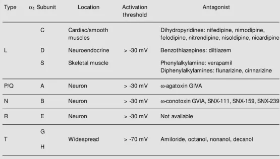
The calcium channels consist of hetero-oligomeric complexes containing at least ß and a2d subunits functionally stabilized by a
central a1 subunit, which forms the ion pore.
Molecular cloning has identified nine cal-cium channel a1 subunit genes (a1A to a1E,
a1S), four ß subunit genes (ß1-ß4), and a
single gene encoding an a2d subunit. The
central a1 subunit carries the channels Ca 2+
selectivity filter, controls its voltage-depend-ent opening and closing via its voltage sen-sors, and also determines its distinct phar-macological properties. The VOCC were classified into low-threshold (or T-type) and high-threshold activated channels. At least four types of high-threshold activated chan-nels have been defined: the L-, N-, P/Q-, and R-types. The localization, possible functions, and sensitivity of VOCC to drugs are sum-marized in Table 1. More recent studies have provided evidence for the involvement of a membrane-delimited G protein (Gßg
subunit)-dependent pathway in the modulation of N-type and P/Q-N-type channels.
The cations Ce3+
, La3+
, Nd3+
, Cd2+
, Co2+
, Ni2+
, Mg2+
and Mn2+
block the Ca2+
-channel pore in a nonselective manner and prevent Ca2+
from entering the cells (see Ref. 2). More selective agents include antagonists of L-type VOCC, which are classified as dihy-dropyridines, benzothiazepines, phenylalkyl-amines and diphenylalkylphenylalkyl-amines; antagonists of N-type VOCC, such as w-conotoxin GVIA (w-CgTX), obtained from the marine snail Conus geographus and its synthetic equiva-lents, and antagonists of P-type VOCC, rep-resented by the funnel web spider toxin, w -agatoxin GIVA (w-AgaTX). Aminoglycoside antibiotics (such as streptomycin, kanamy-cin, neomykanamy-cin, gentamikanamy-cin, and amikacin) have been described as N-type (for a review, see Ref. 3) and P/Q-type (4) antagonists.
Ca2+-channe ls and no cice ptio n
The L-, N- and P/Q-type Ca2+
-channels were demonstrated in the dorsal horn of the spinal cord (for a review, see Ref. 5). The L-type Ca2+
-channels were found in proximal
Table 1 - Classification of voltage-operated Ca2+-channels.
Type a1 Subunit Location Activation Antagonist
threshold
C Cardiac/smooth Dihydropyridines: nifedipine, nimodipine, muscles felodipine, nitrendipine, nisoldipine, nicardipine
L D Neuroendocrine > -30 mV Benzothiazepines: diltiazem
S Skeletal muscle Phenylalkylamine: verapamil
Diphenylalkylamines: flunarizine, cinnarizine
P/Q A Neuron > -30 mV w-agatoxin GIVA
N B Neuron > -30 mV w-conotoxin GVIA, SNX-111, SNX-159, SNX-239
R E Neuron > -30 mV Not available
G
T Widespread > -70 mV Amiloride, octanol, nonanol, decanol
dendrites and cellular bodies of neurons in the CNS, and in the subsynaptic membrane of some glutamatergic synapses. The N-type Ca2+
-channels are concentrated in presynap-tic nerve terminals at the level of the more superficial laminae I and II of the dorsal horn of the spinal cord, a strategic location for a key role of these channels in neurotransmit-ter release from primary afferents. The L-type Ca2+
-channels seem to be more impor-tant for the regulation of cellular calcium-dependent events than for the neurotrans-mitter itself. They participate in the excita-tion-transcription coupling but are not nec-essary for fast synaptic transmission. An-tagonists of the N-type Ca2+-channels block
the release of sensory neuropeptides from primary sensory neurons in culture. Also,
N-and P/Q-type channels mediate fast synaptic transmission at virtually all chemical syn-apses. Thus, N-type channels and probably P-type channels can play a fundamental role in the modulation of nociceptive informa-tion, whereas the involvement of L-type channels in the process seems to be very restricted.
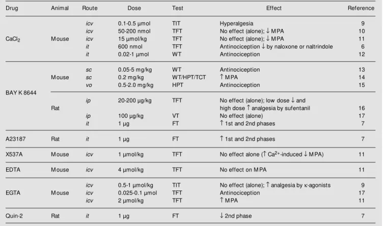
No cice ptio n and Ca2+ availability
There are several lines of evidence for the involvement of Ca2+
in nociception (for references, see Table 2). The intracerebro-ventricular (icv) administration of calcium chloride produces hyperalgesia or has no effect in mouse models of pain. In contrast, intrathecal (it) calcium chloride produced
Table 2 - Effects of Ca2+, a Ca2+ agonist, Ca2+ ionophores or Ca2+ chelators on nociception and opioid-induced antinociception.
icv = Intracerebroventricular; it = intrathecal; sc = subcutaneous; vo = oral; ip = intraperitoneal; FT = formalin test; HPT = hot-plate test; TCT = tail-clamp test; TFT = tail-flick test; TIT = tail-immersion test; VT = vocalization test; WT = w rithing test; M PA = morphine-induced analgesia. = Increase; ¯ = decrease.
Drug Animal Route Dose Test Effect Reference
icv 0.1-0.5 µmol TIT Hyperalgesia 9
icv 50-200 nmol TFT No effect (alone); ¯ M PA 10
CaCl2 M ouse icv 15 µmol/kg TFT No effect (alone); ¯ M PA 11
it 600 nmol TFT Antinociception ¯ by naloxone or naltrindole 6
it 0.02-1 µmol WT Antinociception 12
sc 0.05-5 mg/kg WT Antinociception 13
M ouse sc 0.2 mg/kg WT/HPT/TCT M PA 14
vo 0.5-2.0 mg/kg HPT Antinociception 15
BAY K 8644
ip 20-200 µg/kg TFT No effect (alone); low dose ¯ and
Rat high dose analgesia by sufentanil 16
ip 100 µg/kg VT No effect (alone) 17
it 1 µg FT 1st and 2nd phases 7
A23187 Rat it 1 µg FT 1st and 2nd phases 7
X537A M ouse icv 1 µmol/kg TFT No effect alone ( Ca2+-induced ¯ M PA) 11
EDTA M ouse icv 4 µmol/kg TFT No effect on M PA 11
icv 0.5-1 µmol/kg TIT No effect (alone); analgesia by k-agonists 9
EGTA M ouse icv 0.025-0.1 µmol TFT Antinociception 17
icv 2 µmol/kg TFT M PA 11
naloxone- or naltrindole-sensitive antinoci-ception in the mouse tail-flick or writhing test, an effect imputed to a Ca2+
-induced spinal release of met-enkephalin (for a re-view, see Ref. 6).
Some experiments were conducted using drugs that increase the level of intracellular Ca2+
, such as Ca2+
ionophores (X537A and A23187) or Ca2+
agonists (BAY K 8644), yielding conflicting results. The icv adminis-tration of X537A did not change the noci-ceptive response of mice to thermal noxious stimuli, whereas the it administration of A23187 significantly elevated both phases of the response to formalin in rats. BAY K 8644 was ineffective in the rat tail-flick test following intraperitoneal (ip) administration, but produced antinociception in the mouse hot-plate or writhing test following subcuta-neous (sc) or intravenous (iv) administra-tion, respectively. In contrast, it BAY K 8644 increased the response of rats to forma-lin, thus indicating a critical role of intracel-lular Ca2+
level for the development of per-sistent pain in response to formalin (7). The dose of BAY K 8644 used in each case accounts for the differences (see ahead).
Other studies were conducted using Ca2+
chelators such as EDTA, EGTA or Quin-2. EGTA alone administered icv had no effect or produced dose-dependent antinociception in mice. EDTA alone had a weak or no antinociceptive effect in mice. Quin-2 ad-ministered it reduced the second, but not the first phase of the formalin test in rodents, thus indicating a critical involvement of Ca2+
influx in mediating central sensitization fol-lowing tissue injury, but not in the transmis-sion of inputs in response to brief noxious stimuli (7).
Evidence also exists for the involvement of Ca2+
in peripheral mechanisms mediated at the nociceptor level. The intraplantar ad-ministration of A23187 evokes hyperalgesia in rats that is potentiated by methylxanthines and antagonized by verapamil, La3+
or mor-phine, thus indicating that the hyperalgesic
effect of the Ca2+
ionophore depends on the activity of adenylate cyclase on peripheral nociceptors (8).
Ca2+-channe l antago nists and antino cice ptio n
Several Ca2+
-channel antagonists have been used for the study of the effects of Ca2+
on nociception. Trivalent cations such as La3+
and Ce3+
produce antinociception in both the tail-flick and hot-plate tests follow-ing icv administration to mice (11,17). Intra-thecal La3+
or Nd3+
also produces antinoci-ception in the rat tail-flick and hot-plate tests and blocks both phases of the response to formalin in rats (18). Since the development of the 2nd phase (persistent pain) of the response to formalin depends on the occur-rence of the 1st phase (phasic pain), the effects of the inorganic cations against both phases of the response indicate that VOCC are involved in both the induction and main-tenance of the response to formalin (18). Intrathecal Ni2+
, which preferentially blocks T-type VOCC, was ineffective in the mouse writhing test, thus indicating that T-type chan-nels are not implicated in the spinal process-ing of nociceptive information (12).
The effects of L-type Ca2+
-channel an-tagonists on nociception differ depending on the drug, dosage, and route of administration and algesimetric test used (for references, see Table 3). In general, the antinociception induced by the L-type Ca2+
-channel antago-nists was demonstrated in rodents mainly when models of persistent pain, such as the writhing and formalin tests, were used. How-ever, it diltiazem or verapamil has failed to reduce the persistent hyperalgesia induced by chronic sciatic ligature in rats, also a model of persistent pain. The remaining data on the effects of L-type Ca2+
Table 3 - Effects of L-type Ca2+-channel antagonists on nociception and opioid-induced antinociception.
CSL = Chronic sciatic ligature; DHC = dorsal horn cell electrical activity. For other abbreviations see legend to Table 2.
Drug Animal Route Dose Test Effect Reference
icv 0.5-400 µg/kg WT Antinociception (alone) 20
icv 60-120 µg HPT Antinociception (alone); M PA 21
sc 60-120 µg HPT No effect (alone); M PA 21
sc 15 mg/kg HPT No effect (alone); M PA 22
M ouse sc 10-40 mg/kg WT Antinociception (alone) 13
sc 10-30 mg/kg WT Antinociception (alone) 23
ip 1-30 mg/kg WT/HPT No effect (alone) 24
it 0.5-80 µg WT Antinociception (alone) 12
Diltiazem
sc 20 mg/kg HPT No effect (alone); M PA 25
ip 1-30 mg/kg FT Antinociception (alone) 24
Rat it 3 µg CSL No effect (alone) 26
it 100 µg TFT No effect (alone); M PA 27
it 100 µg FT M inimal antinociception (alone) 18
icv 0.5-400 µg/kg WT Antinociception (alone) 20
icv 15-120 µg HPT Antinociception (alone); M PA 21
icv 25-200 µg HPT Antinociception (alone) 19
sc 20 µg HPT No effect (alone); M PA 12
sc 10-80 mg/kg HPT Weak antinociception 19
M ouse sc 5-20 mg/kg WT Antinociception (alone) 22
sc 2-30 mg/kg WT Antinociception (alone) 24
ip 1-30 mg/kg WT/HPT No effect (alone) 13
it 0.5-80 µg WT Antinociception (alone) 23
it 25-200 µg HPT Antinociception (alone) 19
Verapamil
icv 20 nmol TFT/HPT analgesia of DAM GO; ¯ analgesia of DPDPE 28
sc 10 mg/kg HPT No effect (alone); M PA 25
ip 1-30 mg/kg FT Antinociception (alone) 24
Rat it 5 and 50 µg FT/DHC No effect (alone) 29
it 100 µg FT M inimal antinociception (alone) 18
it 10 µg FT ¯ 2nd phase (alone) 7
it 250 µg CSL No effect (alone) 26
it 50 µg TFT No effect (alone); M PA 27
icv 0.5-400 µg/kg WT Antinociception (alone) 20
sc 15 mg/kg HPT No effect (alone); no change in M PA 22
M ouse sc 5-20 mg/kg WT Antinociception (alone) 13
sc 2-20 mg/kg WT Antinociception (alone) 23
ip 1-30 mg/kg WT/HPT No effect (alone) 24
Nifedipine
ip 1-30 mg/kg FT Antinociception (alone) 24
ip 2 mg/kg TFT M PA 30
Rat it 24 nmol FT M inimal antinociception (alone) 18
it 10 µg FT ¯ 2nd phase (alone) 7
it 0.8-7.0 µg TFT/HPT Antinociception (alone) 31
it 50 and 100 µg TFT No effect (alone) 32
demonstrated that verapamil and flunarizine evoke antinociception in the mouse hot-plate test. Using specific opioid antagonists, they showed that these effects might be due to the agonistic activity of verapamil at µ-, d- and
k3-receptor subtypes. Flunarizine had a mixed
opioid activity, acting as an agonist on µ-receptors and as an antagonist on d- and k -receptor subtypes. In a comparative study, it verapamil or nifedipine was less effective than it Quin-2 in reducing the 2nd phase of the rat response to formalin, results that were interpreted as evidence that Ca2+
influx through channels other than phenylalkyl-amine- and dihydropyridine-sensitive VOCC may be involved in the process (7).
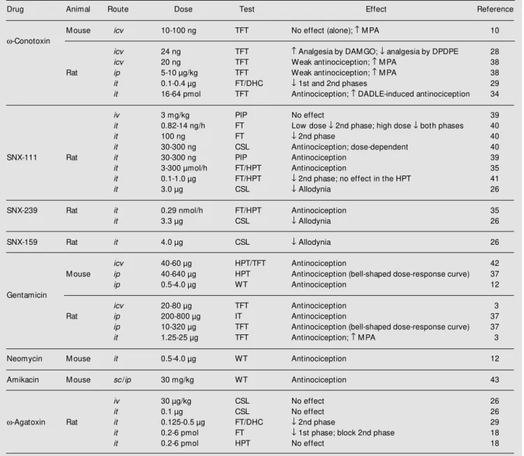
The effects of N-type Ca2+-channel
an-tagonists on nociception may also differ de-pending on the route of administration and pain model used (for references, see Table 4). In general, the conopeptides w-CgTX
and SNX-111 produce weak antinociception in rodent models of phasic pain, but are usually very effective in models of persistent pain. Other conopeptides, SNX-239 and SNX-159, were very effective following it administration in the rat hot-plate or forma-lin tests and significantly reduced the allo-dynia evoked by chronic sciatic ligature. Differently from opiates, the continuous in-fusion of SNX-111 or SNX-239 reduces both phases of the formalin test in rats accompa-nied by no signs of tolerance (35). The site of the antinociceptive effect of N-type antago-nists seems to be within the CNS since they were effective following it, but not systemic or topical application (26).
Aminoglycoside antibiotics have been demonstrated to interact competitively with Ca2+
in several processes including neu-rotransmitter release in peripheral synapses (for a review, see Ref. 36). We have
exam-Drug Animal Route Dose Test Effect Reference
icv 0.5-400 µg/kg WT Antinociception (alone) 20
icv 5 µg TFT No effect (alone); ¯ M PA 10
M ouse vo <100 mg/kg HPT No effect (alone); fentanyl-induced analgesia 15
sc 20 mg/kg WT/HPT M PA 14
ip 1-30 mg/kg WT/HPT Antinociception (alone) 24
Nimodipine
iv <100 mg/kg VT No effect (alone) 15
ip 200 µg/kg TFT No effect (alone); sufentanil-induced analgesia 16
Rat ip 200 µg/kg TFT No effect (alone); ¯ U69593-induced analgesia 33
ip 1-30 mg/kg FT Antinociception (alone) 24
it 50-100 µg/kg TFT No effect (alone) 32
it 60-240 pmol TFT No effect (alone); ¯ antinociception by DAM GO 34
M ouse sc 15-20 mg/kg HPT No effect (alone); M PA 22
it 0.5-80 µg WT Antinociception (alone) 12
Nicardipine
Rat it 20 µg TFT No effect (alone); M PA 27
icv 0.5-400 µg/kg WT Antinociception (alone) 20
icv 1-200 µg HPT Antinociception (alone) 19
Flunarizine M ouse sc 20 mg/kg HPT No effect (alone); M PA 22
sc 5-80 mg/kg HPT Antinociception (alone) 19
it 1-200 µg HPT Antinociception (alone) 19
Cinnarizine Rat ip 6-480 µg/kg TFT Antinociception (alone) 32
it 3-9 µg TFT Antinociception (alone) 32
ined the effects of gentamicin on the re-sponse of rodents to thermal noxious stimuli (tail-flick and hot-plate tests) and to a hyper-algesic stimulus (carrageenan-induced knee incapacitation) (3,37). Gentamicin was anti-nociceptive in all tests when injected by the icv, ip or it route. Marked and dose-depend-ent antinociception was obtained after it ad-ministration, whereas weaker effects and
bell-shaped dose-response curves were obtained following ip or icv administration. The ef-fect of it gentamicin was transitory and dose-dependently reversed by it calcium chloride. Gentamicin, neomycin and kanamycin were also effective in the mouse writhing, hot-plate and tail-flick tests following icv or it administration, and sc kanamycin and ami-kacin were effective in the rat hot-plate test.
Table 4 - Effects of N- or P-type Ca2+-channel antagonists on nociception and opioid-induced antinociception.
IT = Incapacitation test; PIP = postincisional pain. For other abbreviations see legends to Tables 2 and 3.
Drug Animal Route Dose Test Effect Reference
M ouse icv 10-100 ng TFT No effect (alone); M PA 10
w-Conotoxin
icv 24 ng TFT Analgesia by DAM GO; ¯ analgesia by DPDPE 28
icv 20 ng TFT Weak antinociception; M PA 38
Rat ip 5-10 µg/kg TFT Weak antinociception; M PA 38
it 0.1-0.4 µg FT/DHC ¯ 1st and 2nd phases 29
it 16-64 pmol TFT Antinociception; DADLE-induced antinociception 34
iv 3 mg/kg PIP No effect 39
it 0.82-14 ng/h FT Low dose ¯ 2nd phase; high dose ¯ both phases 40
it 100 ng FT ¯ 2nd phase 40
it 30-300 ng CSL Antinociception; dose-dependent 40
SNX-111 Rat it 30-300 ng PIP Antinociception 39
it 3-300 µmol/h FT/HPT Antinociception 35
it 0.1-1.0 µg FT/HPT ¯ 2nd phase; no effect in the HPT 41
it 3.0 µg CSL ¯ Allodynia 26
SNX-239 Rat it 0.29 nmol/h FT/HPT Antinociception 35
it 3.3 µg CSL ¯ Allodynia 26
SNX-159 Rat it 4.0 µg CSL ¯ Allodynia 26
icv 40-60 µg HPT/TFT Antinociception 42
M ouse ip 40-640 µg HPT Antinociception (bell-shaped dose-response curve) 37
ip 0.5-4.0 µg WT Antinociception 12
Gentamicin
icv 20-80 µg TFT Antinociception 3
Rat ip 200-800 µg IT Antinociception 37
ip 10-320 µg TFT Antinociception (bell-shaped dose-response curve) 37
it 1.25-25 µg TFT Antinociception; M PA 3
Neomycin M ouse it 0.5-4.0 µg WT Antinociception 12
Amikacin M ouse sc/ip 30 mg/kg WT Antinociception 43
iv 30 µg/kg CSL No effect 26
it 0.1 µg CSL No effect 26
w-Agatoxin Rat it 0.125-0.5 µg FT/DHC ¯ 2nd phase 29
it 0.2-6 pmol FT ¯ 1st phase; block 2nd phase 18
More recently, sc or ip amikacin was found to be antinociceptive also in the mouse writh-ing test. The effects of gentamicin and neo-mycin were not changed by naloxone, thus indicating that an opioid mechanism is un-likely to be involved in the effects of the antibiotics (12).
The P-type Ca2+
-channel antagonist w -AgaTX was ineffective in the hot-plate test or against the hyperalgesia evoked by chronic sciatic ligature following iv or it administra-tion to rats. However, the it toxin was highly effective in suppressing the 2nd phase of the rat response to formalin, but had no effect or a very modest suppressive effect against the 1st phase of the response to the same test. These results led to the notion that N-type, and possibly P-type, but not L-type VOCC antagonists exert a selective inhibitory effect on nociceptive transmission at the spinal cord level (18). In addition, N-type antago-nists, but not w-AgaTX, were still effective against the 2nd phase of the response to formalin when injected after the 1st phase, thus indicating that N- and P-type VOCC play different roles in nociception (18).
O pio id ago nists and Ca2+
Opioid agonists reduce the intracellular Ca2+
concentration either indirectly, through a µ- or d-receptor-mediated increase in Ca2+
-dependent K+
conductance that leads to nerve cell hyperpolarization and shortening of the action potential duration, or directly through a k-receptor-mediated shortening of Ca2+
action potentials without any change in the resting membrane potential (for a review, see Ref. 19). All the opioid receptors seem to share common mechanisms involving acti-vation of G protein ßg or a subunit-mediated effects (for a review, see Ref. 44). Activation of cloned µ-, d-, and k-opioid receptors in-hibits adenylyl cyclase activity via activa-tion of inhibitory G proteins, thus reducing the Ca2+ influx, inhibits N-type and L-type
VOCC via Go protein, and stimulates
phos-phatidylinositol turnover, thus causing a tran-sient increase in intracellular Ca2+
level. The µ- and d-receptor inhibition of adenylyl cy-clase is mediated by Go and Gi2 proteins,
respectively, while the same effect via k -receptor activation occurs without selectiv-ity toward Gi/Go proteins. The activation of
G proteins selectively modulates a1A and
a1B subunits in a manner identical to that for
native P/Q- and N-type currents, respectively, whereas a1C L-type currents do not exhibit
G-protein sensitivity (for a review, see Ref. 1). Thus, reduction of the intracellular free Ca2+
concentration is the final result of stimu-lating different opioid receptors, but the in-teraction of Ca2+ antagonists with µ- or d
-agonists may be expected to differ from the interaction with k-agonists (27).
spi-nally, to inhibit noxious inputs to dorsal horn neurons. On the other hand, pharmacologi-cal manipulations that interfere with Ca2+
availability can change nociceptive process-ing either supraspinally or spinally. Most of the studies reviewed here emphasize the spi-nal effects of calcium antagonists. To our knowledge, there is only one report showing that administration of Ca2+
into the periaque-ductal gray produces a nonsignificant hyper-algesia in the rat tail-flick test but antago-nizes the antinociceptive effect induced by iv morphine. The same study also demon-strates that administration of EGTA into the same region produces a dose-dependent an-tinociception in the same test (46).
O pio id-induce d antino cice ptio n and Ca2+ availability
Acute administration of opioid agonists to rodents reduces the Ca2+
content in synap-tic vesicles, synaptosomes and several rat brain areas. In contrast, the vesicular content of Ca2+
in synaptosomes, the Ca2+
uptake, and mainly the K+
-stimulated Ca2+
uptake by synaptosomes are increased during the de-velopment of opiate tolerance in rats. Paral-lel to these findings, opiate tolerance was correlated with an increase in the density of dihydropyridine-binding sites and higher basal free intracellular Ca2+
levels in the rat brain (for a review, see Ref. 47). Nimodi-pine, nifedipine and verapamil prevented the development of naloxone-precipitated with-drawal syndrome in rats (48). Previous, but not concurrent administration of nimodipine, prevented the development of tolerance to sufentanil in rats (16).
The literature has also provided several reports of changes produced by substances that interfere with Ca2+
influx or availability in opioid-induced antinociception (for refer-ences, see Tables 2 to 4). Intracerebroven-tricular calcium chloride reduced opioid-induced antinociception in mice. Subcuta-neous BAY K 8644 increased
morphine-induced antinociception in the mouse writh-ing, hot-plate and tail-clip tests. Low doses of BAY K 8644 reduced, whereas high doses enhanced, the antinociception induced by sc opioids in rodents (14,16). The dualistic ef-fect of BAY K 8644 may derive from its Ca2+
agonistic property, which is prominent at low concentrations but declines at higher concentrations (15). Intracerebroventricular EGTA, but not EDTA increases morphine-and k-agonist-induced antinociception in the mouse tail-flick test.
In general, L-type Ca2+
-channel antago-nists potentiate opioid-induced antinocicep-tion in several rodent pain models (for refer-ences, see Table 3). However, there are re-ports showing antagonistic effects of nimo-dipine against the antinociception evoked by morphine in mice (10) or by the k-agonist U69593 in rats (33).
The morphine-induced antinociception was potentiated by w-CgTX following icv administration in mice, and icv or ip injec-tion in rats (38). The icv administration of w -CgTX to rats potentiates the antinociception produced by the µ-agonist DAMGO, and reduces the antinociception evoked by the d -agonist DADLE (28). We found that the antinociceptive effect of it gentamicin was only additive to morphine antinociception in rats (3). The icv Ca2+-induced inhibition of
opiate antinociception seems to result from a Ca2+
influx through N-type VOCC since its effect was prevented by icv w-CgTX but not by icv nimodipine (10).
We have recently shown (34) that itw -CgTX GVIA, but not nimodipine, increased the latency for the rat tail-flick reflex. Nimo-dipine reduced the antinociception evoked by it DAMGO but did not change the effects of DADLE or bremazocine, d- and k-opioid agonists, respectively. In contrast, itw-CgTX GVIA potentiated the antinociceptive effects of DADLE but did not change the effects of DAMGO or bremazocine, thus indicating that the combination of an N-type Ca2+
most effective for the management of pain in that model of phasic pain.
Ca2+
influx is considered to be critical in the transmission of persistent but not brief noxious inputs (for a review, see Ref. 7). Taddese and colleagues (49) have shown that the µ-opioid agonist DAMGO inhibits calcium channels in almost all small noci-ceptors, which mediate persistent pain, but has minimal effects on large nociceptors known to mediate phasic pain. Somatostatin has the opposite specificity, but the effects of both agonists were eliminated by w-CgTX GVIA, thus indicating that the differences between DAMGO and somatostatin are not due to different Ca2+
-channels. However, the antinociceptive effect of N-type Ca2+
-channel antagonists has been shown in both phasic and persistent pain models. There-fore, the interactions between opioid ago-nists and Ca2+
-channel antagonists may in fact differ according to the type of noxious stimuli.
Clinical studie s
The usefulness of Ca2+
-channel antago-nists in the management of clinical pain has also been somewhat controversial, especially regarding the effects of L-type antagonists. An earlier study on this subject indicated that fentanyl-induced analgesia in patients undergoing thoracic surgery was potentiated by the intraoperatory infusion of nimodipine (50). However, the on-demand iv infusion of fentanyl for the control of pain after elective hysterectomy was not significantly different in groups of patients double-blindly assigned to the administration of an additional infu-sion of either placebo or nimodipine (51). Oral administration of slow-release tablets of nifedipine given 18, 9 and 1 h before the beginning of surgery significantly potenti-ated the analgesic effect of morphine slowly infused by the iv route in patients submitted to elective hysterectomy or orthopedic sur-gery (30). However, slow-release nifedipine
given orally 12 and 1 h before surgery did not change significantly the postoperative pain relief produced by epidural fentanyl (52). In our experience, the postoperative pain relief provided by epidural morphine in patients undergoing elective gynecological surgery was significantly enhanced by the sublingual administration of nifedipine (53). In none of these studies did the Ca2+
-channel antagonists have an analgesic effect by them-selves.
Few reports are presently available re-garding the usefulness of Ca2+
-channel an-tagonists in the control of chronic pain. Oral nifedipine at doses increased weekly from 10 mg twice a day to 30 mg twice a day produced complete (7 cases), partial (2 cases) or no relief (1 case) of pain symptoms in patients suffering from reflex sympathetic dystrophy (54). Nimodipine given orally at a dose of 30 mg every 8 h for 3 days did not change the analgesia produced by the con-comitant use of morphine in the earlier phase of treatment for cancer pain (55). In contrast, oral slow release nimodipine reduced the daily dose of oral morphine required for pain management in cancer patients. In this case, the dose of nimodipine was 60 mg on the first day, and was then increased up to 120 mg/day divided into four doses. In addition, the antagonist was introduced only when the patients met the criteria for tolerance to mor-phine (56). A similar design used oral slow-release nifedipine (15 mg twice a day) to show that the antagonist significantly re-duced the daily intake of oral morphine for the adequate management of cancer pain (57), but nifedipine was effective only 3 to 5 days after the beginning of treatment. Alto-gether, these data point to the dependency of the improvement of opioid analgesia on an effective plasma concentration of the Ca2+
-channel antagonist. Moreover, the efficacy of L-type Ca2+
-channel antagonists in the management of chronic pain indicates that pharmacological interference with Ca2+
of opioid analgesics (56).
Based on the effectiveness of N-type Ca2+
-channel antagonists in animal models of pain, recent clinical trials have confirmed that these drugs can provide relief of chronic pain also in human subjects. A dose-dependent anal-gesic effect of continuous it infusion of SNX-111 was demonstrated in one case of intrac-table deafferentation and phantom limb pain secondary to brachial plexus avulsion and subsequent amputation (58). Thirty patients undergoing elective abdominal hysterectomy, radical prostatectomy or total hip replace-ment were treated with an it infusion of SNX-111 (ziconotide, 0.7 to 7.0 µg/h), which was started before the surgical incision and continued for 48 to 72 h postoperatively. It was shown that the daily patient-controlled administration of morphine was significant-ly lower in ziconotide-treated than in pla-cebo-treated patients (59). However, dose-dependent adverse effects such as dizziness, blurred vision, nystagmus and sedation were noticed in both studies. Continuous it infu-sion of ziconotide (0.4 to 5.3 µg/h) in 3 patients suffering from chronic pain, in addi-tion to the aforemenaddi-tioned side effects, also produced potentially serious outcomes, in-cluding bradycardia, orthostatic hypotension, nausea and vomiting, coma, ataxia, dysme-tria, agitation, hallucination, rash, hypogly-cemia, diarrhea, nasal congestion and uri-nary retention (60).
Co nclusio ns
The results reviewed here indicate that Ca2+
plays an important role in regulating endogenous pain systems. In addition, they indicate a close relationship between the analgesic effects of opioids and Ca2+
availa-bility in the CNS. Thus, elevation of extra-cellular free Ca2+
concentration or facilita-tion of its transmembrane flux reduces the opioid antinociception. On the other hand, reduction of extracellular free Ca2+
concen-tration or of its transmembrane flux increases
opioid antinociception or promotes antino-ciception by itself.
The experiments with VOCC antagonists revealed that L-, N-, and P/Q-, but not T-type channels are involved in nociception. Anti-nociception is more frequently obtained with N- or P/Q-type antagonists than with L-type antagonists. Potentiation of opioid-induced antinociception was more frequently seen with L-type antagonists, while N-type an-tagonists were only additive to opioid anti-nociception. In contrast, L-type antagonists prevented the development of opioid toler-ance and were more effective than N-type antagonists in reducing symptoms of opioid withdrawal. The experiments also indicated that the interactions between opioid agonists and Ca2+
-channel antagonists differ accord-ing to the type of noxious stimuli used in each case.
Few clinical studies on the efficacy of Ca2+
development of tolerance to opioid analge-sia. Finally, the observation that N- and P/Q-type channels can participate in different phases of the response to persistent noxious stimulation points to the possible clinical
Re fe re nce s
1. Zamponi GW & Snutch TP (1998). M odu-lation of voltage-dependent calcium chan-nels by G proteins. Current Opinion in Neurobiology,8: 351-356.
2. Barritt GJ (1999). Receptor-activated Ca2+ inflow in animal cells: a variety of path-w ays tailored to meet different intracellu-lar Ca2+ signaling requirements. Bio-chemical Journal,337: 153-169. 3. Rego EM , Corrado AP & Prado WA (1992).
Antinociception induced by intracerebro-ventricular or intrathecal administration of gentamicin in rats. General Pharmacolo-gy,23: 481-485.
4. Pichler M , Wang ZY, Grabner-Weiss C, Reimer D, Hering S, Grabner M , Gloss-mann H & Striessnig J (1996). Block of P/ Q-type calcium channels by therapeutic concentrations of aminoglycoside antibi-otics. Biochemistry,35: 14659-14664. 5. Venegas H & Schaible HG (2000). Effects
of antagonists to high-threshold calcium channels upon spinal mechanisms of pain, hyperalgesia and allodynia. Pain,85: 9-18. 6. Smith FL & Dew ey WL (1992). Evidence that endogenous opioids mediate the anti-nociceptive effects of intrathecally admin-istered calcium in mice. Journal of Phar-macology and Experimental Therapeutics, 262: 995-1003.
7. Coderre TJ & M elzack R (1992). The role of NM DA receptor-operated calcium chan-nels in persistent nociception after forma-lin-induced tissue injury. Journal of Neu-roscience,12: 3671-3675.
8. Ferreira SH & Nakamura M (1979). II -Prostaglandin hyperalgesia: the peripher-al anperipher-algesic activity of morphine, enkepha-lins and opioid antagonists. Prostaglan-dins, 18: 191-200.
9. Ben Sreti M M , Gonzalez JP & Sew ell RDE (1983). Effects of elevated calcium and calcium antagonists on 6,7-benzomor-phan-induced analgesia. European Jour-nal of Pharmacology,90: 385-391. 10. Smith FL & Stevens DL (1995). Calcium
modulation of morphine analgesia: role of calcium channels and intracellular pool
calcium. Journal of Pharmacology and Ex-perimental Therapeutics, 272: 290-299. 11. Harris RA, Loh HH & Way EL (1975).
Ef-fects of divalent cations, cation chelators and an ionophore on morphine analgesia and tolerance. Journal of Pharmacology and Experimental Therapeutics, 195: 488-498.
12. Dogrul A & Yesilyurt O (1998). Effects of intrathecally administered aminoglycoside antibiotics, calcium-channel blockers, nickel and calcium on acetic acid-induced w rithing test in mice. General Pharmacol-ogy, 30: 613-616.
13. Quijada L, Germany A, Hernández A & Contreras E (1992). Effects of calcium channel antagonists and BAY K 8644 on the analgesic response to pentazocine and U 50488H. General Pharmacology, 23: 837-842.
14. Kuzmin AV, Patkina NA & Zvartau EE (1994). Analgesic and reinforcing effects of morphine in mice. Influence of Bay K-8644 and nimodipine. Brain Research, 652: 1-8.
15. Hoffmeister F & Tettenborn D (1986). Cal-cium agonists and antagonists of the di-hydropyridine type: antinociceptive ef-fects, interference w ith opiate-µ-receptor agonists and neuropharmacological ac-tions in rodents. Psychopharmacology, 90: 299-307.
16. Dierssen M , Flórez J & Hurlé M A (1990). Calcium channel modulation by dihydro-pyridines modifies sufentanil-induced an-tinociception in acute and tolerant condi-tions. Naunyn-Schmiedeberg’s Archives of Pharmacology, 342: 559-565. 17. Schmidt WK & Way EL (1980).
Hyperalge-sic effects of divalent cations and antino-ciceptive effects of a calcium chelator in naive and morphine-dependent mice. Journal of Pharmacology and Experimen-tal Therapeutics, 212: 22-27.
18. M almberg AB & Yaksh TL (1994). Volt-age-sensitive calcium channels in spinal nociceptive processing: blockade of N-and P-type channels inhibits
formalin-induced nociception. Journal of Neurosci-ence, 14: 4882-4890.
19. Weissman R, Gestslev V, Pankova IA, Schrieber S & Pick CG (1999). Pharmaco-logical interaction of the calcium channel blockers verapamil and flunarizine w ith the opioid system. Brain Research, 818: 187-195.
20. M iranda HF, Pelissier T & Sierralta F (1993). Analgesic effects of intracerebro-ventricular administration of calcium chan-nel blockers in mice. General Pharmacol-ogy, 24: 201-204.
21. Del Pozo E, Ruiz-Garcia C & Baeyens JM (1990). Analgesic effects of diltiazem and verapamil after central and peripheral ad-ministration in the hot-plate test. General Pharmacology, 21: 681-685.
22. Contreras E, Tamayo L & Amigo M (1988). Calcium channel antagonists increase morphine-induced analgesia and antago-nize morphine tolerance. European Jour-nal of Pharmacology, 148: 463-466. 23. Al-Humayyd M S (1991). Effect of
diltia-zem, nifedipine and verapamil on the anti-nociceptive action of acetylsalicylic acid in mice. General Pharmacology, 22: 121-125.
24. M iranda HF, Bustamante D, Kramer V, Pelissier T, Saavedra H, Paeile C, Fernandez E & Pinardi G (1992). Antinoci-ceptive effects of Ca2+ channel blockers. European Journal of Pharmacology, 217: 137-141.
25. Benedek G & Sziksay M (1984). Potentia-tion of thermoregulatory and analgesic ef-fects of morphine by calcium antagonists. Pharmacological Research Communica-tions, 16: 1009-1018.
26. Chaplan SR, Pogrel JW & Yaksh TL (1994). Role of voltage-dependent calcium chan-nel subtypes in experimental tactile allo-dynia. Journal of Pharmacology and Ex-perimental Therapeutics, 269: 1117-1123. 27. Om ote K, Sonoda H, Kaw am ata M , Iw asaki H & Namiki A (1993). Potentiation of antinociceptive effects of morphine by calcium-channel blockers at the level of
the spinal cord. Anesthesiology, 79: 746-752.
28. Spampinato S, Speroni E, Govoni P, Pistacchio E, Romagnoli C, M urari G & Ferri S (1994). Effect of w-conotoxin and verapamil on antinociceptive, behavioural and t herm oregulat ory responses t o opioids in the rat. European Journal of Pharmacology, 254: 229-238.
29. Diaz A & Dickenson AH (1997). Blockade of spinal N- and P-type, but not L-type, calcium channels inhibits the excitability of rat dorsal horn neurones produced by subcutaneous formalin inflammation. Pain, 69: 93-100.
30. Carta F, Bianchi M , Argenton S, Cervi D, M arolla G, Tamburini M , Breda M , Fantoni A & Penerai AE (1990). Effect of nifedi-pine on morphine-induced analgesia. An-esthesia and Analgesia, 70: 493-498. 31. Wong CH, Dey P, Yarmush J, Wu WH &
Zbuzek VK (1994). Nifedipine-induced an-algesia after epidural injection in rats. An-esthesia and Analgesia, 79: 303-306. 32. Rego EM , Corrado AP & Prado WA (1990).
Antinociceptive effects of calcium chan-nel blockers in the rat. Brazilian Journal of M edical and Biological Research, 23: 297-305.
33. Barro M , Ruiz F & Hurlé M A (1995). k -Opioid receptor mediated antinociception in rats is dependent on the functional state of dihydropyridine-sensitive calcium channels. Brain Research, 672: 148-152. 34. Lia EM & Prado WA (1999). Effects of
intrathecal L- and N-type calcium channel blockers on the antinociception evoked by opioid agonists in the rat tail flick test. Acta Physiologica, Pharm acologica et Therapeutica Latinoamericana, 49: 195-203.
35. M almberg AB & Yaksh TL (1995). Effect of continuous intrathecal infusion of w -conopeptides, N-type calcium-channel blockers, on behavior and antinociception in the formalin and hot-plate tests in rats. Pain, 60: 83-90.
36. Corrado AP, Pimenta de M orais I & Prado WA (1989). Aminoglycoside antibiotics as a tool for the study of the biological role of calcium ions. Acta Physiologica et Phar-macologica Latinoamericana, 39: 419-430. 37. Prado WA, Tonussi CR, Rego EM & Corrado AP (1990). Antinociception in-duced by intraperitoneal injection of gen-tamicin in rats and mice. Pain, 41: 365-371.
38. Basilico L, Parolaro D, Rubino T, Gori E & Giagnoni G (1992). Influence of w -cono-toxin on morphine analgesia and w ith-draw al syndrome in rats. European
Jour-nal of Pharmacology, 218: 75-81. 39. Wang YX, Pettus M , Gao D, Phillips C &
Bow ersox SS (2000). Effects of intrathe-cal administration of ziconotide, a selec-tive neuronal N-type calcium channel blocker, on mechanical allodynia and heat hyperalgesia in a rat model of postopera-tive pain. Pain, 84: 151-158.
40. Bow ersox SS, Gadbois T, Singh T, Pettus M , Wang YX & Luther RR (1996). Selec-tive N-type neuronal voltage-sensiSelec-tive cal-cium channel blocker, SNX-111, produces spinal antinociception in rat models of acute, persistent and neuropathic pain. Journal of Pharmacology and Experimen-tal Therapeutics, 279: 1243-1249. 41. Wang YX, Gao D, Pettus M , Phillips C &
Bow ersox SS (2000). Interactions of intra-thecally administered ziconotide, a selec-tive blocker of neuronal N-type voltage-sensitive calcium channels, w ith mor-phine on nociception in rats. Pain, 84: 271-281.
42. Ocaña M & Baeyens JM (1991). Analge-sic effects of centrally administered ami-noglycoside antibiotics in mice. Neurosci-ence Letters, 126: 67-70.
43. Atamer-Simsek S, Ölmez-Salvarli H, Güç O & Eroglu L (2000). Antinociceptive ef-fect of amikacin and its interaction w ith morphine and naloxone. Pharmacological Research, 41: 355-360.
44. Law PY & Loh HH (1999). Regulation of opioid receptor activities. Journal of Phar-macology and Experimental Therapeutics, 289: 607-624.
45. Besson JM & Chaouch A (1987). Periph-eral and spinal mechanisms of nocicep-tion. Pharmacological Review s, 67: 67-185.
46. Guerrero-M uñoz F & Fearon Z (1982). Opi-oids/opiates analgesic response modified by calcium. Life Sciences, 31: 1237-1240. 47. Díaz A, Flórez J, Pazos A & Hurlé M A (2000). Opioid tolerance and supersensi-tivity induce regional changes in the auto-radiographic density of dihydropyridine-sensitive calcium channels in the rat cen-tral nervous system. Pain,86: 227-235. 48. M ichaluk J, Karolew icz B, Antkiew
icz-M ichaluk L & Vetulani J (1998). Effects of various Ca2+ channel antagonists on mor-phine analgesia, tolerance and depend-ence, and on blood pressure in the rat. European Journal of Pharmacology, 352: 189-197.
49. Taddese A, Nah SY & M cCleskey EW (1995). Selective opioid inhibition of small nociceptive neurons. Science, 270: 1366-1369.
50. Bormann BV, Boldt J, Sturm G, Kling D,
Weidler B, Lohmann E & Hempelmann G (1985). Calciumantagonisten in der Anäs-thesie. Additive Analgesie durch Nimo-dipin w ährend cardiochirurgischer Ein-griffe. Anaesthesist, 34: 429-434. 51. Lehman KA, Kriegel R & Ueki M (1989).
Zur klinischen Bedeutung von Arznei-mittelinteraktionen zw ischen Opiaten und Kalziumantagonisten. Eine randomisierte Doppeblindstudie mit Fentanyl und Nimo-dipin im Rahmen der postoperativen in-travenösen On-Demand Analgesie. Anaes-thesist, 38: 110-115.
52. Garcia LV (1995). Efeito da nifedipina na analgesia induzida pelo fentanil. Doctoral thesis, Faculdade de M edicina de Ribeirão Preto, USP, Ribeirão Preto, SP, Brazil. 53. Pereira IT, Prado WA & Dos Reis M P
(1993). Enhancement of the epidural mor-phine-induced analgesia by systemic ni-fedipine. Pain, 53: 341-355.
54. Prough DS, M cLeskey CH, Poehling GG, Koman LA, Weeks DB, Whitw orth T & Semble EL (1985). Efficacy of oral nifedi-pine in the treatment of reflex sympa-thetic dystrophy. Anesthesiology,62: 796-799.
55. Roca G, Aguilar JL, Gomar C, M azo V, Costa J & Vidal F (1996). Nimodipine fails to enhance the analgesic effect of slow release morphine in the early phases of cancer pain treatment. Pain, 68: 239-243. 56. Santillán R, M aestre JM , Hurlé M A & Flórez J (1994). Enhancement of opiate analgesia by nimodipine in cancer patients chronically treated w ith morphine: a pre-liminary report. Pain, 58: 129-132. 57. Lima ICPR (1996). Efeito da nifedipina na
analgesia de pacientes com dor crônica neoplásica em uso de morfina. M aster’s thesis, Faculdade de M edicina de Ribeirão Preto, USP, Ribeirão Preto, SP, Brazil. 58. Brose W G, Gutlove DP, Luther RR,
Bow ersox SS & M cGuire D (1997). Use of intrathecal ziconotide, a novel, N-type, voltage-sensitive, calcium channel blocker, in the management of intractable brachial plexus avulsion pain. Clinical Journal of Pain,13: 256-257.
59. Atanassoff PG, Hartmannsgruber M WB, Thrasher J, Wermeling D, Longton W, Gaeta R, Singh T, M ayo M , M cGuire D & Luther RR (2000). Ziconotide, a new N-type calcium channel blocker, adminis-tered intrathecally for postoperative pain. Regional Anesthesia and Pain M edicine, 23: 274-278.