Simultaneous changes in the function
and expression of beta 1 integrins
during the growth arrest of poorly
differentiated colorectal cells (LISP-1)
Disciplina de Oncologia, Departamento de Radiologia, Faculdade de Medicina, Universidade de São Paulo, São Paulo, SP, Brasil
R.A. Roela, M.M. Brentani, M.L.H. Katayama, M. Reis and M.H.H. Federico
Abstract
Cells usually lose adhesion and increase proliferation and migration during malignant transformation. Here, we studied how proliferation can affect the other two characteristics, which ultimately lead to invasion and metastasis. We determined the expression of ß1 inte-grins, as well as adhesion and migration towards laminin-1, fibronec-tin, collagens type I and type IV presented by LISP-1 colorectal cancer cells exposed to 2.5% dimethyl sulfoxide(DMSO), an agent capable of decreasing proliferation in this poorly differentiated colorectal cell line. Untreated cells (control), as shown by flow cytometry and monoclonal antibodies, expressed α2 (63.8 ± 11.3% positive cells), α3 (93.3 ± 7.0%), α5 (50.4 ± 12.0%) and α6 (34.1 ± 4.9%) integrins
but not α1, α4, αv or ß4. Cells adhered well to laminin-1 (73.4 ± 6.0%)
and fibronectin (40.0 ± 2.0%) substrates but very little to collagens. By using blocking monoclonal antibodies, we showed that α2, α3 and α6
mediated laminin-1 adhesion, but neither α3 nor α5 contributed to
fibronectin adherence. DMSO arrested cells at G0/G1 (control: 55.0 ± 2.4% vs DMSO: 70.7 ± 2.5%) while simultaneously reducing α5 (24.2
± 19%) and α6 (14.3 ± 10.8%) expression as well as c-myc mRNA
(7-fold), the latter shown by Northern blotting. Although the adhesion rate did not change after exposure to DMSO, α3 and α5 played a major
role in laminin-1 and fibronectin adhesion, respectively. Migration towards laminin-1, which was clearly increased upon exposure to DMSO (control: 6 ± 2 cells vs DMSO: 64 ± 6 cells), was blocked by an antibody against α6. We conclude that the effects of DMSO on LISP-1
proliferation were accompanied by concurrent changes in the expres-sion and function of integrins, consequently modulating adheexpres-sion/ migration, and revealing a complex interplay between function/ex-pression and the proliferative state of cells.
Correspondence
M.H.H. Federico Disciplina de Oncologia Departamento de Radiologia Faculdade de Medicina, USP Av. Dr. Arnaldo, 455, Sala 4112 01246-903 São Paulo, SP Brasil
Fax: +55-11-3082-6580 E-mail: [email protected] Presented at SIMEC 2002 (International Symposium on Extracellular Matrix), Angra dos Reis, RJ, Brazil, October 7-10, 2002. Research supported by PRONEX (No. 4196086900/7038-6). Part of a Master’s thesis presented by R.A. Roela to the Instituto Butantan, USP, São Paulo, SP, Brazil.
Publication supported by FAPESP.
Received December 13, 2002 Accepted July 1, 2003
Key words
•Cell adhesion molecules •Cell migration
•Integrin •Cell cycle •DMSO
Introduction
Normal intestinal epithelial cells exist adherent to a basement membrane composed of laminin-1 and collagen type IV, whereas the interstitial matrix presents collagen type I
integrins has often been implicated in changes in adhesive properties, neoplastic transfor-mation and invasive processes in colon ad-enocarcinoma (2-5). Some reports have as-sociated integrins with cycle status in other cell models (6-8).
Although it is generally assumed that changes in integrins underlie (1,9) changes in cell adhesion and migration (10), and that these changes occur during human colorec-tal differentiation and carcinogenesis (2), we know very little about the relationship be-tween cell cycle and integrin expression or function (11). We do not know whether or not decreased proliferation can, per se, af-fect integrin quantitatively and, if this is true, if changes in integrins displayed by a given cell population are responsible for changes in migration or adhesive properties.
To answer this question, we first investi-gated which agents known to exert an anti-proliferative effect upon colonic cell lines affected the expression and binding affinity of ß1 integrins expressed by LISP-1 cells, a poorly differentiated colorectal cancer cell line derived from a hepatic metastasis. Four different antiproliferative agents described to block proliferation in colorectal cell lines were tested: glucose (12), all-trans retinoic acid (13), 1,25-dihydroxyvitamin D3 (14) and
dimethyl sulfoxide (DMSO) (15,16).
Material and Methods
Cell and culture conditions
LISP-1 cells (17) were donated by the Ludwig Institute (São Paulo Branch, São Paulo, SP, Brazil). Cells were grown in Dulbecco’s modified Eagle’s medium (DMEM, Sigma, St. Louis, MO, USA) supplemented with 10% fetal bovine serum (FBS, Gibco-BRL, Rockville, MD, USA), 10 mM HEPES (Gibco-BRL), and 40 µg/ml gentamicin, at 37ºC, in an atmosphere of 5% CO2, 95% air. Cells were grown in
mono-layer cultures, seeded at 6 x 10³ cells/cm2
and maintained in the absence or presence of 1.25 or 2.5% (v/v) DMSO (Sigma), 50 or 100 mM glucose (Sigma), 500 nM all-trans retinoic acid (Sigma), or 10 nM 1,25-dihy-droxyvitamin D3 (Roche, Mannheim,
Ger-many) for 10 days. The DMSO concentra-tion chosen was the one capable of affecting proliferation but not viability, as determined by the standard Trypan exclusion method.
Antibodies
The following anti-integrin monoclonal antibodies (mAbs) were used for flow cytometric studies and antibody inhibition of adhesion and migration experiments: anti-α1 (clone HP2B6) from Coulter-Immuno-tech (Miami, FL, USA); anti-ß1 (clone P4C10), anti-α2 (clone P1E6), anti-α3 (clone P1B5), anti-α4 (clone P4G9), anti-α5 (clone P1D6), anti-α6 (clone GoH3), anti-ß4 (3E1), and anti-αvß3/ß5, all from Gibco-BRL; flu-orescein isothiocyanate (FITC)-conjugated sheep anti-mouse IgG, FITC-conjugated goat rat IgG, and FITC-conjugated goat anti-rabbit (Sigma).
Determination of DNA content with propidium iodide and flow cytometry
air-cooled 15-mW, 488-nm argon ion laser. We determined the percent of cells in G0/G1, S and G2/M phases of the cell cycle with the aid of ModFit software (Beckton Dickinson).
RNA extraction and Northern blot hybridization
Total RNA from LISP-1 cells after 10 days of DMSO treatment or in standard cul-ture was isolated with TRIzol reagent (Gibco-BRL) according to manufacturer instructions. RNA (10 µg) was electrophoresed on 1% agarose-formaldehyde gels, transferred to Hybond membranes (Amersham-Pharmacia, Little Chalfont, Buckinghamshire, England) and hybridized to the following [32P]-dCTP
(3,000 Ci/mmol, Amersham-Pharmacia) ran-dom-primed (Gibco-BRL) probes: the 1.7-kb fragment of c-myc was a gift from Dr. T.H. Habbitts, Laboratory of Molecular Bi-ology, Cambridge, England (19), and glycer-aldehyde-3-phosphate dehydrogenase was provided by Dr. P. Fort, Centre de Recher-che en Biochimie des Macromolécules, Montpellier, France (20). Hybridizations were performed for 18-24 h at 42ºC.
Integrin expression
Cells were detached from tissue culture plastic dishes using PBS/EDTA and trypsin, incubated with DMEM supplemented with 10% FBS for 2 h at 37ºC, washed in PBS and incubated with specific mAbs at predeter-mined concentrations for 30 min at 4ºC. Cells were then washed in PBS, 1% bovine serum albumin (BSA, Sigma) and incubated with secondary FITC-labeled antibodies for another 30 min. At least 10,000 events were acquired by flow cytometry and analyzed using CellQuest software. In each experi-ment, fluorescence was measured in FL1, and positive cells for each integrin subunit were determined by the percent of positive cells and by mean fluorescence intensity, which was calculated by subtracting from
the mean fluorescence values corresponding to cells incubated with primary and second-ary antibody the corresponding values of cells incubated only with the secondary anti-body.
DNA and integrin dual parameter determination
LISP-1 cells were prepared as described for the determination of integrin expression, and then fixed with 50% cold absolute etha-nol for 15 min on ice and washed twice with PBS. After 15 min at 37ºC with 500 units/ml ribonuclease A, 50 µg/ml isotonic propi-dium iodide was added for an additional 15 min in the dark at room temperature (21). Then, 40,000 events were acquired by FACSCalibur flow cytometry to obtain at least 10,000 events in G0/G1 (nonprolifera-tive phase) or S + G2/M (prolifera(nonprolifera-tive phase) to analyze integrin subunit cell expression.
Cell adhesion and migration assay
The components of the extracellular ma-trix (ECM) used were fibronectin and col-lagen type IV (Sigma) and laminin-1 and collagen type I extracted from mouse Engelbreth Holm Swarm tumor (22) and rat tail (23), respectively. Cells were first la-beled with 10 µCi [35S]-methionine (specific
activity >1,000 Ci/mmol) and washed with DMEM/0.1% BSA. After preincubation with specific anti-integrin mAbs, 104 cells were
added for 1 h at 37ºC in a 5% CO2
and the percent of adherent cells was deter-mined as follows: cell adhesion (%) = 100 x [(adherent cells cpm - adherent cells to BSA cpm)/total cells cpm].
The migratory properties of LISP-1 cells were determined with the aid of a 48-well microchemotaxis chamber assay (Neuro Probe, Laguna Niguel, CA, USA) by count-ing the mean number of cells migratcount-ing directionally through an 8-µm pore polyvi-nyl polycarbonate filter, quenched with 0.5% BSA for 1 h at 37ºC and then precoated with 20 µg/well laminin-1 or 10 µg/well fibronec-tin, collagen type I or IV, for 2 h at 37ºC. Briefly, 5 x 10³ cells resuspended in
serum-free medium containing 0.1% BSA were added to each well, with or without anti-bodies against ß1 integrin receptors. The chamber was incubated for 8 h at 37ºC in a humidified incubator in a 5% CO2
atmos-phere, and the filters were then removed, fixed, stained with 1% toluidine blue in 1% sodium tetraborate and mounted on glass slides. Nonmigrating cells were removed by wiping with a cotton swab, and finally 5 random fields of vision/well (100X objec-tive) were counted for the evaluation of cell migratory properties. All experiments were done in quadruplicate.
Statistical analysis
Results are reported as means ± SD. Data were analyzed statistically by ANOVA, with the level of significance set at P < 0.05.
Results
Kinetic parameters of LISP-1 after DMSO treatment
We initially set out to define which agents, among several known to provoke differen-tiation in colonic cells in vitro, reduced the proliferation of LISP-1 cells. Glucose (50 and 100 mM), 500 nM all-trans retinoic acid and 10 nM 1,25-dihydroxyvitamin D3 were
added independently. None of these treat-ments led to proliferation inhibition (data not shown), except DMSO at 2.5% (v/v). Although LISP-1 doubling time, 20 ± 1 h under standard culture conditions, was weakly affected by 1.25% DMSO treatment (doubling time = 24 ± 3 h), it nearly quadru-plicated when DMSO concentration was raised up to 2.5% (82 ± 3 h; P < 0.05, ANOVA). This optimal effects of 2.5% DMSO over a period of 10 days (from here on considered as standard DMSO treatment) occurred without affecting cell viability, which remained constant at 98% with cells adherent to plastic, without detachment, un-til the end of the experiment.
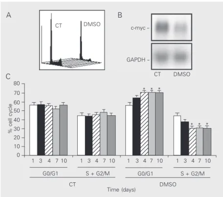
Cell cycle phase distribution, determined at various intervals after 2.5% DMSO treat-ment, provided clear evidence of an accu-mulation of LISP-1 cells at the G0/G1 phase
% cell cycle
80
123 123 123 123 123 123 123 123 123 123 123 123 123
123 123 123 123 123 123 123 123 123 123 123
123 123 123 123 123 123 123 123 123 123 123 123 123 123 123 123 123
12 12 12 12 12 12 12 12
CT DMSO c-myc
GAPDH
-CT DMSO
70
60 50
40 30 20 10 0
1 3 4 7 10 1 3 4 7 10 1 3 4 7 10 1 3 4 7 10
G0/G1 S + G2/M G0/G1 S + G2/M
CT DMSO
* * *
* * *
Time (days)
Figure 1. Dimethyl sulfoxide (DMSO, 2.5%, v/v) inhibits LISP-1 proliferation. A, DNA quantification in control cells (CT) and in cells exposed to DMSO for 10 days. B, c-myc mRNA and GAPDH bands were obtained by Northern blot. C, G0/G1 and S + G2/M, as percent of cells (y-axis), were obtained on different days of DMSO exposure (x-axis). Data are the mean ± SD obtained for 7 independent experiments. GAPDH = glyceraldehyde-3-phosphate dehydrogenase. *P < 0.05 compared to control (ANOVA).
A B
of the cell cycle, which was associated with increased doubling time (Figure 1A). By the 4th day of exposure, the G0/G1 phase frac-tion had significantly changed (control: 55.0 ± 2.4% vs DMSO: 70.7 ± 2.5%; Figure 1C). This difference was maintained until the con-clusion of the experiment at 10 days, as shown in Figure 1C. In parallel, we have observed a steep decrease (7-fold) in the expression of c-myc mRNA (Figure 1B).
Integrin expression
Assessment of ß1 integrin receptors us-ing flow cytometry and anti-integrin mAbs showed that LISP-1 cells display a high per-centage of α3 and ß1 integrin subunit-posi-tive cells, whereas α6 expression was rela-tively low and α2 and α5 were only moder-ately expressed. In contrast, α1, α4, αv and ß4 were not detected at all. DMSO decreased the percent of α5- and α6-positive cells with-out affecting α2 or ß1 subunit expression. Differences in the expression of α3 in LISP-1 cells were only observed at mean fluores-cence intensity level (Table 1).
Although time course analyses indicated that the expression of α3, α5 and α6 was reduced after DMSO exposure, only the de-crease of α5 and α6 integrin expression was simultaneous to G0/G1 cell accumulation. In parallel, the expression of ß1 integrin subunits displayed by control cells, deter-mined at the same times, did not change during the entire experiment. The results obtained for α3, α5 and α6 integrin expres-sion are presented in Figure 2.
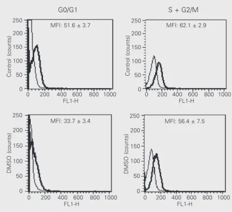
To characterize whether these changes in integrin expression were related to G0/G1 arrest upon DMSO treatment, we compared α3 and α6 integrin expression displayed by cells in G0/G1 and S + G2/M phase fractions after 10 days of DMSO treatment or under standard cell culture conditions. We observed that the fluorescence intensity of α6 integrin was reduced in the G0/G1 phase fraction (control: 51.6 ± 3.7 vs DMSO: 33.73 ± 3.4;
MFI
800
% G0/G1
70
%
α
5
integrin-positive cells
100 600 400 200
0
80
60
40
20
0
100
80
60
40
20
0
%
α
6
integrin-positive cells
60 50 40 30 20 10 0
% G0/G1
70 60 50 40 30 20 10 0
% G0/G1
70 60 50 40 30 20 10 0 1 2 3 4 5 6 7 8 9 10
1 2 3 4 5 6 7 8 9 10
1 2 3 4 5 6 7 8 9 10
* *
* *
*
Time (days)
Control DMSO
Figure 2. Time course of integrin expression and percent cells in G0/G1 during dimethyl sulfoxide (DMSO) treatment of LISP-1. Control cells (filled bars) and DMSO-treated cells (open bars) indicate the variation of mean fluorescence intensity (MFI) (A) and the percent of positive cells for α5 (B) and α6 (C) integrin (left y-axis). The per-cent of G0/G1 fraction (right y-axis) is represented for control cells and DMSO-treated cells. Data are the mean ± SD ob-tained for 5 independent experi-ments. *P < 0.05 compared to control (ANOVA).
P < 0.05), an event that did not occur in the S + G2/M phase (control: 62.1 ± 2.8 vs DMSO: 56.4 ± 7.5; Figure 3). A similar decrease in α3 integrin was observed through G0/G1 (control: 283.9 ± 19.8 vs DMSO: 201.1 ± 7.4) to S + G2/M (control: 280.9 ± 26.4 vs DMSO: 197.2 ± 21.4).
Table 1. Dimethyl sulfoxide (DMSO) modulation of ß1 integrin subunit expression in LISP-1 cells.
mAbs Control DMSO
% MFI % MFI
α2 63.8 ± 11.3 198.3 ± 26.4 56.1 ± 25.1 176.2 ± 0.4
α3 93.3 ± 7.0 348.3 ± 20.4 81.1 ± 19.2 208.7 ± 30.1*
α5 50.4 ± 12.0 170.9 ± 14.1 24.2 ± 19.0* 100.0 ± 28.7*
α6 34.1 ± 4.9 126.5 ± 28.4 14.3 ± 10.8* 111.7 ± 20.0 ß1 99.3 ± 0.5 276.3 ± 40.5 92.2 ± 5.9 226.2 ± 52.9
Cells were incubated with specific monoclonal antibodies (mAbs) and analyzed by flow cytometry as described in Material and Methods. Data are reported as percent of integrin-positive cells and mean fluorescence intensity (MFI). Data are the mean ± SD for 5 experiments.
*P < 0.05 compared to control (ANOVA).
A
B
and moderately to fibronectin (40.0 ± 2.0%), but did not attach to collagens at all. We did not see any gross change in LISP-1 adhesion after DMSO treatment. Adhesion to collagen type IV continued to be below 10% (Figure 4). Adhesion inhibition in the presence of mAbs confirmed that adhesion of LISP-1 cells to laminin-1 was mainly mediated by α2, α3 and α6 integrins. After DMSO expo-sure, the relative contribution of α2 and α6 decreased, turning α3 into the major lami-nin-1 receptor (Table 2). Fibronectin adhe-sion before DMSO exposure was not medi-ated by α3 or by α5 but, after treatment, α5 integrin accounted for 89.9 ± 2.0% of fibro-nectin total adhesion.
Finally we observed how DMSO treat-ment affected migratory activity of LISP-1 cells by using a Neuro Probe chamber assay. Very few cells (6 ± 2 cells) migrated sponta-neously towards laminin-1, whereas 64 ± 6 cells did it after 10 days of exposure to DMSO. This migration towards laminin-1 was inhibited 100% in the presence of the α6-recognizing mAb. On the other hand, we did not observe any LISP-1 migratory activ-ity towards fibronectin or collagens either before or after DMSO treatment.
Discussion
We have shown here that, although resis-tant to several commonly used antiprolifera-tive agents such as 1,25-dihydroxyvitamin D3 and retinoic acid, LISP-1 cell growth was
inhibited by DMSO, a polar solvent which acts as a powerful scavenger of oxygen radi-cals (24). DMSO treatment lengthened LISP-1 doubling time as described previously for other poorly differentiated colonic cells (15,16), increasing the G0/G1 phase fraction as in other two models, i.e., human leukemia and Chinese hamster ovary (CHO) cells (25,26). The corresponding down-regulation of the c-myc mRNA levels already reported in other cell models (27,28) confirmed that the cell cycle of LISP-1 was arrested in
Figure 3. Dimethyl sulfoxide (DMSO) decreased expression of α6 integrin in LISP-1 cells in G0/G1 but not in S + G2/M. Expression of α6 integrin as a function of cell cycle phase was determined as described in Methods. The fine line and thick line correspond to the background and integrin expression, respectively. Fluorescence data are the mean ± SD for 4 independent experiments. MFI = mean fluorescence intensity.
Figure 4. Dimethyl sulfoxide (DMSO) treatment does not af-fect laminin-1 or fibronectin ad-hesion, with only collagen type IV cell adhesion being weakly in-creased. [35 S]-Methionine-la-beled cells were added to sur-faces precoated with 20 µg/well of laminin-1 or 10 µg/well of other ECM components for 1 h at 37ºC, in a 5% CO2
atmos-phere. ECM-attached cells were dissolved with 0.5 N NaOH/1% SDS, and radioactivity was counted in a Beckman ß counter. Each experiment was performed in triplicate wells. Data are the mean ± SD for 5 independent experiments. CI = collagen type I; CIV = collagen type IV; Fn = fibronectin; Ln-1 = laminin-1.
Control (counts)
250
200
150
100
50
0
0 200 400 600 800 1000 FL1-H
0 200 400 600 800 1000 FL1-H
Control (counts)
250
200
150
100
50
0
DMSO (counts)
250
200
150
100
50
0
DMSO (counts)
250
200
150
100
50
0
0 200 400 600 800 1000 FL1-H
0 200 400 600 800 1000 FL1-H
MFI: 51.6 ± 3.7 MFI: 62.1 ± 2.9
MFI: 33.7 ± 3.4 MFI: 56.4 ± 7.5
G0/G1 S + G2/M
Cell adhesion and migration assays
We next compared the rate of adhesion by control and DMSO-treated cells to ele-ments of the ECM, namely, laminin-1, fibro-nectin and collagen type I or IV. LISP-1 cells adhered efficiently to laminin-1 (73.4 ± 6.0%)
% of LISP-1 adhesion
100 80 60 40 20 0
Ln-1 Fn CI CIV
response to DMSO.
Changes in S-phase compartments have been previously reported to be associated with changes in different integrins, namely CD11b, α4 and α2 in hematopoietic and fibroblast cell lines (6-8). How these changes might affect adhesion and migration had never been addressed before in colorectal cancer cells.
In the present report, time course analy-sis indicated that DMSO down-regulated α3, α5 and α6 integrin subunit expression, but only the expression of α5 and α6 integrins was reduced concomitantly with G0/G1 cell cycle arrest. It is also interesting to note that the changes in expression occurred via an alteration of α subunits while the ß1 expres-sion remained constant.
We first asked if the decreased α3, α5 and α6 integrin expression could be attrib-uted to changes in cell density as suggested in previous reports (29). This hypothesis was ruled out since no association between cell density and variation of integrin expres-sion was seen in control cells.
We may speculate that transcription fac-tors such as the above mentioned c-myc, or even AP-1, might play a role by regulating proliferation and consequently integrin ex-pression. A binding site for AP-1 in the promoter region of both α5 and α6 integrin as well as for c-myc in the corresponding α6 integrin promoter region has been previ-ously identified (30,31). Although the data presented here do not demonstrate a corre-sponding reduction in the expression of α6 integrin at the mRNA level, it is possible that the decreased c-myc mRNA levels might underscore the effects of DMSO in reducing α6 integrin protein expression. Adhesion molecules have been shown to be primary DMSO targets in other cell culture models. Increased α6ß1 integrin expression in the LA7 murine mammary cell line (32) and an increase in both α5ß1 and cadherin expres-sion in CHO cells (26) were reported follow-ing DMSO exposure.
ß4 integrin, which was the only integrin previously described as affecting prolifera-tion in colorectal cancer cells (33), was dis-carded since, in our experiments, it was ab-sent in the LISP-1 cell line.
The reduced proliferation of LISP-1 cells grown in the presence of DMSO might also indicate a more “benign” phenotype, in agree-ment with colorectal cell studies showing that the normal mucosa or well-differenti-ated tumors has lower expression of α5 com-pared to less differentiated or metastatic le-sions (9,34).
With all of these changes in integrin ex-pression, we did not expect LISP-1 to retain its adhesion characteristics as far as laminin-1 and fibronectin were concerned after being exposed to DMSO. Maintenance of adhe-sion perhaps reflects changes in integrin af-finity depending on intracellular signaling (35). Effects such as those described for DMSO modulating protein kinase C activity (36) may result in the modulation of integrin phosphorylation and cytoskeletal remodel-ing as reported for in vitro treatment with phorbol myristate acetate (37).
The 10% adhesion increase in collagen type IV substrate that we have observed might be a consequence of changes involv-ing integrin affinity for other ECM. Takinvolv-ing into account that α2 integrin is a laminin/ collagen bifunctional receptor, and that α2
Table 2. Dimethyl sulfoxide (DMSO, 2.5%, v/v) inhibits laminin-1 adhesion to LISP-1 cells.
mAbs Control (%) DMSO (%)
α2 45.3 ± 1.4 15.0 ± 0.8
α3 37.2 ± 2.6 69.1 ± 9.0
α6 22.6 ± 3.0 *
ß1 65.8 ± 7.0 69.3 ± 2.6 α2 + α3 + α6 65.8 ± 2.2 83.1 ± 5.5
expression remained constant, we might sug-gest that a higher collagen type IV adhesion was favored by a diminished participation of this receptor in laminin-1 adhesion (Table 2). Unfortunately, we were unable to test this hypothesis using blocking antibodies, since the magnitude of changes occurring in LISP-1 adhesion to collagen type IV (10%) was too small to be tested.
Although not addressed in colorectal can-cer, changes in affinity may have an impact on other integrin functions, since it is not clear if adhesion and migration on a given substratum depend on the same parameters as those related to integrin-ligand interac-tions and integrin expression (38). In the present study, DMSO treatment favored mi-gration towards laminin-1 without altering adhesion. Our antibody inhibition experi-ments showed that α6 antibody fully
inhib-ited migration towards laminin-1, suggest-ing that this receptor remained active in cell migration, although being replaced by α3 integrin in adhesion.
In summary, we found: i) a close tempo-ral relationship between growth arrest in-duced by DMSO and reduction of c-myc mRNA and of α5 and α6 integrin expres-sion, ii) no correspondence between changes in integrin expression and rates of cell adhe-sion to laminin-1 and fibronectin, and iii) migration towards laminin-1 favored by DMSO, possibly due to an almost complete loss of α6 integrin participation in laminin-1 adhesion. Since our results support the idea that not only expression, but also affinity is important in determining adhesion/migra-tion, future studies addressing integrins as therapeutic targets should take these factors into consideration.
References
1. Bartolazzi A, Cerboni C, Nicotra MR, Mottolese M, Bigotti A & Natali PG (1994). Transformation and tumor progression are frequently associated with expression of α3/ß1 heterodimer in solid tumors.
International Journal of Cancer, 58: 488-491.
2. Beaulieu JF (1999). Integrins and human intestinal cell functions.
Frontiers in Bioscience, 4: 310-321.
3. Ohtaka K, Watanabe S, Iwazaki R, Hirose M & Sato N (1996). Role of extracellular matrix on colonic cancer cell migration and prolifera-tion. Biochemical and Biophysical Research Communications,220: 346-352.
4. Ebert EC (1996). Mechanisms of colon cancer binding to substra-tum and cells. Digestive Diseases and Sciences, 41: 1551-1556. 5. Haier J, Nasralla M & Nicolson GL (1999). Different adhesion
prop-erties of highly and poorly metastatic HT-29 colon carcinoma cells with extracellular matrix components: role of integrin expression and cytoskeletal components. British Journal of Cancer, 80: 1867-1874.
6. Lee W & McCulloch CA (1997). Deregulation of collagen phagocyto-sis in aging human fibroblasts: effects of integrin expression and cell cycle. Experimental Cell Research, 237: 383-393.
7. Yamaguchi M, Ikebuchi K, Hirayama F, Sato N, Mogi Y, Ohkawara J, Yoshikawa Y, Sawada K, Koike T & Sekiguchi S (1998). Different adhesive characteristics and VLA-4 expression of CD34(+) progeni-tors in G0/G1 versus S+G2/M phases of the cell cycle. Blood, 92: 842-848.
8. Folgueira MAAK, Federico MHH, Katayama MLH, Silva MRP & Brentani MM (1999). Expression of vitamin D receptor (VDR) in HL-60 cells is differentially regulated during the process of differentia-tion induced by phorbol ester, retinoic acid and interferon-gamma.
Journal of Steroid Biochemistry and Molecular Biology, 66: 193-201.
9. Streit M, Schmidt R, Hilgenfeld RU, Thiel E & Kreuser ED (1998). Adhesion receptors in malignant transformation and dissemination of gastrointestinal tumors. Journal of Molecular Medicine,74: 253-268.
10. Solimene AC, Carneiro CR, Melati I & Lopes JD (2001). Functional differences between two morphologically distinct cell subpopula-tions within a human colorectal carcinoma cell line. Brazilian Journal of Medical and Biological Research, 34: 653-661.
11. Udagawa T, Hoppwood VL, Pathak S & McIntyre BW (1995). Integrin-mediated entry into S phase of human gastric adenocarci-noma cells. Clinical and Experimental Metastasis, 13: 427-438. 12. Blottiere HM, Zennadi R, Gregoire M, Aillet G, Denis MG, Meflah K
& Le Pendu J (1993). Analysis of the relationship between stage of differentiation and NK/LAK susceptibility of colon carcinoma cells.
International Journal of Cancer, 53: 409-417.
13. Waliszewski P, Waliszewska MK, Gupta M, Milsom JW & Hurst RE (1997). Expression of retinoid-responsive genes occurs in colorectal carcinoma-derived cells irrespective of the presence of resistance to all-trans retinoic acid. Journal of Surgical Oncology, 66: 156-167. 14. Kane KF, Langman MJ & Williams GR (1996). Antiproliferative re-sponses to two human colon cancer cell lines to vitamin D3 are differently modified by 9-cis-retinoic acid. Cancer Research, 56: 623-632.
15. Tsao D, Morita A, Bella JR, Luu P, Rennard JM & Kim YS (1982). Differential effects of sodium butyrate, dimethyl sulphoxide, and retinoic acid on membrane-associated antigen, enzymes, and glyco-proteins of human rectal adenocarcinoma cells. Cancer Research, 42: 1052-1058.
epithelial cell line SW620 in the presence of dimethylsulfoxide.
Journal of Cellular Biochemistry, 48: 316-323.
17. Prado I, Laudanna AA & Carneiro CRW (1995). Susceptibility of colorectal-carcinoma cells to natural-killer-mediated lysis: relation-ship to CEA expression and degree of differentiation. International Journal of Cancer, 61: 854-860.
18. Vindelov LL & Christensen IJ (1990). A review of techniques and results obtained in one laboratory by an integrated system of meth-ods designed for routine clinical flow cytometric DNA analysis.
Cytometry, 11: 753-770.
19. Watt R, Stanton LW, Marcu KB, Gallo RC, Croce CM & Rovera G (1983). Nucleotide sequence of cloned cDNA of human c-myc oncogene. Nature, 3: 725-728.
20. Fort P, Marty L, Piechaczyk M, el Sabrouty S, Dani C, Jeanteur P & Blanchard JM (1985). Various rat adult tissues express only one major mRNA species from the glyceraldehyde-3-phosphate-dehy-drogenase multigenic family. Nucleic Acids Research, 13: 1431-1442.
21. Braylan R, Berson NA, Nourse V & Kruth HS (1982). Correlated analysis of cellular DNA: Membrane antigens and light scatter of human lymphoid cells. Cytometry, 2: 337-343.
22. Timpl R, Rohde H, Robey PG, Rennard SI, Foidart J & Martin GR (1979). Laminin - A glycoprotein from basement membranes. Jour-nal of Biological Chemistry, 254: 9933-9937.
23. Strom SC & Michalopoulos G (1982). Collagen as substrate for cell growth and differentiation. Methods in Enzymology, 82: 544-555. 24. Zhi-wu Y & Quinn PJ (1994). Dimethyl sulphoxide: A review of its
applications in cell biology. Bioscience Reports, 14: 259-281. 25. Teraoka H, Mikoshiba M, Takase K, Yamamoto K & Tsukada K
(1996). Reversible G1 arrest induced by dimethyl sulfoxide in hu-man lymphoid cell lines: Dimethyl sulfoxide inhibits IL-6 induced differentiation of SKW6-CL4 into IGM secreting plasma cells. Ex-perimental Cell Research, 222: 218-224.
26. Fiore M & Degrassi F (1999). Dimethyl sulfoxide restores contact inhibition-induced growth arrest and inhibits cell density-dependent apoptosis in hamster cells. Experimental Cell Research, 251: 102-110.
27. Darling D, Tavassoli M, Linskens MH & Farzaneh F (1989). DMSO induced modulation of c-myc steady-state RNA levels in a variety of different cell lines. Oncogene, 4: 175-179.
28. Srinivas S, Sironmani TA & Shanmugam G (1991). Dimethyl sulfox-ide inhibits the expression of early growth-response genes and arrests fibroblasts at quiescence. Experimental Cell Research, 196: 279-286.
29. Stanley AJ, Banks RE, Southgate J & Selby PJ (1995). Effect of cell density on the expression of adhesion molecules and modulation by cytokines. Cytometry, 21: 338-343.
30. Nishida K, Kitazawa R, Mizuno K, Maeda S & Kitazawa S (1997). Identification of regulatory elements of human alpha 6 integrin subunit gene. Biochemical and Biophysical Research Communica-tions,241: 258-263.
31. Corbi AL, Jensen UB & Watt FM (2000). The alpha2 and alpha5 integrin genes: identification of transcription factors that regulate promoter activity in epidermal keratinocytes. FEBS Letters, 474: 201-207.
32. Zucchi I, Montagna C, Susani L, Montesano R, Affer M, Zanotti S, Redolfi E, Vezzoni P & Dulbecco R (1999). Genetic dissection of dome formation in a mammary cell line: Identification of two genes with opposing action. Proceedings of the National Academy of Sciences, USA, 96: 13766-13770.
33. Clarke AS, Lotz MM, Chao C & Mercurio AM (1995). Activation of the p21 pathway of growth arrest and apoptosis by the beta 4 integrin cytoplasmic domain. Journal of Biological Chemistry, 270: 22673-22676.
34. Gong J, Wang D, Sun L, Zborowska E, Willson JK & Brattain MG (1997). Role of alpha 5 beta 1 integrin in determining malignant properties of colon carcinoma cells. Cell Growth Differentiation, 8: 83-90.
35. Hughes PE & Pfaff M (1998). Integrin affinity modulation. Trends in Cell Biology, 8: 359-364.
36. Cataldi A, Di Pietro R, Centurione L, Grilli A, Centurione G & Miscia S (2000). Phosphatidylinositol-3-kinase activation and atypical protein kinase C zeta phosphorylation characterize the DMSO signalling in erythroleukemia cells. Cellular Signalling, 12: 667-672.
37. Rigot V, Lehmann M, André F, Daemi N & Marvaldi J (1998). Integrin ligation and PKC activation are required for migration of colon carcinoma cells. Journal of Cell Science, 111: 3119-3127. 38. Palecek SP, Loftus JC, Ginsberg MH, Lauffenburger DA & Howitz