ABSTRACT
ORIGINAL AR
cancer cases
Department of Obstetrics and Gynecology, Universidade Estadual de
Campinas (Unicamp), and Institute of Pathology and Molecular Immunology,
Faculdade de Medicina da Universidade do Porto, Porto, Portugal
CONTEXT AND OBJECTIVE: The proteins p63, p-cadherin and CK5 are consistently expressed by the basal and myoepithelial cells of the breast, although their expression in sporadic and familial breast cancer cases has yet to be fully defi ned. The aim here was to study the basal immunopro-fi le of a breast cancer case series using tissue microarray technology.
DESIGN AND SETTING: This was a cross-sectional study at Universidade Estadual de Campinas, Brazil, and the Institute of Pathology and Mo-lecular Immunology, Porto, Portugal.
METHODS: Immunohistochemistry using the antibodies p63, CK5 and p-cadherin, and also estrogen receptor (ER) and Human Epidermal Receptor Growth Factor 2 (HER2), was per-formed on 168 samples from a breast cancer case series. The criteria for identifying women at high risk were based on those of the Breast Cancer Linkage Consortium.
RESULTS: Familial tumors were more frequently positive for the p-cadherin (p = 0.0004), p63 (p < 0.0001) and CK5 (p < 0.0001) than was sporadic cancer. Moreover, familial tumors had coexpression of the basal biomarkers CK5+/ p63+, grouped two by two (OR = 34.34), while absence of coexpression (OR = 0.13) was associ-ated with the sporadic cancer phenotype. CONCLUSION: Familial breast cancer was found to be associated with basal biomarkers, using tissue microarray technology. Therefore, characterization of the familial breast cancer phenotype will improve the understanding of breast carcinogenesis.
KEY WORDS: Breast neoplasms. Biological markers. Genetic markers. Immunohistochemistry. Inborn genetic diseases.
INTRODUCTION
The understanding of breast biology and pathology is currently based on a concept of two cell types: glandular or luminal cells and myoepithelial cells.1 These cells have
a common origin, arising from totipotent progenitor cells located in a suprabasal com-partment between the myoepithelium and the luminal layer.2 Recent studies on breast
cancer genetic expression using microarrays of complementary DNA (cDNA) have made it possible to distinguish two principal classes of tumor: one with the characteristics of basal cells and the other with the characteristics of luminal cells.3-6
The majority of sporadic breast cancer cases originate from luminal epithelial cells, and this fi nding is supported by morphologi-cal, biochemical and molecular evidence.4,5,7,8
A previous study carried out by our group showed that it is possible to characterize a basal breast cancer phenotype using the fol-lowing markers: p63, cytokeratin 5 (CK5) and p-cadherin (p-cad).9 Essentially, cytokeratins
5 (CK5), 6 and 14 are recognized as basal forms of cytokeratin, whereas cytokeratins 8, 18 and 19 are expressed by glandular or luminal cells.10,11
The protein p63 is a homologous nuclear transcription factor of p53 and is necessary for breast development, as shown in experimental studies with knockout rats. The Tp63 gene encodes at least six differ-ent isoforms, and one of these (∆Np63) is
expressed in the basal cell population of the epithelium.12 Immunohistochemical studies
have shown that p63 protein expression takes place in the nuclei of normal adult epithelial progenitor/basal cells, and that the predominant isoform is ∆N-p63α.
Expression of this protein is lost with dif-ferentiation of the progenitor cells into luminal cells.13,14
p-cadherin is a glycoprotein that, in breast ducts and ductal-terminal units, is only expressed by myoepithelial and basal cells.15,16 Some studies have shown an
associa-tion between p-cadherin expression in breast carcinomas and a myoepithelial embryonic and stem-cell-like phenotype.17-19
Thus, p63, p-cad and CK5 proteins are consistently expressed by the basal and myoep-ithelial cells of the breast,10-13,20,21 although the
expression of these proteins in sporadic and familial breast cancer has yet to be fully de-fi ned. Immunohistochemical prode-fi les in such cases have become easier to determine with the advent of tissue microarray technology.
OBJECTIVE
The objective of this study was to evaluate the expression of basal biomarkers such as p63, p-cadherin and CK5, as well as estrogen re-ceptor (ER) and Human Epidermal Rere-ceptor Growth Factor 2 (HER2), in a series of familial breast cancer cases, using tissue microarray technology (TMA).
MATERIALS AND METHODS
Informed consent
Informed consent
Clinical information, pathology reports, slides and paraffi n blocks were obtained with the informed consent of patients, under the guidelines and approval of the Medical Research Ethics Committee of the School of Medical Sciences, Universidade Estadual de Campinas (Unicamp), and the National Commission for Research Ethics (Comissão Nacional de Ética em Pesquisa, Conep).
Patient selection
Patient selection
de Atenção Integral à Saúde da Mulher, CA-ISM/Unicamp) were identifi ed and invited to participate in this study. The criteria for identifying women at high risk were based on those of the Breast Cancer Linkage Consor-tium,22,23 as follows: early onset (less than 45
years old) and/or bilaterality; more than three cases of breast cancer and more than one case of ovarian cancer in the family; more than two fi rst-degree relatives involved and cases of male breast cancer.
Tissue microarray (TMA)
Tissue microarray (TMA)
construction construction
The TMA was constructed by acquiring 2.0 mm biopsy cores from representative areas of 168 tumors (TMA builder ab1802, Abcam®, Cambridge, United Kingdom).
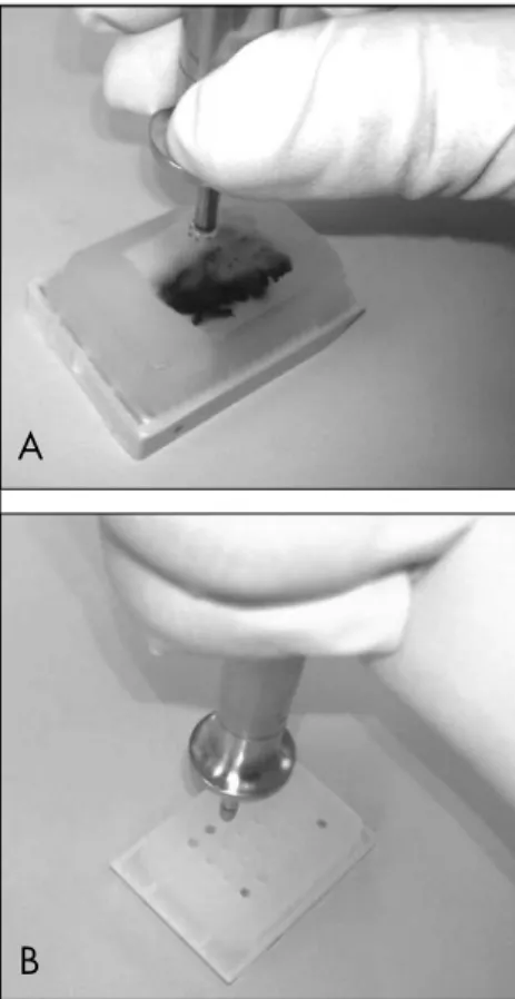
Eleven TMA blocks (Figures 1A and 1B) were constructed, each containing 24 tissue cores arranged in a 4 x 6 sector. In each TMA block, non-neoplastic breast tissue cores were also included as controls. After construction, 2 µm tissue sections were cut and placed on Superfrost® Plus glass slides. One hematoxy-lin and eosin-stained 4 µm section from each block was reviewed to confi rm the presence of morphologically representative areas of the original lesions.
Immunohistochemistry
Immunohistochemistry
Immunohistochemical staining was performed using the streptavidin-biotin-peroxidase technique (Lab Vision Corpora-tion, Fremont, California, United States) on each set of 11 glass slides comprising the TMAs. Antigen retrieval was performed by incubating the TMA sections in an antigen unmasking solution at pH 6.0 (Vector Labo-ratories, Inc., Burlingame, California, United States), or in ethylenediaminetetracetic acid (EDTA), pH 8 (Lab Vision) at 98º C. The
antigen retrieval time, antibodies, dilutions and suppliers are listed in Table 1. After washing in phosphate buffer solution (PBS), endogenous peroxidase activity was blocked by incubating the slides in a 3% hydrogen peroxide solution in methanol (Merck, Germany). The slides were incubated with blocking serum (Lab Vision) for 10 min-utes and then incubated with the specifi c antibody. Immunostaining was performed overnight at 4º C (p-cadherin and CK5) or
for one hour at room temperature (HER2, ER, and p63). After washing, the slides were incubated with biotinylated secondary anti-body, followed by streptavidin-conjugated peroxidase (Lab Vision). Diaminobenzidine was used as a chromogen. The tissues were
then counterstained with hematoxylin, and cover slips were attached using a permanent mounting solution (Zymed, San Francisco, California, United States).
The immunoreactions were classifi ed by estimating the percentage of tumor cells showing characteristic staining. In non-neo-plastic breast tissues, p63 showed nuclear positivity in myoepithelial cells. p-cadherin presented distinctive membranous and oc-casionally cytoplasmic immunoreactivity in non-neoplastic myoepithelial cells. CK5 staining was present in the myoepithe-lial cells of breast lobules and ducts. Two pathologists (RD and FS) evaluated the immunohistochemical staining. Because non-neoplastic mammary secretory cells do not express p-cadherin, either membra-nous or cytoplasmic immunoreactivity was considered positive when more than 10% of the neoplastic cells expressed this marker.12,24
Similarly, we adopted the same cutoff value for nuclear p63 and ER reactivity.
To evaluate HER2, the percentage of cells with membranous staining and the in-tensity of the staining were assessed. HER2 was evaluated according to the four-category system (0-3+) and was considered positive when 3+ was attributed. We compared our HER2 results with fl uorescence in situ
hybridization (FISH) information that had previously been obtained. Out of the 73 tu-mor cases with FISH information available, 27 were simultaneously positive and 39 were simultaneously negative. Only seven tumors presented discordant information, of which fi ve were HER2-positive and FISH-negative, and two were HER2-negative and FISH-positive. In cases of discordance, the FISH results were deemed to prevail.
Statistical analysis
Statistical analysis
Data entry was carried out in the Excel software program (Microsoft) and then the data were exported to the StatView statistical analysis program, version 5.0 (SAS Institute Inc., Cary, North Carolina, United States). The relationships between the expression patterns of the molecular biomarkers were evaluated by constructing contingency tables
and consequently applying the chi-squared test. The associations were considered statisti-cally signifi cant when p < 0.05.25
RESULTS
TMA validation
TMA validation
To validate the immunohistochemical analysis of the TMA, the ER expression for the sporadic cancers that was obtained in this study was compared with the data in the patients’ clinical records. Out of the 118 cases studied, 111 were concordant (86 were simul-taneously positive and 25 were simulsimul-taneously negative). Only three cases presented discord-ant information and four could not be inter-preted. The high percentage of concordance (97.4%) justifi ed the subsequent analysis.
B
Table 1. Antibodies used in immunohistochemical study of breast cancer tumors
Biomarker Antibody Clone Dilution Origin
CK5 Mmab XM26 1:80 Neomarkers, USA
p-cadherin Mmab 56 1:50 BDTransduction, USA
p63 Mmab 4A4 1:150 Neomarkers, USA
Erα Rmab SP-1 1:20 Neomarkers, USA
HER2 MMab NCL-L-CB11 1:60 Novocastra, UK
Figure 1A and 1B. Construction of tissue microarray blocks, each containing 24 tissue biopsy cores.
A
Immunohistochemical profi le
Immunohistochemical profi le
of familial and sporadic of familial and sporadic
breast cancer cases breast cancer cases
Immunohistochemical analysis was con-ducted on each group of 11 slides that made up the TMAs for p63, p-cadherin, CK5, ER and HER2. The results are presented in Table 2 and Figures 2 and 3.
The familial breast cancer phenotype was more frequently positive for the basal biomar-kers p-cadherin (p = 0.0004), p63 (p < 0.0001) and CK5 (p < 0.0001) than was the sporadic cancer phenotype. To evaluate the association between coexpression of basal biomarkers and the type of cancer, whether familial or sporadic breast cancer, the tumors were divided into six groups combining coexpression or lack of coexpression of the p63, p-cadherin and CK5 proteins. The presence of coexpression of the basal biomarkers CK5+/p63+ (odds ratio, OR = 34.34), grouped two by two, was associated with the familial breast cancer phenotype, while the absence of coexpression of the basal biomarkers /p63- and CK5-/p-cadherin- (OR = 0.13) was associated with the sporadic cancer phenotype. All the cases of basal phenotype (ER-/HER2-) were familial breast cancers (Table 3).
DISCUSSION
The objective of this study was to evalu-ate the expression of basal biomarkers such as p63, p-cadherin and CK5 in a series of familial breast cancer cases, using TMA. Familial breast cancer was characterized by the expression of these biomarkers. On the other hand, basal biomarker expression was signifi cantly lower in cases of the sporadic cancer phenotype.
The studies with cDNA microarrays carried out by Perou et al.3 and de Sorlie
et al.6 involved more than 8,000 genes
from which different phenotypes of breast cancer were characterized. This scenario points towards the existence of strong gene interaction in the carcinogenic process, al-though this technology is not yet applicable in clinical practice. It is necessary, however, to identify markers that represent these genetic spectrums, using techniques that are currently available and are applicable in clinical practice.
This study therefore makes a contribution in the sense that it shows that the proteins associated with the basal cell phenotype are much more frequently present in familial breast cancer phenotype, and that they are much more frequently absent in the cancers that are considered sporadic and which follow the luminal pattern.
Table 2. Results from immunohistochemical staining on tissue microarray of breast cancer tumors
Biomarker Interpretable biopsy cores n
Positive staining n (%)
Negative staining n (%)
p-cadherin 166 121 (73%) 45 (27%)
CK5 149 33 (21%) 116 (78%)
p63 154 31 (20%) 123 (78%)
ER 166 121 (73%) 45 (27%)
HER2 162 56 (34%) 106 (65%)
Table 3. Immunohistochemical profi les of familial (FH+) and sporadic (FH-) breast cancer phenotypes, as seen on tissue microarray
FH (+) n (%)
FH (-)
n (%) OR (95% CI)
p (chi-squared test)
Only p63+ 19 (40%) 12 (11%) 5.37 (2.16-13.53) < 0.0001
Only CK5+ 23 (49%) 10 (9.8) 8.42 (3.43-23.15) < 0.0001
Only p-cad+ 25 (51%) 21 (22%) 3.72 (1.67-8.36) 0.0004
p-cad+ and p63+ 12 (25%) 3 (3%) 9.89 (2.39-47.23) < 0.0001 p-cad- and p63- 15 (33%) 63 (69%) 0.22 (0.10-0.51) < 0.0001 CK5+ and p63+ 12 (26%) 1 (1%) 34.2 (4.33-731.14) < 0.0001 CK5- and p63- 17 (36%) 80 (80%) 0.13 (0.06-0.31) < 0.0001
p-cad+ and CK5+ 14 (30%) 6 (7%) 6.46 (2.07-20.9) 0.0003
p-cad- and CK5- 14 (29%) 68 (76%) 0.13 (0.05-0.30) < 0.0001
Only ER- 19 (38%) 26 (22%) 0.45 (0.21-0.99) 0.03
Only HER2 negative 37 (77%) 42 (42%) 0.22 (0.09-0.52) < 0.0001
Total number of cases (n) 50 118
FH (+): familial history positive; FH (-): familial history negative.
Positive cases were those that confi rmed the profi le under analysis. All others were considered negative.
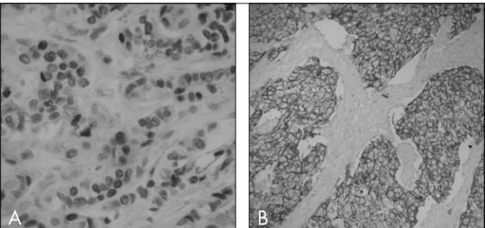
Figure 2. Sporadic breast carcinoma profi le. A) ER positivity; B) HER2 positivity.
Figure 3. Familial breast carcinoma profi le. A) p-cadherin positivity; B) p63 positivity.
A
B
1. Deugnier MA, Teuliere J, Faraldo MM, Thiery JP, Glukhova MA. The importance of being a myoepithelial cell. Breast Cancer Res. 2002;4(6):224-30.
2. Birnbaum D, Bertucci F, Ginestier C, Tagett R, Jacquemier J, Charafe-Jauffret E. Basal and luminal breast cancers: basic or luminous? (review). Int J Oncol. 2004;25(2):249-58. 3. Perou CM, Sorlie T, Eisen MB, et al. Molecular portraits of
human breast tumours. Nature. 2000;406(6797):747-52. 4. DiRenzo J, Signoretti S, Nakamura N, et al. Growth factor
require-ments and basal phenotype of an immortalized mammary epithelial cell line. Cancer Res. 2002;62(1):89-98. 5. van’t Veer LJ, Dai H, van de Vijver MJ, et al. Gene expression
profi ling predicts clinical outcome of breast cancer. Nature. 2002;415(6871):530-6.
6. Sorlie T, Wang Y, Xiao C, et al. Distinct molecular mechanisms underly-ing clinically relevant subtypes of breast cancer: gene expression analyses across three different platforms. BMC Genomics. 2006;7:127. 7. Ali S, Coombes RC. Endocrine-responsive breast cancer
and strategies for combating resistance. Nature Rev Cancer. 2002;2(2):101-12.
8. Callagy G, Cattaneo E, Daigo Y, et al. Molecular classifi cation of breast carcinomas using tissue microarrays. Diagn Mol Pathol. 2003;12(1):27-34.
9. Matos I, Dufl oth R, Alvarenga M, Zeferino LC, Schmitt F. p63, cytokeratin 5, and P-cadherin: three molecular markers to distinguish basal phenotype in breast carcinomas. Virchows Arch. 2005;447(4):688-94.
10. Bocker W, Moll R, Poremba C, et al. Common adult stem cells in the human breast give rise to glandular and myoepithelial cell lineages: a new cell biological concept. Lab Invest. 2002;82(6):737-46. 11. Boecker W, Buerger H. Evidence of progenitor cells of glandular
and myoepithelial cell lineages in the human adult female breast epithelium: a new progenitor (adult stem) cell concept. Cell Prolif. 2003;36(Suppl 1):73-84.
12. Reis-Filho JS, Simpson PT, Martins A, Preto A, Gartner F, Schmitt FC. Distribution of p63, cytokeratins 5/6 and cytokeratin 14 in 51 normal and 400 neoplastic human tissue samples using TARP-4 multi-tumor tissue microarray. Virchows Arch. 2003;443(2):122-32.
13. McKeon F. p63 and the epithelial stem cell: more than status quo? Genes Dev. 2004;18(5):465-9.
14. Westfall MD, Pietenpol JA. p63: Molecular complexity in devel-opment and cancer. Carcinogenesis. 2004;25(6):857-64. 15. Peralta Soler A, Knudsen KA, Salazar H, Han AC, Keshgegian
AA. P-cadherin expression in breast carcinoma indicates poor survival. Cancer. 1999;86(7):1263-72.
16. Paredes J, Milanezi F, Reis-Filho JS, Leitão D, Athanazio D, Schmitt F. Aberrant P-cadherin expression: is it associated with estrogen-independent growth in breast cancer? Pathol Res Pract. 2002;198(12):795-801. 17. Han AC, Soler AP, Knudsen KA, Salazar H.Distinct cadherin
profi les in special variant carcinomas and other tumors of the breast. Hum Pathol. 1999;30(9):1035-9.
18. Gamallo C, Moreno-Bueno G, Sarrió D, Calero F, Hardisson D, Palacios J. The prognostic signifi cance of P-cadherin in infi ltrat-ing ductal breast carcinoma. Mod Pathol. 2001;14(7):650-4. 19. Madhavan M, Srinivas P, Abraham E, et al.Cadherins as
predic-tive markers of nodal metastasis in breast cancer. Mod Pathol. 2001;14(5):423-7.
20. Palacios J, Honrado E, Osorio A, et al. Immunohistochemical characteristics defi ned by tissue microarray of hereditary breast cancer not attributable to BRCA1 or BRCA2 mutations: differences from breast carcinomas arising in BRCA1 and BRCA2 mutation carriers. Clin Cancer Res. 2003;9(10 Pt 1):3606-14.
21. Palacios J, Honrado E, Osorio A, Diez O, Rivas C, Benítez J. Re: Germline BRCA1 mutations and a basal epithelial phenotype in breast cancer. J Natl Cancer Inst. 2004;96(9):712-4. 22. Pathology of familial breast cancer: differences between breast
cancers in carriers of BRCA1 or BRCA2 mutations and sporadic cases. Breast Cancer Linkage Consortium. Lancet. 1997;349(9064):1505-10.
23. Ford D, Easton DF, Stratton M, et al. Genetic heterogeneity and penetrance analysis of the BRCA1 and BRCA2 genes in breast cancer families. The Breast Cancer Linkage Consortium. Am J Hum Genet.1998;62(3):676-89.
24. Reis-Filho JS, Milanezi F, Paredes J, et al. Novel and classic myoepi-thelial/stem cell markers in metaplastic carcinomas of the breast. Appl Immunohistochem Mol Morphol. 2003;11(1):1-8.
25. Altman DG. Practical statistics for medical research. London: Chapman & Hall; 1991.
26. Foulkes WD. BRCA1 functions as a breast stem cell regulator. J Med Genet. 2004;41(1):1-5.
27. Warnberg F, Nordgren H, Bergkvist L, Holmberg L. Tumour markers in breast carcinoma correlate with grade rather than with invasiveness. Br J Cancer. 2001;85(6):869-74. 28. Aubele MM, Cummings MC, Mattis AE, et al. Accumulation of
chromosomal imbalances from intraductal proliferative lesions to adjacent in situ and invasive ductal breast cancer. Diagn Mol Pathol. 2000;9(1):14-9.
29. Farabegoli F, Champeme MH, Bieche I, et al. Genetic pathways in the evolution of breast ductal carcinoma in situ. J Pathol. 2002;196(3):280-6.
30. Lee JS, John EM, McGuire V, et al. Breast and ovarian cancer in relatives of cancer patients, with and without BRCA mutations. Cancer Epidemiol Biomarkers Prev. 2006;15(2):359-63. 31. Lakhani SR, Reis-Filho JS, Fulford L, et al. Prediction of BRCA1
status in patients with breast cancer using estrogen receptor and basal phenotype. Clin Cancer Res. 2005;11(14):5175-80. 32. Foulkes WD, Stefansson IM, Chappuis PO, et al. Germline
BRCA1 mutations and a basal epithelial phenotype in breast cancer. J Natl Cancer Inst. 2003;95(19):1482-5.
33. Tischkowitz MD, Foulkes WD. The basal phenotype of BRCA1-related breast cancer: past, present and future. Cell Cycle. 2006;5(9):963-7.
Sources of funding:This study was funded by: Coordenação
de aperfeiçoamento de pessoal de nível superior (Capes) [BEX 244802-5], Brazil; Fundo de Apoio ao Ensino e Pesquisa (Faep) da Universidade Estadual de Campinas (Unicamp), Brazil; Fundação Luso-Americana para o Desenvolvimento (Flad) [refL-V-172/2002], Portugal.
Confl icts of interest: None
Date of fi rst submission: October 15, 2006
Last received: June 20, 2007
Accepted:June 20, 2007
REFERENCES
The estrogens that bind to the ER located in the cell nucleus, and the growth factors that bind to the HER2 protein located in the cell membrane represent two different routes for cell proliferation stimuli originating outside the cell. Therefore, it is possible to hypothesize that, when these lesions do not express ER or HER2 protein, the stimuli for cell proliferation would be determined by factors inside the cell or at least would be less dependent on external stimuli. Thus, the development of ductal carcinoma in situ (DCIS) would be more heavily dependent on external factors modulated by ER and HER2, while the progression to invasive cancer would be less dependent on this route, possibly because of the progressive accumulation of genetic altera-tions that occur in the malignant cells.26-29
Perou et al.3 characterized basal-subtype
breast cancers as ER and HER2-negative. Therefore, these would be the cancers in which cell proliferation would depend less on external factors. Several studies have shown that women with the breast cancer 1 (BRCA1) gene mutation present breast cancer with a genetic expression
pattern that is compatible with the basal sub-type.5,30-33 However, the multistage process of
carcinogenesis in the breast epithelium of women with genetic abnormalities who are highly susceptible to carcinogenesis would not depend on ER or HER2 protein expression, since the stimuli required for cell proliferation would be determined by the abnormalities present in the cell genome. Obviously, this explanation does not exclude the possibility that these cells may also have ER and HER2 protein expression.
Foulkes and colleagues26,32 studied this
phenotype and found that around 40% of basal cancers presented a mutation in the BRCA1 gene, thus suggesting that this gene has a regula-tory function in the progenitor cells of the breast and that it promotes the orderly transition of the cells to the glandular epithelial phenotype. Mutation of this gene would lead to interruption of the differentiation process, and the cells would remain stagnant in a primitive basal phenotype with no proliferation control.
The results from our study are in agreement with this theory on carcinogenesis, since they
show that the familial breast cancer phenotype is more frequently ER (-) and HER2 protein (-) than are sporadic cancers. Moreover, the p63, p-cadherin and CK5 biomarkers showed that fa-milial breast cancer cases more frequently present expressions compatible with the basal pheno-type. As a rule, the sporadic cancer phenotype presents lower frequency of expression of these biomarkers. The remainder of our results are in full accordance with those in the literature.
CONCLUSIONS
AUTHOR INFORMATION
Rozany Mucha Dufl oth, MD, PhD. Department of Pathology,
Universidade Federal de Santa Catarina, Florianópolis, Santa Catarina, Brazil.
Irina Matos, MSc. Institute of Pathology and Molecular
Immu-nology, Universidade do Porto, Porto, Portugal.
Fernando Schmitt, MD, PhD. Institute of Pathology and
Mole-cular Immunology, Universidade do Porto and Faculdade de Medicina da Universidade do Porto (FMUP), Porto, Portugal.
Luiz Carlos Zeferino, MD, PhD.Department of Obstetrics
and Gynecology, Universidade Estadual de Campinas, Campinas, São Paulo, Brazil.
Address for correspondence:
Rozany Mucha Dufl oth
Rodovia Amaro Antônio Vieira, 2.463 — Apto. 506 C Residencial Solar de Francavilla — Itacorubi Florianópolis (SC) — Brasil — CEP 88034-102 Tel. (+55 48) 331-9473
Fax. (+55 48) 3331-9542 E-mail: [email protected]
Copyright © 2007, Associação Paulista de Medicina
RESUMO
Tissue microarrays para testar as proteínas basais no câncer de mama familiar
CONTEXTO E OBJETIVO: As proteínas p63, p-cad e CK 5 são expressas em células basais/mioepiteliais da mama. Entretanto a expressão dessas proteínas no câncer esporádico e familiar ainda não é bem conhecida. O objetivo do estudo foi estudar essas proteínas no câncer de mama, utilizando a técnica de
tissue microarray, assim como ER e HER2.
TIPO DE ESTUDO E LOCAL: Estudo transversal, realizado no Centro de Atenção Integral à Saúde da Mulher, Universidade Estadual de Campinas, Brasil, e no Instituto de Patologia e Imunologia Molecular da Univer-sidade do Porto, Portugal.
MÉTODOS: O estudo analisou a expressão das proteínas p63, CK 5, p-cad, ER e HER2 numa série de 168 casos de câncer de mama. Os critérios utilizados para identifi car as mulheres com alto risco foram os do Breast Cancer Linkage Consortium.
RESULTADOS: A série de câncer familiar foi freqüentemente mais positiva para as proteínas basais p-cadherin (p = 0,0004), p63 (p < 0,0001) e CK 5 (p < 0,0001) que o câncer esporádico. A presença da co-expressão das proteínas basais CK 5+/p63+, agrupados dois a dois, foi associada com o fenótipo do câncer familiar (odds ratio, OR = 34,34), enquanto que sua ausência foi com o câncer esporádico (OR = 0,13).
CONCLUSÕES: O câncer da mama familiar está associado aos marcadores de células basais proteínas p63, p-cad e CK 5, utilizando-se a técnica de tissue microarray. Por fi m, parece legítima a interpretação destes resultados como mais uma evidência que suporta a hipótese da existência de células precursoras do câncer familiar da mama. O conhecimento dos perfi s de expressão destas células, bem como das vias de sinalização envolvidas, benefi ciarão o entendimento da carcinogênese mamária.