712
Rev Soc Bras Med Trop 50(5):712-714, September-October, 2017 doi: 10.1590/0037-8682-0057-2017
Case Report
Corresponding author: Dr. Nader Shakibazad.
e-mail: [email protected]
Received 14 February 2017
Accepted 20 June 2017
Colonic basidiobolomycosis with liver involvement
masquerading as gastrointestinal lymphoma:
a case report and literature review
Omid Reza Zekavat
[1], Babak Abdolkarimi
[2], Gholamreza Pouladfar
[3],
Gholamreza Fathpour
[1], Maral Mokhtari
[4]and Nader Shakibazad
[1][1]. Hematology Research Center, Shiraz University of Medical Sciences, Shiraz, Iran. [2]. Department of Pediatric Hematology/Oncology, School of Medicine, Lorestan University of Medical Sciences, Khorramabad, Iran. [3]. Professor Alborzi Clinical Microbiology Research Center,
Shiraz University of Medical Sciences, Shiraz, Iran. [4]. Department of Pathology, Shiraz University of Medical Sciences, Shiraz, Iran.
Abstract
Basidiobolomycosis is an unusual fungal skin infection that rarely involves the gastrointestinal tract. This study reported a 5-year-old boy with gastrointestinal basidiobolomycosis that had been misdiagnosed as gastrointestinal lymphoma. He was treated by surgical resection and a combination of posaconazole and amphotericin B deoxycholate with an acceptable response and no recurrence.
Keywords: Basidiobolomycosis. Lymphoma. Gastrointestinal.
INTRODUCTION
Basidiobolomycosis (BM) is a rare infection caused by the fungus Basidiobolus ranarum (B. ranarum). BM is an environmental saprophyte found throughout the world and often infects immunocompetent patients. Patients with B. ranarum
infection may present with subcutaneous, gastrointestinal, or systemic lesions1,2.
Diagnosis of gastrointestinal BM is dificult, and its clinical presentation is nonspeciic, with no identiiable risk factors. An
optimal treatment regimen for this uncommon infection has not yet been established. This study presents a boy with colonic BM involving the liver, masquerading as gastrointestinal lymphoma.
CASE REPORT
A 5-year-old boy, living in Bushehr province in the south
of Iran, was referred to an oncology center afiliated with
Shiraz University of Medical Sciences. He had a 2-month history of diffuse abdominal pain, non-bilious vomiting, poor appetite, weight loss, and a detectable mass on abdominal sonography. He had no fever, diarrhea, constipation, jaundice,
or lower GI bleeding. He also had no recent history of travel abroad.
In his physical examination, he seemed ill and emaciated, his b o d y temperature was 39°C, and his liver was tender on palpation 3cm inferior to the costal margin. His laboratory results are shown in Table 1.
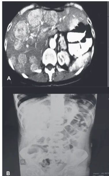
Abdominopelvic CT (Computerized Tomography)scan showed multiple heterogeneous densities in the liver and a significantly thickened edematous intestinal wall with
stratiication of the ascending colon throughout the hepatic lexure, the adjacent part of the transverse colon, the cecum,
and the terminal ileum. A mass measuring about 3.5 × 5cm
iniltrated to the cecum and ascending colon as well as the terminal ileum, extending to the hepatic lexure in direct contact
with the gallbladder. Multiple lymphadenopathies were detected. The mass was similar to Castleman's disease, lymphoma, or tuberculosis (Figure 1). Accordingly, gastrointestinal lymphoma was suspected.
Colonoscopy with multiple biopsies was performed,
revealing acute and chronic inlammation with numerous
eosinophils and poorly shaped granulomas. The granulomas were surrounded with giant cells, morphologically consistent with fungal elements, and periodic acid-Schiff and Gomori-methenamine silver stains were both positive. These symptoms are indicative of BM (Figure 2).
713 Zekavat OR et. al -Basidiobolomycosis masquerading as lymphoma
A
B
FIGURE 1 - A: Abdominal computed tomography scan shows multiple homogenous parenchymal densities in the liver. B: Plain abdominal radiogram, upright.
FIGURE 2 - Histopathology of surgically resected abdominal mass: hematoxylin and eosin stain (× 100), demonstrating granulomatosis inlammation with central necrosis and numerous eosinophils.
Variable Value
Hb 12g/dL
WBC 23,900/µL (EOS: 11% NEU: 59%)
Platelet 507,000/µL
Prothrombin time 12 sec
PTT 32 sec
LDH 3996U/L
Total bilirubin 3.5mg/dL
Direct bilirubin 2.5mg/dL
ALT 55IU/L
AST 65IU/L
Gamma-GT 23U/L
Alkaline phosphatase 220U/L
BUN 12mg/dL
Creatinine 0.4mg/dL
CRP 90mg/L
ESR 35mm/h
G6PD Suficient
HBSAg Negative
Anti-HCV Negative
Anti-HIV Negative
EBV-VCA-IgM Negative
CMV-IgM Negative
Blood culture Negative
Immunoelectrophoresis Normal
TABLE 1
Laboratory results.
Hb: hemoglobin; WBC: white blood cell; EOS: eosinophil; NEU: neutrophil;
PTT: partial thromboplastin time; LDH: lactate dehydrogenase; ALT: alanine aminotransferase; AST: aspartate aminotransferase; GT: glutamyl transferase;
BUN: blood urea nitrogen; CRP: C-reactive protein; ESR: erythrocyte sedimentation rate; G6PD: glucose-6-phosphate dehydrogenase; HBSAg:
hepatitis B virus surface antigen; HCV: hepatitis C virus; HIV: human
immunodeiciency virus; EBV-VCA-IgM: Epstein-Barr virus-viral capsid
antigen-immunoglobulin M; CMV-IgM: cytomegalovirus-immunoglobulin M.
internal oblique and transversus abdominis muscle, and there was a small area of peritoneal ulceration, which was excised by a surrounding margin of healthy tissue. The specimen was sent for histopathological evaluation and culture, which revealed the presence of fungal hyphae with large width and thin walls, surrounded by eosinophils with multinucleated giant cells, lymphocytes, and histiocytes. A biopsy specimen from the omentum showed fat necrosis. Culture results of the mass indicated white to grey colonies with radiated folds that were consistent with gastrointestinal basidiobolomycosis. The results
714
Rev Soc Bras Med Trop 50(5):712-714, September-October, 2017
dihydrorhodamine (DHR) low cytometric test, immunoglobulin
and anti-tetanus antibody levels, and CH50 levels were in the normal range. HIV serology also was negative.
Treatment with amphotericin B (intravenous 1mg/kg/day for 2 months) and posaconazole (200mg by mouth four times per day) was started. The patient was followed closely by means of physical exams and abdominal computed tomography scans, which revealed no recurrence 6 months after starting therapy with posaconazole. Written informed consent was obtained from the parents.
DISCUSSION
Basidiobolomycosis is an unusual fungal infection with skin manifestation and rarely involves other systems. It is caused by
B. ranarum, which infects immunocompromised patients and is an opportunistic pathogen in immunocompetent individuals2,
such as the healthy, immunocompetent 5 year-old patient in the present case. Most similar cases with cutaneous involvement have been reported from tropical areas where the climate is warm and humid, such as Iran. In recent years, several cases of gastrointestinal BM in Iran have been reported3-6.
It is unclear how the fungus is introduced into the host’s gastrointestinal tract, but it may occur through ingestion of contaminated soil, food, or exposure to animal feces4.
Alternatively, it may be a zoonotic fungus7,8.
The clinical manifestations of gastrointestinal BM include abdominal pain, fever, vomiting, weight loss, and abdominal mass, which can be found during abdominal examination, by imaging, or during laparotomy, as it was in the current patient. The presence of an abdominal mass may be misdiagnosed as malignancy, especially Burkitt’s lymphoma or as an
inlammatory process such as appendicular mass, inlammatory
bowel disease, or other infectious diseases including intestinal tuberculosis, sarcoidosis, and amebiasis9. Lack of awareness
or facilities to diagnose such a rare disease contributes to missing an early diagnosis and an increased risk of morbidity. Leukocytosis, marked eosinophilia, and an elevated erythrocyte sedimentation rate and C-reactive protein level were present in the current case, as well as in prior reports10.
Although there have been some reports of clinical improvement with antifungal therapy alone, most patients with disseminated abdominal infection have received a combination of surgical and medical therapies. An optimal treatment regimen for this uncommon fungal infection has not yet been established. The best choice of antifungal agent is not clear, but itraconazole has been used with success in many reports9,11. A study regarding
the use of antifungal agents showed that itraconazole has been used most frequently for BM (73%), followed by amphotericin (22%), ketoconazole (8%), and voriconazole (5%); in addition, potassium iodide and trimethoprim/sulfamethoxazole may also
have some clinical eficacy12. Generally, antifungal therapy is
used for 8 months, and overall survival is estimated at 80%. Clinical failure has been described with amphotericin B1,11,12.
The use of posaconazole in the present case had several potential advantages. For example, there was no need for dose
adjustment in hepatic or renal insuficiency. Moreover, it was
well tolerated with limited side effects, and its absorption was
not inhibited by medications that affect gastric acidity12.
However, the limitationsof using posaconazole include cost, the lack of an intravenous preparation for hospitalized patients, and the need for therapeutic monitoring due to variable absorption. Gastrointestinal BM is an emerging disease in the south of Iran, so special attention should be given to patients exhibiting an abdominal mass with localized eosinophilia. Moreover, the present case demonstrates the importance of antifungal therapy. This study introduces posaconazole as an effective single agent treatment with minimum complications during a prolonged treatment plan.
Conlict of interest
The authors declare that there is no conlict of interest.
REFERENCES
1. Zabolinejad N, Naseri A, Davoudi Y, Joudi M, Aelami MH. Colonic basidiobolomycosis in a child: report of a culture-proven case. Int J Infect Dis. 2014;22:41-3.
2. Ejtehadi F, Anushiravani A, Bananzadeh A, Geramizadeh B. Gastrointestinal basidiobolomycosis accompanied by liver involvement: a case report. Iran Red Crescent Med J. 2014;16(9):e14109.
3. Geramizadeh B, Foroughi R, Keshtkar-Jahromi M, Malek-Hosseini SA, Alborzi A. Gastrointestinal basidiobolomycosis, an emerging infection in the immunocompetent host: a report of 14 patients. J Med Microbiol. 2012;61(Pt 12):1770-4.
4. Geramizadeh B, Heidari M, Shekarkhar G. Gastrointestinal basidiobolomycosis, a rare and under-diagnosed fungal infection in immunocompetent hosts: a review article. Iran J Med Sci. 2015;40(2):90-7.
5. Seyedmousavi S, Guillot J, Tolooe A, Verweij PE, de Hoog GS. Neglected fungal zoonoses: hidden threats to man and animals. Clin Microbiol Infect. 2015;21(5):416-25.
6. Van den Berk GE, Noorduyn LA, van Ketel RJ, van Leeuwen J, Bemelman WA, Prins JM. A fatal pseudo-tumour: disseminated basidiobolomycosis. BMC Infect Dis. 2006;6:140.
7. Bittencourt AL, Andrade JA, Carvalho EM. Basidiobolomicose: Registro de um caso com aspectos incomuns. Rev Inst Med Trop Sao Paulo. 1987;29(6):381-4.
8. Pasha TM, Leighton JA, Smilack JD, Heppell J, Colby TV, Kaufman L. Basidiobolomycosis: an unusual fungal infection mimicking inlammatory bowel disease. Gastroenterology. 1997;112(1):250-4.
9. Mathew R, Kumaravel S, Kuruvilla S, Varghese RG, Shashikala MD, Srinivasan S, et al. Successful treatment of extensive basidiobolomycosis with oral itraconazole in a child. Int J Dermatol. 2005;44(7):572-5.
10. Rabie ME, El Hakeem I, Al-Shraim M, Al Skini MS, Jamil S. Basidiobolomycosis of the colon masquerading as stenotic colon cancer. Case Rep Surg. 2011;(2011):Article ID 685460.
11. Maani AS, Paul G, Jardani A, Nayar M, Lawati F, Al-Baluishi S, et al. Gastrointestinal basidiobolomycosis: irst case report from Oman and literature review. Sultan Qaboos Univ Med J. 2014; 14(2):e241-4.