Atypical presentation of a common opportunistic infection in advanced AIDS
Texto
Imagem


Documentos relacionados
Technological developments in imaging, such as elastography, magnetic resonance imaging-guided biopsy, magnetic resonance imaging-ultrasonography fusion, can revolutionize the
Spectrum of indings on magnetic resonance imaging of the brain in patients with neurological manifestations of dengue fever..
The role of diffusion magnetic resonance imaging in Par - kinson’s disease and in the differential diagnosis with
The role of diffusion magnetic resonance imaging in Par- kinson’s disease and in the differential diagnosis with
Figure 3 - Magnetic resonance imaging scan of the head revealing a large osteolytic lesion in the hard palate and in the superior alveolar arcade, forming an
Objectives: To evaluate the use of magnetic resonance imaging in patients with β -thalassemia and to compare T2* magnetic resonance imaging results with
Magnetic resonance imaging of the abdomen showed multiple solid, nodular, hypervascular lesions primarily in the right liver lobe, and a dense, heterogeneous, 10.1 x 6.2cm
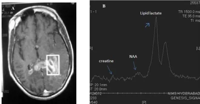
Proton magnetic resonance spectroscopy (MRS) of the human brain has proven to be a useful technique in several neurological and psychiatric disorders and benefits from higher
