Fisioter Mov. 2016 Apr/June;29(2):411-9 )SSN -Fisioter. Mov., Curitiba, v. , n. , p. - , Apr./June
Licenciado sob uma Licença Creative Commons DO): http://dx.doi.org. . / - . . .AO
[T]
Head and neck lymphedema: what is the physical
therapy approach? A literature review
[)]
Linfedema de cabeça e pescoço: qual a abordagem
da i sioterapia? Uma revisão de literatura
A]Lunara Basqueroto Della Justina [a], Mirella Dias [b]*
[a] Universidade do Planalto Catarinense UN)PLAC , Lages, SC, Brazil [b] Universidade do Sul de Santa Catarina UN)SUL , Palhoça, SC, Brazil
[RAbstract
Introduction: (ead and neck lymphedema is considered a chronic and complex complication with potential
to cause physical, functional, emotional and social impairment. Objective: To identify the approaches to
physi-cal therapy used to treat head and neck cancer-related lymphedema. Method: A bibliographic search was
con-ducted in February and March in books and electronic databases, L)LACS, MEDL)NE, SC)ELO, Cochrane, PEDro, and BDTD using the following keywords: lymphedema, treatment, head and neck cancer, and physical therapy connected by the Boolean operator AND without a speci ic time frame. Results and discussion: Early
diagnosis and assessment is key to properly managing and effectively treating lymphedema. Diagnosis is re-ached through clinical history and physical assessment by measuring the distance between two anatomical landmarks, circumference measures, and lymphedema rating scales. Complex decongestive therapy, which includes manual lymph drainage, compressive bandaging, kinesiotherapy and skin care, is the technique most frequently used and currently considered to be the gold standard. Conclusions: No consensus is reported in
the literature in regard to a standard procedure to assess and treat head and neck cancer-related lymphedema. Assessments and treatments described in the literature are mainly restricted to the limbs; therefore, further studies are needed to support effective clinical actions in the physical therapy approach to this condition.
Keywords: Lymphedema. (ead and neck neoplasms. Physical therapy modalities. Therapeutics.
Resumo
Introdução: O linfedema de cabeça e pescoço é considerado uma complicação crônica e complexa que pode provocar prejuízos ϔísicos, funcionais, emocionais e sociais importantes. Objetivo: Identiϔicar as abordagens ϔisioterapêuticas utilizadas no linfedema pós-tratamento de câncer de cabeça e pescoço. Método: Para o de-lineamento dessa revisão, foi realizado levantamento bibliográϔico nos meses de fevereiro e março de 2012, em livros e nas bases de dados eletrônicas LILACS, MEDLINE, SCIELO, Cochrane, PEDro, BDTD utilizando as palavras-chave: linfedema, tratamento, câncer de cabeça e pescoço, ϔisioterapia ligadas pelo operador boleano AND, sem estabelecer data limite para a publicação. Resultados e discussão: O diagnóstico precoce e a ava-liação são fundamentais para o manejo adequado e tratamento efetivo do linfedema. O diagnóstico é realizado através da história clínica e do exame ϔísico pela medida da distância entre dois pontos anatômicos, das medi-das de circunferência e escalas de intensidade do linfedema. A ϔisioterapia complexa descongestiva ou linfote-rapia que inclue: drenagem linfática manual, bandagem compressiva, cinesiotelinfote-rapia e cuidados com a pele, é a técnica mais utilizada na intervenção do linfedema, considerada padrão ouro atualmente. Conclusões: De acordo com a literatura revisada, não há consenso sobre o procedimento padrão para avaliação e tratamento do linfedema após o câncer de cabeça e pescoço. A avaliação e o tratamento para linfedema descritos na lite-ratura estão restritos, principalmente, a membros, sendo assim, novos estudos devem ser realizados a ϔim de subsidiar ações clínicas efetivas na abordagem ϔisioterapêutica desta sequela.
Palavras-chave: Linfedema. Neoplasias de cabeça e pescoço. Modalidades de ϔisioterapia. Tratamento.
Introduction
Estimates from the National Cancer )nstitute of Brazil – )NCA indicate an incidence of approxi-mately , cases of cancer in and in Brazil. Of these, , are new cases of oral can-cer and , are new cases of laryngeal cancer . These two types of cancer are part of the group of malignancies of the head and neck .
Malignant neoplasms of the head and neck repre-sented by tumors located in the upper aero-digestive tract present signi icant morbidity and mortality . Because it is in an anatomically complex area, head and neck cancer can lead to signi icant changes in vital structures, resulting in aesthetic and functional sequelae, especially in the face, temporo-mandibular joint, shoulder girdle and lungs , .
The therapeutic methods used to treat head and neck neoplasms include surgical resection, radio-therapy, and chemoradio-therapy, or a combination of these , , . Even though many of these treat-ments are effective in eradicating the tumor and/ or generate better results in regard to survival, local control and preservation of function, these interven-tions usually involve co-morbidities . The follow-up of patients after cancer treatment is important because there may be some after-effects, such as lymphedema , , .
(ead and neck lymphedema that results from thera-pies used in the treatment of head and neck cancer is considered a chronic and complex complication that manifests as a feeling of heaviness or tightness and per-manent discomfort in the affected area; visible swallow-ing may or may not exist . )t may, however, become evident and result in important physical, functional , , emotional and social impairment, negatively im-pacting the quality of life of these individuals , . Complex decongestive therapy, which includes manual lymph drainage, compressive bandaging or elastic bandages, kinesiotherapy, and skin care, is currently acknowledged as the most ef icacious therapeutic method to treat lymphedema .
Given the need for further clari ication regarding approaches to physical therapy in the treatment of lymphedema for patients with head and neck cancer, and also due to a lack of scienti ic studies addressing the topic, this review’s aim was to identify the physi-cal therapy approaches used to treat head and neck cancer-related lymphedema.
Method
Fisioter Mov. 2016 Apr/June;29(2):411-9 Head and neck lymphedema
413 identify publications addressing the topic using the
keywords lymphedema, treatment, head and neck neoplasms, and physical therapy and their equivalent in Portuguese and Spanish, connected by the Boolean operator AND, without establishing a time frame.
A sample from the result of the bibliographic search using this strategy was selected using the following inclusion criteria: full texts with no re-striction for study design or time frame; written in English, Portuguese, or Spanish; target population being adults; and studies presenting information con-cerning the topic under study. Studies identi ied in more than one database were considered only once. Analysis of papers included reading titles, abstracts and full texts after selection.
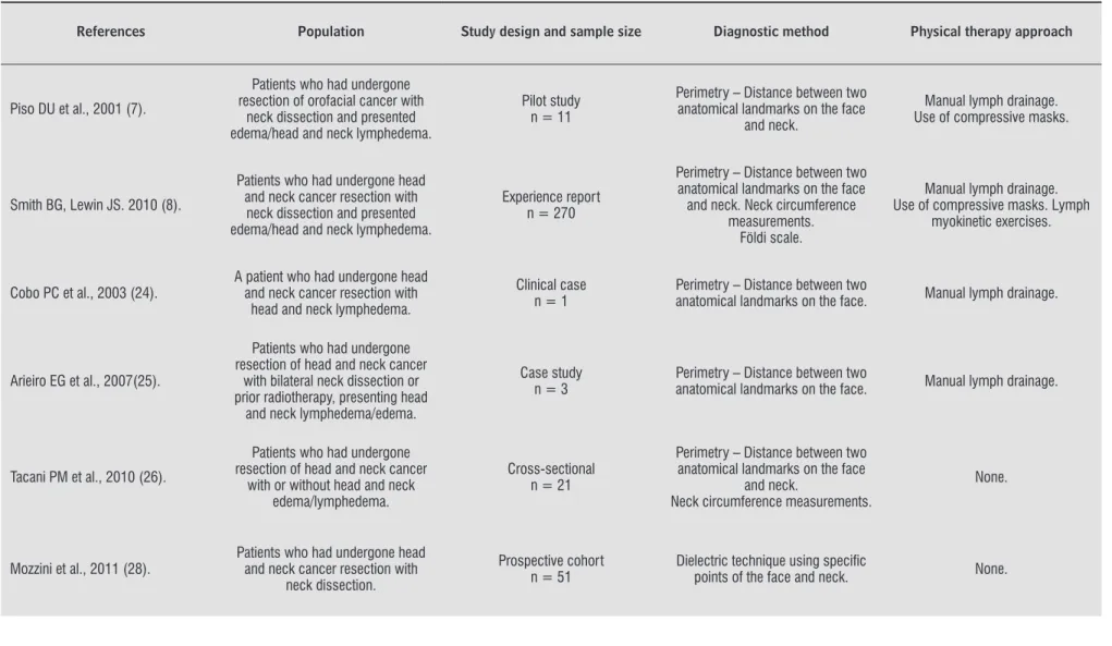
The search resulted in studies. After applying inclusion criteria and disregarding repeated papers, a total of six studies were ultimately included. Table presents the characteristics of the studies included in the review with respective physical therapy ap-proaches used to treat head and neck cancer-relat-ed lymphcancer-relat-edema.
Therefore, the studies selected were organized in order to clarify the general aspects of head and neck lymphedema, the instruments and techniques used to measure head and neck lymphedema in the physical therapy approach and inally, the physical therapy procedures used for individuals with head and neck lymphedema.
Results and Discussion
General aspects of head and neck lymphedema
(ead and neck lymphedema, one of the conse-quences of head and neck cancer treatment, occurs due to a dysfunction in cervicofacial lymphatic drain-age that is caused by surgical procedures in which some structures are removed, such as the neck lymph nodes, and due to radiotherapy applied to the area, which predisposes the patient to the development of lymphedema , , , .
Lymphedema, considered a chronic and pro-gressive pathological condition, involves excess interstitial luid with high protein concentration that originates from an inef icient lymphatic sys-tem. Lymphedema develops due to an imbalance between lymphatic demand and the system’s abil-ity to drain the lymph; that is, drainage abilabil-ity is
impaired due to the destruction of or an obstruction in the lymphatic route. (ence, as a consequence, or-gans and tissues are compressed, leading to pain and functional changes. )n addition to physical changes, the lymphedema can cause psychological and social problems accruing from aesthetic changes, affecting self-esteem and social acceptability and consider-ably reducing patient quality of life - .
Lymphedema may manifest in patients with head and neck cancer as visible edema in the face and neck area with alterations in the skin’s mechanical properties, especially elasticity and viscosity, and sensory changes , . Additionally, it may af-fect aerodigestive routes, such as in the oral cavity
tongue , pharynx and larynx - . )mpairment in these areas may affect communication, feeding, breathing, in some cases even impeding walking if sight is impaired. Reduced amplitude of cervical movement is common, as well as dysfunctions in the shoulder girdle - .
Most patients with head and neck cancer are di-agnosed in the advanced stages of the disease . The late diagnosis of these neoplasms is related to a worse prognosis, raising the likelihood of sequelae and deformities. When a head and neck cancer diag-nosis is delayed, the expected outcome is worse, with more invasive and mutilating therapeutic interven-tions that affect the lymphatic system and increase the risk of lymphedema , .
The presence of lymphedema among patients after head and neck cancer treatment is usually neglected and under-recognized; it is less per-ceived than lymphedema of the extremities . The overall prevalence of lymphedema among head and neck cancer patients ranges from % and % , , . This discrepancy may be explained by a lack of a standard diagnosis and universal assessment criteria, as well as differ-ences among the therapeutic procedures used to treat this type of cancer , , .
Deng et al conducted a study in the United States to verify the prevalence of lymphedema in a sample composed of patients after head and neck cancer treatment. A total of . % of the patients presented lymphedema. The study reports a prevalence of external lymphedema, internal lymphedema, and combined lymphede-ma concomitantly internal and external lymph-edema with a prevalence of . %, . % and
Fi
si
o
te
r
Mo
v.
2
0
1
6
A
p
r/
J
u
n
e
;2
9
(2
):4
1
1
-9
De
lla
J
u
sti
n
a
LB
, Di
a
s
M.
Table 1 - Characteristics of studies addressing physical therapy approaches to address head and neck cancer-related lymphedema
References Population Study design and sample size Diagnostic method Physical therapy approach
Piso DU et al., 2001 (7).
Patients who had undergone resection of orofacial cancer with
neck dissection and presented edema/head and neck lymphedema.
Pilot study n = 11
Perimetry – Distance between two anatomical landmarks on the face
and neck.
Manual lymph drainage. Use of compressive masks.
Smith BG, Lewin JS. 2010 (8).
Patients who had undergone head and neck cancer resection with
neck dissection and presented edema/head and neck lymphedema.
Experience report n = 270
Perimetry – Distance between two anatomical landmarks on the face and neck. Neck circumference
measurements. Földi scale.
Manual lymph drainage. Use of compressive masks. Lymph
myokinetic exercises.
Cobo PC et al., 2003 (24).
A patient who had undergone head and neck cancer resection with
head and neck lymphedema.
Clinical case n = 1
Perimetry – Distance between two
anatomical landmarks on the face. Manual lymph drainage.
Arieiro EG et al., 2007(25).
Patients who had undergone resection of head and neck cancer
with bilateral neck dissection or prior radiotherapy, presenting head
and neck lymphedema/edema.
Case study n = 3
Perimetry – Distance between two
anatomical landmarks on the face. Manual lymph drainage.
Tacani PM et al., 2010 (26).
Patients who had undergone resection of head and neck cancer
with or without head and neck edema/lymphedema.
Cross-sectional n = 21
Perimetry – Distance between two anatomical landmarks on the face
and neck.
Neck circumference measurements.
None.
Mozzini et al., 2011 (28).
Patients who had undergone head and neck cancer resection with
neck dissection.
Prospective cohort n = 51
Dielectric technique using specifi c
Fisioter Mov. 2016 Apr/June;29(2):411-9 Head and neck lymphedema
415
Instruments and techniques used to assess head and neck lymphedema
An early diagnosis and assessment to verify its physiopathology is essential for the appropriate management and effective treatment of lymphedema. Among the techniques described in the literature, diagnosis is reached through examination of clinical history and physical assessment. The quantitative assessment of lymphedema includes measuring skin folds, perimetry, compression techniques, and water displacement techniques. Most of these techniques, considered to be indirect methods, are related to the assessment of limbs and are dif icult to apply to the head and neck area because they are reproduced through volume and circumference measurement. )n addition to these techniques, other devices can be used, such as image exams: ultrasound, magnetic resonance and computed tomography. The high cost of image techniques, however, hinders their use in the routine assessment of lymphedema , , - .
To date, there is no standardized technique to mea-sure head and neck lymphedema. The methods identi-ied in the literature , , - include the distance between two anatomical landmarks, circumference measured with a tape and the dielectric technique. Piso et al measured lymphedema after surgical resection of the head and neck using the following: angle of jaw to the inner corner of eye; angle of jaw to the outer corner of eye; tip of chin to the outer corner of eye; and angle of jaw to the tip of chin. Cobo et al veri ied facial lymphedema through the mandibu-lar angle as passing by the chin, mandibumandibu-lar angles as passing by the mid-line of the oral commissures and earlobe passing through the upper lip.
The measurements performed by Arieiro et al to assess lymphedema in patients who had undergone head and neck cancer surgery resection associated with bilateral neck dissection or prior radiotherapy involved the following anatomical landmarks: angle of the jaw to the outer corner of the eye; angle of the jaw to the inner corner of the eye; tip of chin to the outer corner of eye; tip of chin to the jaw angle; earlobe to upper lip area; jaw angles by the midline of the oral commissures; angle of mouth to earlobe; tip of chin to earlobe; and tip of chin to nasal sidewall.
Tacani et al proposed a protocol to assess head and neck lymphedema developed with measures of distances between anatomical landmarks comprising the face and neck region and neck circumference: angle
of jaw to the outer corner of eye; angle of jaw to the inner corner of eye; jaw angles through the midline of the oral commissures; earlobe through upper lip re-gion; earlobe to tip of chin; earlobe to lip angle; earlobe through submental area; tip of chin to the outer corner of eye; tip of chin to the jaw angle; tip of chin to the nasal sidewall; neck circumference, eight centimeters below earlobe. According to the results reported by Tacani et al , the protocol they proposed was ac-curate, and presented inter- and intra-reproducibility, enabling its used in clinical practice.
Smith & Lewin described a protocol to assess lymphedema in patients with head and neck can-cer that is used in the M. D. Anderson Cancan-cer Center
MDACC , University of Texas, USA. The protocol consists of two measures of head circumference diagonal and submental ; three measures of neck circumference bottom, middle and upper neck and distance between anatomical landmarks. The follow-ing anatomical landmarks were part of the proto-col: distance between jaw angles; distance between landmarks located in the tragus; tragus to the tip of chin; tragus to the lip angle; angle of jaw to the nose sidewall; angle of jaw to the eye inner corner; angle of jaw to the eye outer corner; tip of chin to the eye inner corner; tip of chin to the angle of jaw.
The dielectric method, considered to be non-invasive, reproducible and easy to apply, is used to measure local water alterations in skin and subcu-taneous fat in any area of the body. Central and pe-ripheral edemas, luid retention and swollen tissue can be measured. The dielectric technique consists of an electromagnetic high-frequency wave generated by the control unit and transmitted through a probe placed under the skin and then transmitted into the subcutaneous tissue, where energy absorption oc-curs by water enabling a localized, exact, quantitative and objective assessment of the luid volume existing below the probe .
Smith & Lewin report the use of the MDACC scale, which was developed by the M.D. Anderson Cancer Center, based on the Földi’s scale, to classify head and neck cancer-related lymphedema.
Assessment is essential for the effective treatment of lymphedema, however, most studies addressing the topic report that the instruments used to mea-sure head and neck cancer-related lymphedema are insuf icient because the procedures used to treat this type of cancer are mutilating and involve the resec-tion of lymph nodes and other structures, reducing tissue volume in the compromised area, hindering the effective use of circumferential techniques or veri ication of the distance between two landmarks
, , , , , .
Physical therapy approaches used in the treatment of head and neck lymphedema
Currently, complex decongestive therapy stands out among lymphedema-related interventions and is composed of two phases intensive and maintenance that including the following procedures: manual lymph drainage, compression bandaging or elastic bandage, lymph myokinetic exercises and skin care .
Manual lymph drainage is a technique commonly used by physical therapists to treat lymphedema, with the objective to drain excess interstitial luid, luid in tissue and vessels, removing substances that result from cell metabolism and maintaining water balance in interstitial spaces. This technique is composed of slow, rhythmic and gentle strokes that follow the di-rection of the physiological lymph drainage. )t is initi-ated with evacuation, a process intended to transport and remove the lymph, followed by collection, which absorbs edema at the level of initial lymph vessels in the area compromised by lymphedema , .
Compression bandaging works through modifying the venous capillary, lymphatic and tissue dynamics, increasing the ef icacy of muscle and joint pumping. )t is used to maintain and increase the effects of manual lymphatic drainage .
Kinesiotherapy applied through lymph myokinetic exercise is aimed to stimulate muscle activity and recover joint amplitude. Skin care prevents infections and improves skin condition .
The studies addressing physical therapy in the treat-ment of head and neck lymphedema investigate the ef-icacy of manual lymph drainage , , as well as its ef icacy associated with compressive bandaging , . between the nasal sidewall and the tragus to the left
to the lower edge of the left jaw; starting from mid-point of the nasal sidewall and tragus to the right to the bottom edge of mandible; chin; area of the hyoid bone; starting from the midpoint between the na-sal sidewall and tragus to the right to align with the hyoid bone to the right; starting from the midpoint between the nasal sidewall and tragus to the left to align with the hyoid bone to the left. The areas most frequently affected by edema were the mandibular and neck areas.
)n addition to quantitative assessment, a lymph-edema can also be classi ied in regard to its inten-sity. A commonly used scale, Földi’s scale, was based on the experience of its authors who treated more than , patients with lymphedema. The scale, which classi ies lymphedema into phases, is not spe-ci ic for patients with head and neck cancer-related lymphedema but can be used to understand the physiopathological process and consequent clinical changes Table .
Table 2 - Classification and staging of lymphedema
Phase I
Lymphedema is reversible with a small increase in the interstitial lymph and a certain stasis in the lymphatic vessels. Reduces easily in response to lymphatic circulation stimuli.
Phase II
Lymphedema is irreversible, presenting fi brosis of the interstitial fl uid in some parts of the affected area and increased skin fi rmness, with a certain degree of collector- and capillary-stagnant lymph. A therapeutic approach is required.
Phase III
Severe lymphedema with a high level of static lymph and severe stagnation of lymph in vessels and capillaries, the affected area considerably increases in volume; skin becomes dry, brittle, with dark and orange peel aspect, becoming more vulnerable to infections such as erysipelas, lymphangitis, while the region affected by lymphedema is deformed.
Phase IV
The most severe of all. It presents all the alterations of Phase III with greater severity. Lymph vessels are impaired because they are
stretched by stasis; valve insuffi ciency leads to lymphatic refl ux causing accumulation of interstitial lymph and consequent leak to the skin
through lymphatic fi stulas and lymph cysts.
Fisioter Mov. 2016 Apr/June;29(2):411-9 Head and neck lymphedema
417 in the study conducted by Smith and Lewin . The treatment combines outpatient treatment and maintenance at home using manual lymph drainage, compressive bandage masks and lymph myokinetics. This model of treatment is based on the experience of treating more than patients with head and neck cancer-related lymphedema. The authors re-port the need for further clari ication and scienti ic evidence regarding the procedures to treat head and neck lymphedema; however, as suggested by their experience, the model developed by the MD Anderson Cancer Center improves patients’ condition, when comparing patients who adhered to the treatment with those who did not.
The limitations of this study include the limited number of scienti ic studies addressing this topic and a lack of methodological rigor among the studies found.
Conclusions
This literature review suggests that the physical therapy approach to the treatment of lymphedema in head and neck cancer-related lymphedema is es-sential to preventing and minimizing physical, func-tional, emotional and social impairment resulting from this complication.
The literature shows there is no consensus regard-ing a standard procedure to assess and treat head and neck lymphedema. Assessments and treatments pro-vided for lymphedema as described in the literature are mainly restricted to treatment of the limbs. The studies addressed in this review reinforce the impor-tance of assessing conditions necessary for treatment of head and neck lymphedema to be effective and sug-gest the employment of less expensive and measure-ments and instrumeasure-ments that are easy-to-apply, such as the distance between landmarks, circumference measurements and rating scales commonly used in clinical practice. )n regard to the therapeutic approach to lymphedema, complex decongestive therapy is the modality that has the irmest scienti ic support, while manual lymph drainage is the treatment most frequently used to treat head and neck lymphedema and is considered bene icial to preventing and mini-mizing the consequences of this condition.
Further studies with optimized methodological designs should be conducted to support safe, ap-propriate and effective clinical actions in physical therapy approaches to head and neck lymphedema. The treatment of head and neck lymphedema is
more complex than the treatment provided to lymph-edema in the upper limbs caused by breast cancer. Patients affected by head and neck lymphedema may present external or internal lymphedema or even a combination of both .
According to studies, external lymphedema can be treated with manual lymph drainage and compres-sive bandage masks; however, the ef icacy of these resources for the treatment of internal lymphedema is unknown. Additionally, because the lymphedema is located on the face and neck, compressive bandage masks are not always well-tolerated by patients and usually need to be customized, that is, build accord-ing to the anatomy of each patient’s head and neck. Additionally, if these masks are misused, they may interfere with blood circulation in the area - .
Piso et al investigated the ef icacy of manual lymph drainage associated with the use of com-pressive bandage masks to treat head and neck lymphedema. Eleven patients received sessions of manual lymphatic drainage, performed accord-ing to the Vodder method, which lasted between and minutes. The patients used the mask for ap-proximately four weeks. The authors concluded that the use of manual lymph drainage associated with compressive bandage masks is ef icacious to reduce lymphedema in this area.
Other studies have also investigated the ef icacy of manual lymph drainage to treat head and neck cancer-related lymphedema. Cobo et al veri ied the ef icacy of manual lymph drainage in a case study of face lymphedema. They veri ied that sessions of minutes each, on average, was ef icacious. The inal assessment using the distance between anatomi-cal landmarks revealed that the measures were de-creased and facial lymphedema was reduced. Arieiro et al studied three inpatients who developed lymphedema after head and neck cancer surgery. The study protocol was composed of ten -minute sessions of manual lymph drainage applied on each hemiface, based on Camargo and Marx’s method. The lymphedema was measured before and after the tech-nique was applied and analysis of results suggested that manual lymph drainage was ef icacious in the sample under study to reduce facial lymphedema after head and neck cancer surgery.
. Büntzel J, Glatzel M, Mücke R, Micke O, Bruns F. )n lu-ence of amifostine on late radiation-toxicity in head and neck cancer: a follow-up study. Anticancer Res.
; : - .
. Leduc A, Leduc O. Drenagem linfática: teoria e prática. . ed. São Paulo: Manole; .
. Mortimer PS. The pathophysiology of lymphedema. Cancer. ; : - .
. Rockson SG, Miller LT, Senie R, Brennan MJ, Casley-Smith JR, Földi E, et al. American Cancer Society Lymphedema Workshop.Workgroup ))): Diagnosis and management of lymphedema. Cancer. ;
Supl : - .
. Zimmermann T, Leonhardt (, Kersting S, Albrecht S, Range U, Eckelt U. Reduction of postoperative lymph-edema after oral tumor surgery with sodium selenite. Biol Trace Elem Res. ; : - .
. Bruns F, Büntzel J, Mücke R, Schönekaes K, Kisters K, Micke O. Selenium in the treatment of head and neck lymphedema. Med Prin Pract. ; : - . . Murphy BA, Gilbert J. Dysphagia in head and neck
cancer patients treated with radiation: assessment, sequelae, and rehabilitation. Semin Radiat Oncol.
; : - .
. Ridner S(. Lymphedema of the head and neck: an overview. NLN Lymph Link. ; : - .
. Chen M(, Chang PM, Chen PM, Tzeng C(, Chu PY, Chang SY, et al. Prolonged facial edema is an indica-tor of poor prognosis in patients with head and neck squamous cell carcinoma. Support Care Cancer. ;
: - .
. )nternational Society of Lymphology. The diagnoses and treatment of peripheral lymphedema. Consensus document of the )nternational Society of Lymphology. Lymphology. ; : - .
. Cobo PC, Díaz PLM, Molina DR, Garcia EV, Vázquez AS, Fernandez Vega V. Drenaje linfático manual en el linfedema facial. Rehabilitación. ; : - . . Arieiro EG, Machado KS, Lima VP, Tacani RE, Diz
AM. A e icácia da drenagem linfática manual no pós-operatório de câncer de cabeça e pescoço. Rev Bras Cir Cabeça Pescoço. ; : - .
References
. Ministério da Saúde. )nstituto Nacional de Câncer. Estimativa : incidência de câncer no Brasil. Rio de Janeiro: )NCA; .
. Dobrossy L. Epidemiology of head and neck cancer: magnitude of the problem. Cancer and Metastasis Rev.
; : - .
. Argiris A, Karamouzis MV, Raben D, Ferris RL. (ead and neck cancer. Lancet. ; : - . . Marur S, Forastiere A. (ead and neck cancer:
chag-ing epidemiology, diagnosis and treatment. Mayo Clin Proc. ; : - .
. Sigler BA. Nursing care for head and neck tumor pa-tients. )n: Thawley SE, Panje WR, Batsakik JG, Lindberg RD. Comprehensive management of head and neck tu-mors. . ed. Philadelphia: Saunders; .v. , p. - . . Prince MEP, Ailles LE. Cancer stem cells in head and
neck squamous cell cancer. J Clin Oncol. ; : - .
. Piso DU, Eckardt A, Liebermann A, Gutenbrunner C, Schäfer P, Gehrke A. Early rehabilitation of head-neck edema after curative surgery for orofacial tumors. Am J Phys Med Rehabil. ; : - .
. Smith BG, Lewin JS. Lymphedema management in head and neck cancer. Curr Opin Otoryngol (ead Neck Surg. ; : - .
. Deng J, Ridner S(, Dietrich MS, Wells N, Wallston KA, Sinard RJ, et al. prevalence of secondary lymphedema in patients with head and neck cancer. J Paim Symp-tom Manage. ; : - .
. Micke O, Bruns F, Mücke R, Schäfer U, Glatzel M, DeVries AF, et al. Selenium in the treatment of radia-tion-associated secondary lymphedema. )nt J Radiat Oncol Biol Phys. ; : - .
. Penner JL. Psychosocial care of patients with head and neck cancer. Semin Oncol Nurs. ; : - . . Murphy BA, Gilbert J, Ridner S(. Systemic and global
toxicities of head and neck treatment. Expert Rev An-ticancer Ther. ; : - .
Fisioter Mov. 2016 Apr/June;29(2):411-9 Head and neck lymphedema
419
. Tacani PM, Santos APR, Poscolere DD, Padilha QCSV, Amatu TK), Montezello D, et al. Protocolo de avaliação de linfedema de cabeça e pescoço. Rev Bras Cir Cabeça Pescoço. ; : - .
. Lahtinen T, Nuutinen J, Alanen E. Dieletric Properties of the skin. Phy Med Biol. ; : - .
. Mozzini CB. Edema na face e no pescoço após esvazia-mento cervical com ou sem ressecção da veia jugular interna [Thesis]. São Paulo: Faculdade de Medicina da Universidade de São Paulo; . p.
Received: / /
Recebido: 24/05/2016
Approved: / /