Authors
Luis Alberto Batista Peres1
Leandro Pelegrini de Almeid2
Luana Bertinato Bolson2
Mariana de Freitas Brites2
Juliano Maximiano David2
Leandro Tazima2
1 Ph.D; Adjunct Professor.
2 Universidade Estadu-al do Oeste do Paraná (UNIOESTE) – Campus Cascavel.
Submitted on: 08/12/2010 Approved on: 29/03/2011
Correspondence to:
Luis Alberto Batista Peres Rua São Paulo, 769 – apto. 901, Centro
Cascavel – PR – Brazil Zip code: 85801-020 E-mail: [email protected]
This paper was carried out at UNIOESTE.
The authors declare there is no conlict of interest.
R
ESUMOIntrodução: Nefrolitíase é comum e tem al-ta al-taxa de recorrência. Objetivos: Avaliar a prevalência das principais alterações me-tabólicas e anatômicas e a análise química do cálculo encontrado em pacientes com nefrolitíase na região Oeste do Paraná.
Métodos: Foi realizado um estudo retros-pectivo em 681 pacientes adultos com nefrolitíase. A investigação laboratorial incluiu pelo menos duas amostras de uri-na de 24 horas, com dosagens de cálcio, ácido úrico, citrato, oxalato, sódio e crea-tinina; cistinúria qualitativa, pH urinário após 12 horas de jejum e restrição hídrica, urocultura e análise química do cálculo, quando disponível. Técnicas de imagem renal incluíram pelo menos ultrassono-grafia e uroultrassono-grafia excretora. Resultados:
As alterações metabólicas mais frequen-temente encontradas foram: hipercalci-úria (51,8%), hiperuricoshipercalci-úria (27,6%) e hipocitratúria (23,5%). A análise química dos cálculos mostrou oxalato de cálcio em 85,7% dos casos. As alterações anatô-micas mais frequentes foram: cisto renal, duplicação pieloureteral e obstrução da junção pieloureteral. Conclusões: Este tra-balho serviu de base para o conhecimento das características de pacientes com ne-frolitíase na região Oeste do Paraná.
Palavras-chave: nefrolitíase, hipercalciúria, oxalato de cálcio.
A
BSTRACTIntroduction: Nephrolithiasis is com-mon and has a high rate of recurrence.
Objectives: To assess the prevalence of the main metabolic and anatomical changes and the chemical analysis of stone found in patients with nephrolithiasis in the West region of Paraná. Methods: Retrospective study with 681 adult patients with ne-phrolithiasis. The laboratory investigation included at least two samples of 24-hour urine test with doses of calcium, uric acid, citrate, oxalate, sodium and creatinine; qualitative cystinuria, urinary pH follo-wing 12-hour fast and water restriction, urine culture and chemical analysis, when the stones were available. Renal imaging techniques included at least renal ultra-sound and excretory urogram. Results:
The metabolic changes most frequen-tly found were: hypercalciuria (51.8%), hyperuricosuria (27.6%), and hypocitra-turia (23.5%). Chemical analysis of sto-nes showed calcium oxa late in 85.7% of the cases. The most frequently anatomical changes were renal cyst, duplicated ureter, and ureteropelvic junction obstruction.
Conclusions: This paper served as a base for knowing the characteristics of patients with nephrolithiasis in the West area of Paraná.
Keywords: nephrolithiasis, hypercalciuria, calcium oxalate.
Investigation of nephrolithiasis in the West of Paraná
Investigação de nefrolitíase no Oeste do Paraná
I
NTRODUCTIONNephrolithiasis is a prevalent and recur-rent condition, considered as one of the most common urinary tract diseases. It affects from 5 to 15% of the world popu-lation, and has a great impact on economy
and health worldwide1-3. In spite of the
Many factors are related to the susceptibility to this disease, such as: age, gender, sedentary lifestyle, occupation, geographic and climatic aspects, heredity, and anatomic and metabolic alterations7,8.
The initial purpose of the diagnosis of renal lithiasis must be to identify metabolic alterations. Hypercalciuria, hyperuricosuria, hypocitraturia, hy-peroxaluria, cystinuria and urinary infection are the main causes of calculus formation9,10.
Calcium oxalate is the chemical compound most commonly found in kidney stones. Studies suggest that it is present in approximately 80% of the cases11.
Anatomic factors contribute to the formation of calculus, for example, with the high insertion of the ureter into the renal pelvis and the ureteropelvic junction obstruction, and they may also contribute to calculus formation by means of deficient drainage with urinary stasis and a higher incidence of infec-tion. Patients with anatomically abnormal kidney stones must undergo metabolic evaluation to identify risk factors, start the medical preventive therapy, and reduce the risk of relapse12.
The objective of the present study was to demon-strate the main characteristics of patients with neph-rolithiasis in the West region of Paraná.
M
ETHODSA retrospective study carried out with 681 patients in the Nephrology Service of Ambulatório Geral do Hospital Universitário do Oeste do Paraná, diagno-sed with nephrolithiasis from 1995 to 2010. Inclusion criteria were: spontaneous, endoscopic or surgical kidney stone elimination, and/or radiological confir-mation of its presence in the urinary tract in the pre-vious six months. The 24-hour urine test information of patients was registered with more than one sample, family history, clinical presentation, analysis of kid-ney stones and imaging exams.
Laboratory investigation considered two or more blood and urine samples from the 24-hour tests, in-cluding calcium, uric acid, citrate, sodium, creatinine and urinary oxalate; and calcium, uric acid, creati-nine and parathormone in the blood. Qualitative cystinuria, urinary pH after a 12-hour fast and fluid restriction uroculture and kidney stone analysis were performed.
Laboratory methods and reference values adopted for the 24-hour urine test samples were: calcium, atomic absorption spectrophotometry (< 4.0 mg/kg), uric acid, enzymatic uricase method (> 15 mg per kg), citrate, enzymatic method of citrate-lyase (> 320 mg),
sodium, ion selective method (<150 mEq), creatinine, alkaline picrate method (> 1,000 mg) and urine vol-ume, volumetric measurement in a Becker collector for visual analysis.
For the plasma dosage, the methods of choice were: calcium, colorimetric method (8.5 to 10.5 mg/ dL), uric acid, colorimetric method with uricase (2.0 to 7.0 mg/dL), creatinine, alkaline picrate method (0.7 to 1.4 mg/dL) and parathormone, intact mole-cule assay. For the examinations with isolated urine samples, the methods were: qualitative cystinuria, sodium nitroprusside test, and measurement of uri-nary pH by means of reactive strips with red methyl and blue bromothymol indicator systems. Decreased urine volume was considered when at least one of the samples presented less than 15 mL/kg/day9,13. To
perform chemical analysis, the colorimetric method was used3,11.
Anatomical changes were considered as being fully investigated in cases in which kidney imaging exams were performed, including renal ultrasound and ex-cretory urography.
Fisher’s exact test and χ2 were used to compare the variables, being considered as statistically signifi-cant p<0.05. This study was approved by the Human Research Ethics Committee of Universidade Estadual do Oeste do Paraná (UNIOESTE).
R
ESULTSOut of the 1,450 medical files studied (mean age of 39.5 ± 12.9 years), 781 patients were female (54.7% with a positive family history); 681 concluded meta-bolic investigation (mean age of 39.1 ± 10.6 years; 54.9% were female); 388 met the inclusion criteria for anatomical change (mean age of 39.0 ± 24.0 ye-ars; 53.9% were female); and 126 were submitted to kidney stone chemical analysis (mean age of 40.6 ± 10.9 years; 50% were female).
Calcium oxalate calculi were found in 85.7% of the cases. Hypercalciuria and hyperuricosuria were the most associated metabolic disorders in patients with calcium oxalate and uric acid (60%). Table 2 shows the chemical analysis performed when the kid-ney stones were available.
Anatomical changes were found in 33.5% of the 388 analyzed patients. Renal cyst, complete or incom-plete ureteral duplication and ureteropelvic junction obstruction were the most frequent ones. Table 3 de-scribes these alterations.
D
ISCUSSIONNephrolithiasis is a highly prevalent and recurrent disease, and also one of the most common urinary tract disorders12. Besides, it is an avoidable cause of
morbidity and represents a high cost to society14.
Men present a greater risk of having kidney stones, with rates up to four times higher when compared to women. The condition affects mainly youngsters. In this study, men are prevalent (61.2%), with mean
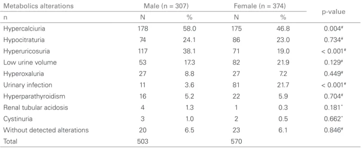
Table 1 METABOLICALTERATIONSFOUNDIN 681 PATIENTSSUFFERINGFROMNEPHROLITHIASISACCORDINGTOGENDER
Metabolics alterations Male (n = 307) Female (n = 374)
p-value
n N % N %
Hypercalciuria 178 58.0 175 46.8 0.004#
Hypocitraturia 74 24.1 86 23.0 0.734#
Hyperuricosuria 117 38.1 71 19.0 < 0.001#
Low urine volume 53 17.3 82 21.9 0.129#
Hyperoxaluria 27 8.8 27 7.2 0.449#
Urinary infection 11 3.6 81 21.7 < 0.001#
Hyperparathyroidism 16 5.2 22 5.9 0.704#
Renal tubular acidosis 4 1.3 1 0.3 0.181*
Cystinuria 3 1.0 2 0.5 0.662*
Without detected alterations 20 6.5 23 6.1 0.846#
Total 503 570
# χ2, * Fisher’s exact test
Table 2 CHEMICALANALYSISOFURINARY
CALCULUSIN 126 PATIENTS
Chemical composition N %
Calcium oxalate 108 85.7
Calcium carbonate 55 43.7 Uric acid 27 21.4
Ammonia 20 15.9
Calcium phosphate 10 7.9
Cystine 1 0.79
Magnesium 3 2.4
Total of chemical compounds 224
Table 3 ANATOMICALCHANGESIN 153 PATIENTS
Anatomical changes Total %
Renal cyst 55 35.9 Pyeloureteral duplication 28 18.3
UPJ Stenosis 15 9.8
Single kidney 11 7.2 Atrophic kidney 9 5.9
Medullary sponge kidney 10 6.5 Polycystic kidneys 6 3.9
Pelvic kidney 4 2.6 Neurogenic bladder 3 1.9
Renal ptosis 3 1.9 Horseshoe kidney 4 2.6
Bad renal rotation 2 1.3 Calyceal clubbing 1 0.7
Dilatation of the ureter 1 0.7 Stenosis of the distal ureter 1 0.7
Polycystic horseshoe kidney 1 0.7 Kidney tumor 1 0.7
Total of alterations 155
age of 32.2 years, which is in accordance with litera-ture15-17. The prevalence of caucasians is also
obser-ved (85%), but this is in accordance with the general population of the West of Paraná. In 96.5% of the patients at least one metabolic disorder was found. The most frequent metabolic changes were hypercal-ciuria (51.8%), hyperexcretion of uric acid (27.6%) and hypocitraturia (23.5%), result that is similar to other studies18,19.
The decrease in urine volume is considered to be a cause of nephrolithiasis. In countries with hot cli-mate, extrarenal losses and low fluid ingestion may also contribute to the formation of calculi. In the pres-ent study, a decrease in urine volume was observed in 13.8% of the patients, result that is much lower than the rates found in the countryside of the state of Sao Paulo, where the climate is warmer; in this region, the index was 77%20.
Hypercalciuria was the cause of more than 50% of metabolic disorders in adults, and from 53 to 75% in children21. It is possible that there is a strong genetic
component, with autosomal dominant interaction22.
It is a result of the mutations in genes that are direct or indirectly involved with the renal tubular transport of calcium, among them, CLCN5, CLCNKB and WNK kinases. CLCN5 gene mutations are related to the so called Dent’s Disease. Hypercalciuria may be associated with hypophosphatemia due to mutations in phosphorus /sodium cotransporter proteins, encod-ed by the NPT2a gene23-25. In children with
hypercal-ciuria, the prevalence of renal lithiasis in the family ranges from 46 to 49%26-28.
Idiopathic hypercalciuria is a heterogeneous dis-order that includes absorptive, renal and resorptive forms29-31. A hypersodic diet must be taken into
ac-count for the pathogenesis of hypercalciuria32. In this
study, hypercalciuria was the prevalent metabolic change. In the West of Paraná, the ingestion of milk and dairy products is not expressive; meanwhile, the ingestion of salt and protein is frequent, which proba-bly contributes to the occurrence of hypercalciuria10.
Hypocitraturia is a metabolic abnormality found in 20 to 60% of the patients with nephrolithiasis28,33,34.
In this study, such change was observed in 23.5% of the metabolic disorders. Tefekli et al.35 referred to
hy-pocitraturia as the most prevalent metabolic risk fac-tor in children and adults with renal lithiasis (60.6%). Since the etiology of hypocitraturia is multifactorial and directly related to the consumption of animal protein, promoting acid overload, its incidence varies in the different studied regions36.
Hyperuricosuria results from the frequent inges-tion of purines or high endogenous producinges-tion. Low fluid ingestion and urinary pH below 5.5 favor the precipitation in acidic urine28,37. Hyperuricosuria was
detected in 27.6% of the metabolic disorders observed in patients. It is believed that the hyperprotein diet in the region should be a risk factor. National literature observes such disorder in 18 to 76% of the cases20.
In this study, hyperuricosuria was more frequent in male patients.
Hyperoxaluria is a rare disorder, and it was ob-served in approximately 1% of the studied patients5.
In this study, such disorder was found in 8% of the patients. Infected calculi are a consequence of the microbial proliferation that changes the chemicals found in urine. Urease positive microorganisms pro-duce ammonia and bicarbonate that can cause stru-vite precipitation, forming Staghorn calculus in the collector system2. In the present study, urinary tract
infection was more frequent in female patients; how-ever, there is no information about the incidence of Staghorn calculus.
Daudon et al.38 found a male prevalence among
patients with calcium oxalate calculi and uric acid; a female preponderance for patients with calcium phos-phate and struvite calculi; and an increasing preva-lence of uric acid calculi with age in both genders. Chemical analyses demonstrated that calcium oxalate is the most common component found in kidney stones39. Calcium oxalate calculi were also found in
85.7% of the cases in this study.
Calculus is more common in patients with ana-tomic disorders. The prevalence of kidney stones in patients with renal cyst, autosomal dominant poly-cystic kidney disease, medullary sponge kidney, ure-teropelvic junction stenosis and pyeloureteral dupli-cation exceeds that of kidney stones in the general population, which suggests that malformation disor-ders favor calculus formation. Urine stasis with delay in carrying crystals increases the risk of urinary tract infections40,41. The most frequent anatomical changes
in the present study were renal cysts and reteropelvic junction obstruction in ureteral duplication.
This paper was the base for the knowledge of the metabolic profile of patients with lithiasis of the West region of Parana, Brazil. The most frequent metabolic alterations were hypercalciuria, hypocitraturia and hyperuricosuria.
R
EFERENCES1. Sakhaee K. Pharmacology of stone disease. Adv Chronic Kidney Dis 2009; 16:30-8.
2. Moe OW. Kidney stones. Pathophysiology and medical management. Lancet 2006; 367:333-4.
3. Amaro CR, Goldberg J, Amaro JL, Padovani CR. Metabolic assessment in patients with urinary lithiasis. Int Braz J Urol 2005; 31:29-33.
4. Pearle MS. Prevention of nephrolithiasis. Curr Opin Nephrol Hypertens 2001; 10:203-9.
5. Wilkinson H. Clinical investigation and management of patients with renal stones. Ann Clin Biochem 2001; 38:180-7.
6. Alpay H, Ozen A, Gokce I, Biyikli N. Clinical and metabolic features of nephrolithiasis and microlithiasis in children. Pediatr Nephrol 2009; 24:2203-9.
7. Ekane S, Wildschutz T, Simon J, Schulman CC. Urinary lithiasis: epidemiology and physiopathology. Acta Urol Belg 1997; 65:1-8.
8. Pak CY, Resnik MI, Preminger GM. Ethnic and geographic diversity of stones disease. Urology 1997; 50:504-7.
9. Colella J, Kochis E, Galli B, Munver R. Urolithiasis/ nephrolithiasis: what’s it all about? Urol Nursing 2005; 25:427-75.
10. Peres LAB, Molina AS, Galles MHL. Metabolic investigation of patients with urolithiasis in a specific region. Int Braz J Urol 2003; 29:217-20.
11. Evan A, Lingeman JE, Coe FL, Shao Y, Parks H, Bledsoe SB et al. Crystal associated nephropathy in patients with brushite nephrolithiasis. Kidney Int 2005; 67:576-91. 12. Raj GV, Auge BK, Assimos D, Preminger GM. Metabolic
abnormalities associated with renal calculi in patients with horseshoe kidneys. J Endourol 2004; 18:157-61. 13. Heilberg IP, Schor N. Renal stone disease: causes,
evaluation and medical treatment. Arq Bras Endocrinol Metab 2006; 4:823-31.
14. Worcester EM, Coe FL. Nephrolithiasis. Prim Care Office Pract 2008; 35:369-91.
15. Lotan Y. Economics and cost of cost of care of stone disease. Adv Chronic Kidney Dis 2009; 16:5-10. 16. Robertson WG, Peacock M, Baker M, Marshall DH,
Pearlman B, Speed R et al. Studies on the prevalence
and epidemiology of urinary stone disease in men in Leeds. Br J Urol. 1983; 55:595-8.
17. Hughes P. Kidney stones epidemiology. Nephrol 2007; 12:S26-30.
18. Pak CYC, Resnick MI. Medical therapy and new approaches to management of urolithiasis. Urol Clin North Am 2000; 27:243-53.
19. Low RK, Stoller ML. Uric acid related nephrolithiasis. Urol Clin North Am 1997; 24:135-49.
20. Ayusso LL, Schor N. Evaluation of patients with renal lithiasis in tropical region. Br J Nefrol 2001; 23:205-12.
21. Levy FL, Adams-Huet B, Pak CYC. Ambulatory evaluation of nephrolithiasis: an update of a 1980 protocol. Am J Med 1995; 98:50-8.
22. Coe FL, Parks JH, Moore ES. Familial idiopathic hypercalciuria. N Engl J Med 1979; 300:337-40. 23. Langman CB. The molecular basis of kidney stones.
Curr Opin Pediatr 2004; 16:188-93.
24. Raja KA, Schurman S, D’Mello DG, Blowey D, Goodyer
P, Why SV et al. Responsiveness of hypercalciuria to
thiazide in Dent’s disease. J Am Soc Nephrol 2002; 13:293338-44.
25. Prie D, Huart V, Bakouh N, Planelles G, Dellis O, Gerard B. Nephrolithiasis and osteoporosis associated with hypophosphatemia caused by mutations in the type 2a sodium-phosphate cotransporter. N Engl J Med 2002; 347:983-91.
26. Spivacow FR, Negri AL, del Valle EE, Calviño I, Fradinger E, Zanchetta JR. Metabolic risk factors in children with kidney stone disease. Pediatr Nephrol 2008; 23:1129-33.
27. Polito C, La Manna A, Cioce F, Villani J, Nappi B, Di Toro R. Clinical presentation and natural course of idiopathic hypercalciuria in children. Pediatr Nephrol 2000; 15:211-14.
28. Curhan GC, Taylor EN. 24-h uric acid excretion and the risk of kidney stones. Kidney Int 2008; 73:489-96. 29. Pak CY, Sakhaee K, Pearle MS. Detection of absorptive
hypercalciuria type I without the oral calcium load test. J Urol 2011; 185: 915-9.
30. Sayer JA. Renal stone diasease. Nephrol Physiol 2011; 118:35-44.
31. Stechman MJ, Loh NY, Thakker RV. Genetic causes of hypercalciuria nephrolithiasis. Pediatr Nephrol 2009; 24:2321-32.
32. Pak CYC, Resnick MI. Medical therapy and new approaches to management of urolithiasis. Urol Clin North Am 2000; 27:243-53.
33. Pak CYC. Etiology and treatment of urolithiasis. Am J Kidney Dis 1991; 18:624-37.
34. Zuckerman JM, Assimos DG. Hypocitraturia: pathophysiology and medical management. Rev Urol 2009; 11:134-44.
35. Tefekli A, Esen T, Ziylan O, Erol B, Armagan A, Ander H et al. Metabolic risk factors in pediatric and adult calcium oxalate urinary stone formers: is there any difference? Urol Int 2003; 70:273-7.
36. Karabacak OR, Ipek B, Ozturk U, Demirel F, Saltas H, Altuq U. Metabolic evaluation in stone disease metabolic differences between the pediatric and adult patients with stone disease. Urology 2010; 76:238-41.
37. Low RK, Stoller ML. Uric acid related nephrolithiasis. Urol Clin North Am 1997; 24:135-49.
38. Daudon M, Doré JC, Jungers P, Lacour B. Changes in stone composition according to age and gender of patients: a multivariate epidemiological approach. Urol Res 2004; 32:241-7.
39. Grampsas SA, Chandhoke PS, Fan J, Glass MA,
metabolic risk factors for nephrolithiasis in patients with autosomal dominant polycystic kidney disease. Am J Kidney Dis 2000; 36:53-7.
40. Gambaro G, Fabris A, Puliatta D, Lupo A. Lithiasis in cystic kidney disease and malformations of the urinary tract. Urol Res 2006; 34:102-7.
41. Romero V, Akpinar H, Assinos DG. Kidney Stones: a global picture of prevalence, incidence, and associated risk factors. Rev Urol 2010; 12:86-96.