Oral angioleiomyoma: case report and a review of
current findings
Angioleiomioma oral: relato de um caso e revisão dos achados atuais
Luiz Arthur Barbosa da Silva1, Ana Miryam Costa de Medeiros1, Patrícia Teixeira de Oliveira1, Éricka Janine Dantas da Silveira1, Márcia Cristina da Costa Miguel1
*
Abstract
Angioleiomyoma is a benign neoplasm that was considered a tumor of smooth-muscle origin until the most recent (2013) WHO classiication of soft tissue tumors, in which it was reclassiied as a tumor of perivascular origin. Angioleiomyomas rarely occur in the oral cavity. hese lesions are treated surgically with good prognosis. his article presents a review of reports of oral angioleiomyoma in the literature from the last 5 years and describes the case of a 44-year-old man who presented with an asymptomatic nodule in the upper lip that had developed over a 6-month period. Diagnostic hypotheses of pleomorphic adenoma or canalicular adenoma were raised. Biopsy of the lesion, histopathological and immunohistochemical analysis (S100, CD34, H-caldesmon, and desmin) conirmed a diagnosis of angioleiomyoma. It is noteworthy that immunohistochemistry is an important auxiliary method for diferential diagnosis of angioleiomyoma from other tumors, particularly myopericytoma.
Keywords: angioleiomyoma; diagnoses; immunohistochemistry.
Resumo
O angioleiomioma é uma neoplasia benigna que, a partir da nova classiicação da OMS (2013) para os tumores de tecidos moles, deixou de ser considerado um tumor de origem muscular lisa, passando a ser considerado um tumor de origem perivascular. Raramente os angioleiomiomas ocorrem na cavidade oral. A lesão é tratada cirurgicamente, com prognóstico considerado favorável. Este trabalho revisa os casos de angioleiomioma oral relatados na literatura nos últimos 5 anos e descreve esse tumor em um homem de 44 anos que apresentou um nódulo assintomático localizado em lábio superior, com evolução de 6 meses. As hipóteses diagnósticas foram de adenoma pleomórico e adenoma canalicular. A lesão foi submetida a biópsia e análise histopatológica e imuno-histoquímica (S100, CD34, α-SMA, H-caldesmon e desmina) conirmaram o diagnóstico de angioleiomioma. Destacamos a imuno-histoquímica como um importante método auxiliar no diagnóstico diferencial do angioleiomioma com outras lesões e, principalmente, com o miopericitoma.
Palavras-chave: angioleiomioma; diagnóstico; imuno-histoquímica.
1Universidade Federal do Rio Grande do Norte – UFRN, Programa de Pós-graduação em Patologia Oral, Natal, RN, Brazil. Financial support: None.
Conlicts of interest: No conlicts of interest declared concerning the publication of this article. Submitted: January 24, 2017. Accepted: March 07, 2017.
INTRODUCTION
Angioleiomyoma is a benign neoplasm that had been considered a tumor of smooth-muscle origin until the most recent (2013) World Health Organization
(WHO) classiication of soft tissue tumors, when it was reclassiied as a tumor of perivascular origin.1
Its etiology remains uncertain, but hypotheses involving minor traumas, venous stasis, hormone dysfunctions, and genetic alterations have been raised.2,3
Angioleiomyomas of the oral cavity are rare, the lips are most frequent site, followed by the palate, mucosa of the cheek, and tongue.4-6 Generally, patients
diagnosed with angioleiomyoma are middle-aged adults and it has a preference for males.4,7,8
Clinically, oral angioleiomyoma is characterized as a submucosal nodule, with a irm consistency, slow growth and, in the majority of cases, a size of 2 cm in diameter or less.8,9
Microscopically, it is a well-delimited lesion, with vascular spaces of different sizes and shapes and smooth muscle cells with varying morphology, arranged in disorganized bundles interspersed with collagen ibers.10 Differential diagnosis for angioleiomyoma
should focus on ruling out myoibroma, neuroibroma, neurilemmoma, leiomyosarcoma and, primarily, myopericytoma. Morphological indings are useful for differentiation when considered together with immunohistochemical results.5,11,12 The most indicated
treatment is conservative surgical excision.6,13
The aim of this study was to describe the clinical, morphological, and immunohistochemical indings of a case of oral angioleiomyoma and compare them with indings described in case reports published during the last 5 years in the specialized scientiic literature, identiied using the PubMed database.
CASE DESCRIPTION
A male, 44-year-old, melanoderm patient sought care at an oral diagnosis service, presenting an asymptomatic increase in volume, with a lobulated surface, normochromic mucosal, ibrous consistency, located on the right side of the upper lip, and with duration of approximately 6 months (Figure 1).
His medical and family histories were not relevant to the case. After clinical examination, the diagnostic hypotheses were canalicular adenoma and pleomorphic adenoma. Under local anesthesia, an excisional biopsy of the lesion was conducted with no complications. The specimen removed was ixed in 10% formol and sent to a pathology laboratory. Examination of histological sections revealed a benign neoplasm of mesenchymal origin, located in a subepithelial
region and characterized by proliferation of cells
with a variety of morphologies (ovoid, fusiform, and ondulated), arranged in disorganized bundles that surrounded multiple blood vessels of several
calibers (Figure 2A). The clinical hypotheses
of benign salivary gland neoplasms (adenoma pleomorphic or canalicular adenoma) were rejected. Immunohistochemical analyses were conducted to conirm the diagnosis (Table 1). The tumor cells were
negative for S100 (Figure 2B), and positive staining
for CD34 was restricted to the walls of blood vessels
(Figure 2C). The tumor cells had intense staining for
α-SMA (Figure 2D), and were immunopositive for H-caldesmon (Figure 2E) and desmin (Figure 2F).
A diagnosis of oral angioleiomyoma was conirmed based on these indings. Fifteen months later the patient is still being followed-up clinically and there have been no signs of relapse.
DISCUSSION
According to the WHO, angioleiomyomas are
benign dermal or subcutaneous tumors made up of
well-differentiated smooth muscle cells that organize
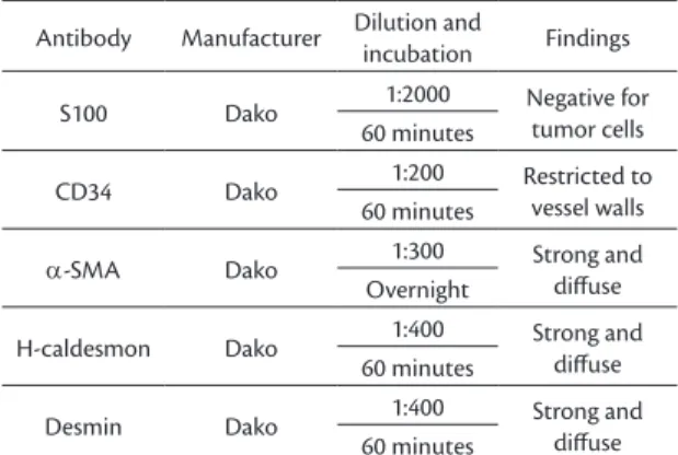
Table 1. Immunohistochemical panel.
Antibody Manufacturer Dilution and
incubation Findings
S100 Dako 1:2000 Negative for
tumor cells 60 minutes
CD34 Dako 1:200 Restricted to
vessel walls 60 minutes
α-SMA Dako 1:300 Strong and
difuse Overnight
H-caldesmon Dako 1:400 Strong and
difuse 60 minutes
Desmin Dako 1:400 Strong and
difuse 60 minutes
around a variety of vascular structures.14 Their etiology
remains to be conirmed. Although genetic studies related to the origin of perivascular lesions are still limited, mutations to the BRAF, NF1, NOTCH2 and NOTCH3 genes are under investigation.1 Additionally, estrogen
receptor and progesterone receptor expression have been investigated with relation to angioleiomyomas,
on the basis that there is a possibility that hormonal
changes participate as an etiologic factor.2
Angioleiomyomas can arise on any part of the body, but in the majority of cases they occur in extremities, primarily the lower limbs, followed by the head and trunk.8 High incidences are also reported in the uterus,
gastrointestinal tract, and skin.4,6 Angioleiomyomas are
rare in the oral cavity, since smooth muscle is scarce in this region.9 These tumors appear to arise from the
tunica media of small vessels, from arteriovenous anastomoses, or the smooth muscle cells of the circumvallate papillae of the tongue.5,12 Although
the lips are described as the most common site of
angioleiomyomas within the oral cavity,4-6 according to
reports in the specialized literature published over the
last 5 years (summarized in Table 2), the gingiva was
the most common anatomical site in 29.4% of cases.
Oral angioleiomyoma is more common in male
patients,4,8 with a male:female ratio of 3:1.3 Normally,
the peak incidence of this tumor is between the 4th and
6th decades of life;8,10 but there are cases in pediatric
patients.7 In the review summarized in Table 2, it was
observed that 76.4% of the tumors were in males, and patients’ ages ranged from 9 to 85 years, with a mean age of 47.3 years. The patient in the present study was a 44-year-old male.
The clinical indings in our case corroborate those described in previous literature. Angioleiomyomas present as asymptomatic submucosal nodules with slow growth, that are well-deined, mobile, occasionally with a bluish color and intact surface, and generally measure 2 cm in diameter.7,9
The immunohistochemical proile described here conirms the muscular origin of the neoplastic cells in this tumor, since strong and diffuse immunostaining was observed for myogenic proteins, such as α-SMA, H-caldesmon, and desmin. The most important
differential diagnosis for angioleiomyoma is
myopericytoma, because these two types of lesion have overlapping histopathological indings.9 In one
study evaluating 122 cases of angioleiomyoma and 12 cases of myopericytoma, Matsuyama, Hisaoka and
Hashimoto11 attempted to determine the characteristic
immunohistochemical proiles of these two tumors, observing that α-SMA, HHF-35, and H-caldesmon exhibited the same immunostaining proile, whereas desmin was negative in 75% of myopericytoma cases and just 17.1% of angioleiomyoma cases. Therefore, desmin appears to be a useful marker for differentiation between these two lesions. From an immunohistochemical point of view, desmin may
also be useful for differentiating angioleiomyomas
from myoibromas, since the neoplastic cells in myoibromas are negative for this marker.9
The profile of immunostaining for the S100
protein is also useful for differentiating a diagnosis of angioleiomyoma from neoplasms such as
neuroibroma and neurilemmoma, since in addition to the morphological proile of these lesions, the
fact that the tumor cells of angioleiomyomas fail to
react to this protein is entirely divergent from what is observed with lesions of neural origin.15
Histopathological differentiation between angioleiomyoma and low grade leiomyosarcoma may be dificult. Among other parameters, leiomyosarcomas can be identiied by mitosis counts ranging from 5 to 10 per ield, cell nuclei with blunt ends, and foci of necrosis. Therefore, careful long-term follow-up is necessary in view of the diagnostic uncertainty between these two entities.3,15
The treatment of choice for oral angioleiomyoma
is conservative surgical excision. Rare recurrence has been described, probably the result of incomplete surgical excision. There are no reports of malignant
transformation and patients’ prognosis is considered
excellent.6,16 The patient described here has been in
follow-up for 15 months with no evidence of relapse. Awareness of the WHO14 reclassification of
angioleiomyoma as a tumor of perivascular origin
is important and more genetic studies are needed to
elucidate their true etiology. Immunohistochemical studies are an important method to aid in arriving at the correct diagnosis of angioleiomyoma, particularly
Table 2. Reports of oral angioleiomyoma cases reported in the literature over the last 5 years (2011-2016).
Authors N Sex Age (Years) Anatomic site Symptomology
Present study (2016) 1 Male 44 Lower lip Asymptomatic
Arpağ et al.5 2 Male 25 Gingiva Asymptomatic
Female 55 Gingiva
Bajpai et al.13 1 Male 39 Gingiva Asymptomatic
Ishikawa et al.6 1 Male 51 Tongue Asymptomatic
Inaba et al.2 1 Female 45 Mucosa of cheek Asymptomatic
Osano et al.3 1 Male 45 Mucosa of cheek Asymptomatic
Ranjan and Singh9 1 Female 45 Gingiva Asymptomatic
Tsuji et al.4 1 Male 79 Hard palate Asymptomatic
Eley et al.8 1 Male 39 Hard palate Asymptomatic
Menditti et al.15 1 Male 14 Gingiva Asymptomatic
Gueiros et al.10 3
Male 53 Upper lip Asymptomatic
Male 54 Lower lip Asymptomatic
Male 66 Upper lip Asymptomatic
Mahima et al.12 1 Male 57 Retromolar area Asymptomatic
Reddy et al.7 1 Male 9 Mandible Asymptomatic
Vidaković et al.16 1 Female 85 Parotid Asymptomatic
with respect to differential diagnosis from other lesions, especially myopericytoma.
REFERENCES
1. Fletcher CD. The evolving classification of soft tissue tumours - an update based on the new 2013 WHO classification. Histopathology. 2014;64(1):2-11. PMid:24164390. http://dx.doi.org/10.1111/ his.12267.
2. Inaba T, Adachi M, Yagisita H. A case of angioleiomyoma in the buccal space. Odontology. 2015;103(1):109-11. PMid:23907201. http://dx.doi.org/10.1007/s10266-013-0128-z.
3. Osano H, Ioka Y, Okamoto R, et al. Angioleiomyoma of the cheek: a case report. J Oral Sci. 2015;57(1):63-6. PMid:25807911. http:// dx.doi.org/10.2334/josnusd.57.63.
4. Tsuji T, Satoh K, Nakano H, Kogo M. Clinical characteristics of angioleiomyoma of the hard palate: report of a case and an analysis of the reported cases. J Oral Maxillofac Surg. 2014;72(5):920-6. PMid:24480770. http://dx.doi.org/10.1016/j.joms.2013.11.008. 5. Arpağ OF, Damlar I, Kılıç S, Altan A, Taş ZA, Özgür T. Angioleiomyoma
of the gingiva: a report of two cases. J Korean Assoc Oral Maxillofac Surg. 2016;42(2):115-9. PMid:27162753. http://dx.doi.org/10.5125/ jkaoms.2016.42.2.115.
6. Ishikawa S, Fuyama S, Kobayashi T, Taira Y, Sugano A, Iino M. Angioleiomyoma of the tongue: a case report and review of the literature. Odontology. 2016;104(1):119-22. PMid:25238675. http:// dx.doi.org/10.1007/s10266-014-0175-0.
7. Reddy B, Rani BS, Anuradha C, Chandrasekhar P, Shamala R, Lingamaneni K. Leiomyoma of the mandible in a child. J Oral Maxillofac Pathol. 2011;15(1):101-4. PMid:21731289. http://dx.doi. org/10.4103/0973-029X.80015.
8. Eley KA, Alroyayamina S, Golding SJ, Tiam RN, Watt-Smith SR. Angioleiomyoma of the hard palate: report of a case and review of the literature and magnetic resonance imaging findings of this rare entity. Oral Surg Oral Med Oral Pathol Oral Radiol. 2012;114(2):e45-9. PMid:22769421. http://dx.doi.org/10.1016/j. oooo.2012.01.014.
9. Ranjan S, Singh KT. Gingival angioleiomyoma-infrequent lesion of oral cavity at a rare site. J Oral Maxillofac Pathol. 2014;18(1):107-10. PMid:24959048. http://dx.doi.org/10.4103/0973-029X.131928. 10. Gueiros LA, Romañach MJ, Pires-Soubhia AM, Pires FR,
Paes-de-Almeida O, Vargas PA. Angioleiomyoma affecting the lips: report of 3 cases and review of the literature. Med Oral Patol Oral Cir Bucal. 2011;16(4):e482-7. PMid:20526260. http://dx.doi. org/10.4317/medoral.16.e482.
11. Matsuyama A, Hisaoka M, Hashimoto H. Angioleiomyoma: a clinicopathologic and immunohistochemical reappraisal with special reference to the correlation with myopericytoma. Hum Pathol.
2007;38(4):645-51. PMid:17270242. http://dx.doi.org/10.1016/j. humpath.2006.10.012.
12. Patil K, Mahima VG, Srikanth HS. Recurrent oral angioleiomyoma. Contemp Clin Dent. 2011;2(2):102-5. PMid:21957385. http:// dx.doi.org/10.4103/0976-237X.83071.
13. Bajpai M, Pardhe N, Kumar M. Angioleiomyoma of gingiva masquerading as pyogenic granuloma. J Coll Physicians Surg Pak. 2016;26(7):631-2. PMid:27504561.
14. Hisaoka M, Quade B. Angioleiomyoma. In: Fletcher CD, Bridge JA, Hagendoorn PCW, Martens F, editors. WHO classification of tumor of soft tissue and bone. Lyon: IARC; 2013. p. 120-1. 15. Menditti D, Laino L, Nastri L, Caruso U, Fiore P, Baldi A. Oral
angioleiomyoma: a rare pathological entity. In Vivo. 2012;25(1):161-3. PMid:22210732012;25(1):161-3.
16. Vidaković B, Knezević AK, Manojlović S, Knezević G. Angiomyoma of the cheek. Coll Antropol. 2011;35(1):207-9. PMid:21661373.
*
Correspondence
Márcia Cristina da Costa Miguel Universidade Federal do Rio Grande do Norte - UFRN,
Pós-graduação em Patologia Oral Avenida Salgado Filho, 1787 - Lagoa Nova CEP 59056-000 - Natal (RN), Brazil Tel.: +55 (84) 3215-4138 E-mail: [email protected]
Author information
LABS - PhD student in Oral Pathology, Programa de Pós-graduação em Patologia Oral, Universidade Federal do Rio Grande do Norte. AMCM - PhD in Oral Pathology from Universidade Federal do Rio Grande do Norte; full professor, Universidade Federal do Rio Grande do Norte (UFRN). PTO - PhD in Dentistry from Pontifícia Universidade Católica do Rio Grande do Sul; adjunct professor, Universidade Federal do Rio Grande do Norte (UFRN). EJDS and MCCM - PhDs in Oral Pathology from Universidade Federal do Rio Grande do Norte (UFRN); adjunct professors, UFRN.
Author contributions
Conception and design: LABS Analysis and interpretation: EJDS Data collection: AMCM Writing the article: LABS, PTO Critical revision of the article: EJDS Final approval of the article*: LABS, AMCM, PTO, EJDS, MCCM Statistical analysis: N/A. Overall responsibility: MCCM