DOI: 10.5935/2359-4802.20170086
33
International Journal of Cardiovascular Sciences. 2018;31(1)33-46
ORIGINAL ARTICLE
Mailing Address: Carlos José Oliveira de Matos
Rua Orlando Magalhães Maia, 1330, Res. Eduardo Abreu, apto, 802. Postal Code: 49025-530, Jardins, Aracaju, SE – Brazil. E-mail: [email protected]; [email protected]
Overview with Meta-analysis of Systematic Reviews of the Diagnostic and Prognostic
Value of Coronary Computed Tomography Angiography in the Emergency Department
Irlaneide da Silva Tavares,1 Carlos José Oliveira de Matos,1 Marco Antonio Prado Nunes,1 Antonio Carlos Sobral Sousa,1,2 Divaldo Pereira de Lyra Júnior,1 Joselina Luzia Menezes Oliveira1,2
Universidade Federal de Sergipe (UFS);1 Centro de Ensino e Pesquisa e Laboratório de Ecocardiografia (ECOLAB) do Hospital e Fundação São Lucas,2 Aracaju, SE – Brazil
Manuscript received March 04, 2017; revised July 01, 2017; accepted July 07, 2017.
Abstract
Background: The high prevalence of CAD, as well as your impact on health expenditure and the various treatment options to reduce morbidity and mortality related to CAD, comes to develop a diagnostic tool precis and with important findings in the Emergency Department.
Objetive:To conduct an overview with meta-analysis to compile evidence from multiple systematic reviews (SR) on the diagnostic and prognostic value of coronary computed tomography angiography (CCTA) to assess acute chest pain in the emergency department (ED).
Methods:We included SR of primary studies that evaluated the diagnostic and prognostic value of CCTA ≥ 64 channels in the ED. The studies were conducted in patients at low and intermediate risk for coronary artery disease
(CAD). Quality assessment was performed using PRISMA and approved reviews that scored ≥ 80%. Two authors
independently extracted data using a standardized form. Spearman correlation test, Chi-square test, Cochran’s Q test or Higgins and Thompson statistical I2 were used. For meta-analysis, “mada” package statistical software R
Core Team, 2015, was used. The significance level adopted was 95%.
Results: Four reviews were eligible for inclusion in this overview, resulting in 13 articles after applying the exclusion criteria, and only 10 of these were used for meta-analysis, adding up to a total of 4831 patients (mean age, 54 ± 6 years; 51% male), of whom 46% were hypertensive, 32% had dyslipidemia, 13% had diabetes and 26% had a family history of premature CAD. In the meta-analysis, 9 studies defined CCTA positive in the presence of luminal lesions ≥ 50%, while 1 study defined it as luminal lesions ≥ 70%. Sensitivity ranged from 77% to 98%, and specificity, from 73% to 100%. The univariate analysis showed homogeneity of diagnostic odds ratio (DOR) [Q = 8.5 (df = 9), p = 0.48 and I2 = 0%]. The pooled mean DOR for CCTA in primary analyses was 4.33 (95% CI: 3.47 - 5.18).
The area under the curve (AUC) was 0.982 (95% CI: 0.967 - 0.999). There was no death, 29 (0.6%) infarcts, 92 (1.9%) revascularizations and 312 (6.4%) invasive coronary angiographies. The diagnosis of acute coronary syndrome occurred in 7.3% of the 1655 patients included in the meta-analysis.
Conclusions: The use of CCTA as a tool for stratification of patients at low or intermediate cardiovascular risk, who are in the ED with chest pain, has high accuracy, safety, reduces length of hospital stay and probably the
costs, producing an early diagnosis and more effective decision making. (Int J Cardiovasc Sci. 2018;31(1)33-46)
Keywords: Coronary Artery Disease; Tomography, X-Ray Computed; Chest Pain; Emergency Medicine;
Meta-Analysis as Topic.
Introduction
In 2010, in the United States of America, nearly 6 million patients with chest pain visited the emergency departments (ED); this is the second most frequent
reason for visits to this unit, although only a minority receives the diagnosis of acute coronary syndrome (ACS).1 North American statistics show that in 2011 coronary artery disease (CAD) was responsible for
Coronary artery disease is responsible for a substantial impact on the use of health care, with an estimated
cost of US$ 21.9 billion in 2011. Between 2013 and 2030 the costs are estimated to increase ≈ 100%.3 Due to the high prevalence of CAD, as well as its impact on health expenditure and the various treatment options to reduce morbidity and mortality related to CAD, accurate diagnosis is essential.
A high precision effective diagnostic test to exclude acute CAD could reduce the cost of the USA health care system by billions of dollars. The advent of coronary computed tomography angiography (CCTA), a noninvasive method to study the coronary anatomy,
with tomography scanner ≥ 64 channels, reducing
artifacts as well as increasing spatial and temporal resolution, has given rise to a quick test, effective to reliably exclude ACS.4 Although invasive coronary angiography (ICA) is the "gold standard" for CAD detection, it is not appropriate for extensive use because it is invasive, not routinely available, and has high cost and increased risk of complications. Furthermore, the immediate and future probability of cardiac events in patients without CAD or with minimal CAD is low for patients with chest pain in the ED.5,6
Systematic reviews (SR) are studies with the highest level of evidence (higher in the hierarchy of evidence-based research) and rigorous methodological quality.7 Due to the rapid expansion of the literature and the presence of a relatively high number of SR on this topic, the purpose of this study was to conduct an overview of meta-analyses to compile evidence from multiple SR related to the diagnostic and prognostic value of CCTA in the assessment of acute chest pain in the ED.
Methods
Literature search
The search was conducted from January 2005 (the first year of published studies from 64-slice scanners) to July 2015. The strategy was developed through the Medical Subject Heading (MeSH) terms: “coronary artery disease”,
“computed tomography”, “chest pain” and “emergency department”. The electronic databases researched were
MEDLINE and COCHRANE LIBRARY. This overview
included SR on the diagnostic and prognostic value of
CCTA in the ED. Only studies reported in English were
eligible and had their references checked.
We analyzed all studies of SR, excluding duplicates,
performed with CCTA < 64 channels, with at least 30 patients. In the presence of more than one study with
the same database, the oldest was deleted.
Ethics approval was not required for this overview.
Quality assessment
All eligible SR were assessed using the PRISMA quality assessment tool(Preferred Reporting Items for Systematic Reviews and Meta-Analysis),8 and those
scoring ≥ 80% were approved.
Data extraction
Two authors independently extracted data using a standardized data extraction form including study characteristics (design, inclusion and exclusion criteria),
characteristics of the intervention (at least 64-slice
computed tomography, use and timing of cardiac enzymes relative to CCTA, follow-up duration), patients characteristics (age, sex, cardiac risk factors), outcomes
[death, nonfatal myocardial infarction (MI), repeated
ED chest pain evaluation, repeated hospitalization for ACS, ICA, revascularization by percutaneous coronary intervention (PCI)/coronary artery bypass
graft (CABG)], hospital length of stay (LOS), and
cost. Disagreements were resolved by consensus or consultation with a third individual.
Data synthesis and statistical analysis
Numerical variables were described as mean and standard deviation and categorical variables, as simple and relative frequencies. The sensitivity and specificity
were described as estimates with 95% confidence
interval (CI), rounded to the nearest integer. Using true positive (TP), false positive (FP), true negative (TN), and false negative (FN), we derived sensitivity, specificity, positive and negative likelihood ratios (posLR and negLR, respectively), and positive and negative predictive values (VP+ and VP-, respectively) for each study.
35
Matos et al. Overview of the CCTA in the Emergency Int J Cardiovasc Sci. 2018;31(1)33-46
Original Article
the overall variation among the studies, stating, as the null hypothesis, that the studies that make up the meta-analysis are homogeneous. The I2 evaluates the estimate of the variance due to heterogeneity, rather than chance, and is based on traditional statistical variance defined as Cochran’s Q.9 Significant heterogeneity was set to I2 > 50%. Data were used with a significance level
of 95%. Data analysis was performed with R-package “mada” for meta-analysis (R Core Team, 2015) that
presents some approaches for diagnostic studies, such as descriptive statistics and graphs. In the data analysis, in 2 x 2 tables, cells with zeros often lead to statistical
artifacts, since certain reasons can ‘not exist’; so the package “mada” uses the value of 0.5 as a correction of
continuity "standard". This package does not calculate the aggregated value of sensitivity and specificity. It is not appropriate analytical indicator.10
In presence of publication bias, the funnel plot, method known to assess publication bias, is unlikely to be useful to detect the effect of sample size because these parameters will vary depending on the cut-off values and random error.11 Meta-regression was not performed, since its purpose is to evaluate the causes of heterogeneity
and the diagnostic odds ratio (DOR) was homogeneous.
Results
The literature search generated a total of 4 SR that
evaluated the diagnostic and prognostic value of
CCTA ≥ 64 channels in the ED,25-28 containing 91 primary
studies. From these, 13 articles meeting the inclusion
criteria were included in the qualitative analysis. Due to
absence of quantitative dates, only 10 studies were used
in the meta-analysis. The main reasons for exclusion
of the primary studies were: not performed in the ED; duplicated studies; and CCTA of 4 or 16 channels. Figure 1 shows a flow chart of study exclusion.
A total of 4831 patients were included (mean age of 54 ± 6 years, 51% male), of whom 46% were hypertensive, 32% had dyslipidemia, 13% had diabetes and 26% had a
family history of premature CAD. The primary studies included and their clinical characteristics are outlined
in Table 1. In general, patients with atrial fibrillation,
ventricular arrhythmias, enzymatic changes, renal failure, hemodynamic instability, allergy to contrast, and pregnant women were excluded from studies.
The studies were conducted in patients at low and intermediate risk for CAD (except Ueno et al.,19 2009, that
includes high-risk patients) with normal cardiac enzymes
and nonischemic initial ECG.
In the meta-analysis, 9 studies defined positive CCTA when in the presence of luminal lesions ≥ 50%, while 1 study defined it when luminal lesion ≥ 70%. A total of 1655 patients
were included. Descriptive statistics for diagnostic test
(CCTA) performance are described in Tables 2 and 3.
The study by Rubinstein et al.16 reported the highest sensitivity
(S = 98%), while the study by Johnson et al.22 reported the
highest specificity (E = 100%). The largest study was that
by Hollander et al.,18 2009, which included 568 patients, and
reported a 94% sensitivity and a 92% specificity.
All studies showed high positive likelihood ratio (the highest in the study by Johnson et al.,22 2008), and low negative likelihood ratio (the lowest in the study by Rubinstein et al.,16 2007).
To assess the effect of different cut-offs for “significant” luminal obstruction on analysis, we performed diagnostic threshold analyses. We found a Spearman correlation
coefficient of 0.045, p > 0.05 (95% CI -0.602 to 0.656), which
means ‘very weak’ correlation or no significant correlation.
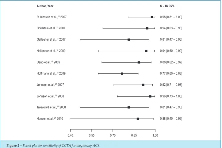
The equality test for sensitivity showed homogeneity
across studies [χ2 = 8.4 (df = 9), p = 0.5] and heterogeneity
for specificity [χ2 = 55.5 (df = 9), p < 0.001], confirmed
with the forest plot (Figures 2 and 3).
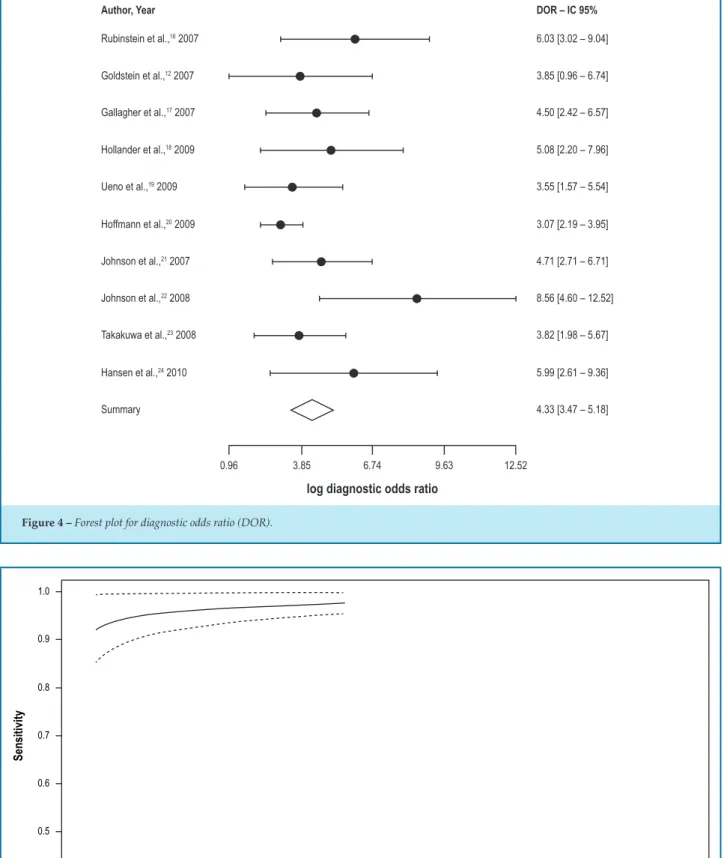
The univariate analysis showed homogeneity of DOR [Q = 8.5 (df = 9), p = 0.48 and I2 = 0%]. Figure 4 shows estimate of the synthesis.
The χ2 test did not reject the hypothesis of homogeneity
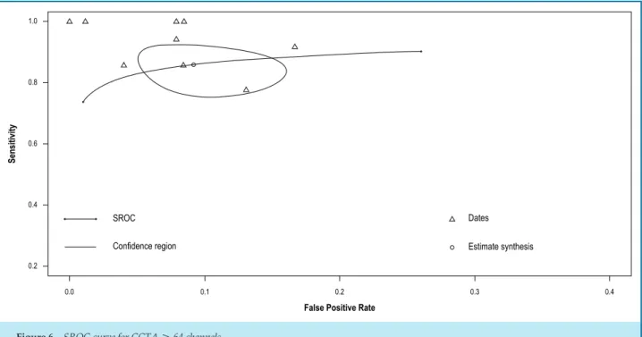
for the model [χ2 = 10.14 (df = 1), p = 0.34, θ (theta) = 0.018 (CI95%: 0.0014 to 0,0246)] and so we opted for the fixed effects model. The area under the curve (AUC) was 0.982 (95% CI: 0.967-0.999).
As for the events, there was great variability between studies in the outcomes assessed. A summary of the compound events in all studies, with their specific
characteristics, is described in Table 4. In most studies, the events investigated were death, MI, CABG and ICA. Others evaluated the diagnosis of ACS, and, in three
studies, the technique of "triple rule-out" was used (in addition to investigating CAD, pulmonary embolism and aortic dissection).
Figure 7 shows the main events: 29 (0.6%) MI, 92 (1.9%) CRM, and 312 (6.4%) ICA. There was no death. The diagnosis of ACS occurred in 7.3% of the 1655 patients
1042 studies identified through database
997 excluded by title and summary
42 systematic reviews to full text analysis
37 not systematic reviews in emergency department
5 systematic review in emergency department
1 excluded post-PRISMA (< 0.8)
4 systematic reviews = 91 primary studies
78 primary studies excluded: * 4-16 channels = 7 studies
* no emergency department = 49 studies * duplicates = 17 studies
* same database = 2 studies
* ≤ 30 patients = 3 studies
13 studies included in qualitative synthesis
3 excluded due to absence of data for quantitative analysis
10 studies included in quantitative synthesis
Figure 1 – Flow diagram describing the process of study inclusion
Although analyzed heterogeneously, the four randomized clinical trials (RCTs) also evaluated the
hospital LOS and costs. Compared to usual care, the use of CCTA reduced the hospital LOS in all studies, and the
costs, in three studies.
Discussion
This study had the purpose to evaluate the diagnostic and prognostic value of CCTA in the evaluation of acute
chest pain in the ED. We included 4 SR, totaling 13 studies.
After primary analysis of the exclusion criteria, we used
10 studies for quantitative analysis (meta-analysis).
We found that CCTA has high sensitivity and specificity for CAD detection in patients with chest pain in the ED in all studies, and showed high positive likelihood ratio and low negative likelihood ratio. The distribution of sensitivity was homogeneous, while that of specificity was heterogeneous. We found weak correlation on the effect of the different cut-offs for the diagnosis of significant luminal obstruction.
The DOR was homogeneous and significant. There was
variability on the number and type of events between the studies. The clinical trials reported decreased
37
Table 1 – Primary studies included and their clinical characteristics
Authors Type
of study Year
Type of CCTA (Channels)
Centers Patients (N) Age
(Mean ± SD) Men (%)
BMI, (kg/m2
± DP)
HTN N (%)
HL N (%)
DM N (%)
FH of premature CAD N (%)
Smoker N (%)
Goldstein et al.12 RCT 2007 64 Single center 197 50 ± 12 50 29 ± 5 75/38 70(36) 20(10) 82(42) 35(18)
CT-STAT13 RCT 2011 64 a 320 Multicenter 699 50 ± 10 46 28 ± 5 259/37 234(33) 48(7) 212(30) 157(22)
ACRIN-PA14 RCT 2012 ≥ 64 Multicenter 1370 49 ± 9 47 - 695/51 367(27) 194(14) 394(29) 447(33)
ROMICAT II15 RCT 2011 64 Single center 1000 54 ± 8 53 29 ± 5 541/54 454(45) 173(17) 271(27) 492(49)
Rubinstein et al.16 Cohort 2007 64 Single center 58 56 ± 10 64 - 33/57 32(55) 12(21 9(16) 22(38)
Gallagher et al.17 Cohort 2007 64 Single center 85 49 ± 11 53 - 31(36,5) 23(27) 8(9) 50(59) 22(25,9)
Hollander et al.18 Cohort 2009 64 Single center 568 47 ± 9 44 - 251(44) 108(19) 77(14) 104(18) 200(35)
Ueno et al.19 Cohort 2009 64 Single center 36 66 ± 12 53 - 17(47) 19(53) 9(25) 8(22) 13(36)
Hoffmann et al.20 Cohort 2009 64 Multicenter 368 53 ± 12 61 29 ± 6 145(39) 135(37) 40(11) 180(49)
Johnson et al.21 Cohort 2007 64 Single center 55 67 ± 10 64 - - - - -
-Johnson et al.22 Cohort 2008 64 Single center 109 63 ± 14 72 - - - - -
-Takakuwa et al.23 Cohort 2008 64 Single center 197 49 ± 11 72 - 92(46,7) 53(26,9) 29(15) 62(31) 62(31,5)
Hansen et al.24 Cohort 2010 64 Single center 89 56 ± 9 63 - 35(39) 37(42) 7(8) 29(33) 39(44)
CCTA: coronary computed tomography angiography; SD: standard deviation; BMI, body mass index; HTN: hypertension; HL: hyperlipidemia; DM: diabetes mellitus; FH: family history; CAD: coronary artery disease; RCT: randomized clinical trial
M
at
os et al
.
O
ver
view of the C
C
TA in the Emer
genc
y
In
t J C
ar
dio
vasc S
ci. 2018;31(1)33-46
O
riginal A
Table 2 – Test performance characteristics of CCTA in the studies included
Year Authors N TP FN FP TN S IC95% E IC95%
2007 Goldstein et al.12 99 8 0 24 67 0.94 0.63 - 0.99 0.73 0.64 - 0.81
2007 Rubinstein et al.16 58 20 0 3 35 0.98 0.81 - 1.00 0.91 0.78 - 0.97
2007 Gallagher et al.17 85 6 1 3 72 0.81 0.47 - 0.96 0.95 0.88 - 0.98
2009 Hollander et al.18 568 7 0 47 508 0.94 0.60 - 0.99 0.92 0.89 - 0.94 2009 Ueno et al.19 36 11 1 4 20 0.89 0.62 - 0.97 0.82 0.63 - 0.92 2009 Hoffmann et al.20 368 24 7 44 293 0.77 0.60 - 0.88 0.87 0.83 - 0.90
2007 Johnson et al.21 55 16 1 3 35 0.92 0.71 - 0.98 0.91 0.78 - 0.97 2008 Johnson et al.22 109 13 0 0 96 0.96 0.73 - 1.00 1.00 0.95 - 1.00
2008 Takakuwa et al.23 197 6 1 16 174 0.81 0.47 - 0.96 0.91 0.87 - 0.95 2010 Hansen et al.24 89 3 0 1 85 0.88 0.40 - 0.99 0.98 0.93 - 1.00
CCTA: coronary computed tomography angiography; TP: true positive; FN: false negative; FP: false positive; TN: true negative; S: sensibility; CI: confidence interval; E: specificity.
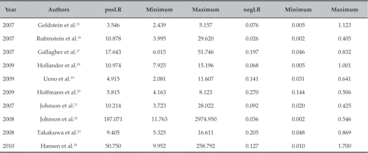
Table 3 – Descriptive analysis of the likelihood ratio
Year Authors posLR Minimum Maximum negLR Minimum Maximum
2007 Goldstein et al.12 3.546 2.439 5.157 0.076 0.005 1.123
2007 Rubinstein et al.16 10.878 3.995 29.620 0.026 0.002 0.405
2007 Gallagher et al.17 17.643 6.015 51.746 0.197 0.046 0.832
2009 Hollander et al.18 10.974 7.925 15.196 0.068 0.005 1.001 2009 Ueno et al.19 4.915 2.081 11.607 0.141 0.031 0.641 2009 Hoffmann et al.20 5.815 4.163 8.123 0.270 0.144 0.506 2007 Johnson et al.21 10.214 3.723 28.022 0.092 0.020 0.425
2008 Johnson et al.22 187.071 11.763 2974.950 0.036 0.002 0.546
2008 Takakuwa et al.23 9.405 5.325 16.611 0.205 0.048 0.869 2010 Hansen et al.24 50.750 9.952 258.792 0.127 0.010 1.700
posLR: positive likelihood ratios; negLR: negative likelihood ratios.
The quality of cardiac image by CCTA is directly related to the evolution of tomography. Current technical developments of CT scanners and the software are intended to improve the spatial and temporal resolution of cardiac CT images while reducing the radiation dose received from a typical examination. They include wider detector arrays that allow a higher number of simultaneously acquired image slices, faster x-ray tube rotation, and use of alternative image
reconstruction techniques.29 Its use in patients with
early biomarkers and negative ECG for myocardial
ischemia is already included in the algorithm of chest pain evaluation in several emergency centers, a strategy that is supported by the current Appropriate Use Criteria for Cardiac Computed Tomography30 and
Focused Update of the Guidelines for the Management
39
Hansen et al.,24 2010 Author, Year
Rubinstein et al.,16 2007
Goldstein et al.,12 2007
Gallagher et al.,17 2007
Hollander et al.,18 2009
Ueno et al.,19 2009
Hoffmann et al.,20 2009
Johnson et al.,21 2007
Johnson et al.,22 2008
Takakuwa et al.,23 2008
0.88 [0.40 – 0.99] S – IC 95%
0.98 [0.81 – 1.00]
0.94 [0.63 – 0.96]
0.81 [0.47 – 0.96]
0.94 [0.60 – 0.99]
0.88 [0.62 – 0.97]
0.77 [0.60 – 0.88]
0.92 [0.71 – 0.98]
0.96 [0.73 – 1.00]
0.81 [0.47 – 0.96]
0.40 0.55 0.70 0.85 1.00
Figure 2 – Forest plot for sensitivity of CCTA for diagnosing ACS.
0.63 0.72 0.82 0.91 1.00
Hansen et al.,24 2010 0.98 [0.93 – 1.00]
Author, Year
Rubinstein et al.16, 2007
Goldstein et al.,12 2007
Gallagher et al.,17 2007
Hollander et al.,18 2009
Ueno et al.,19 2009
Hoffmann et al.,20 2009
Johnson et al.,21 2007
Johnson et al.,22 2008
Takakuwa et al.,23 2008
E – IC 95%
0.91 [0.78 – 0.97]
0.73 [0.64 – 0.81]
0.95 [0.88 – 0.96]
0.91 [0.89 – 0.94]
0.82 [0.63 – 0.92]
0.87 [0.83 – 0.90]
0.91 [0.78 – 0.97]
0.99 [0.95 – 1.00]
0.91 [0.87 – 0.95]
Figure 3 – Forest plot for specificity of CCTA for diagnosing ACS.
Matos et al. Overview of the CCTA in the Emergency Int J Cardiovasc Sci. 2018;31(1)33-46
Summary 4.33 [3.47 – 5.18]
0.96 3.85 6.74 9.63 12.52
log diagnostic odds ratio Author, Year
Rubinstein et al.,16 2007
Goldstein et al.,12 2007
Gallagher et al.,17 2007
Hollander et al.,18 2009
Ueno et al.,19 2009
Hoffmann et al.,20 2009
Johnson et al.,21 2007
Johnson et al.,22 2008
Takakuwa et al.,23 2008
Hansen et al.,24 2010
DOR – IC 95%
6.03 [3.02 – 9.04]
3.85 [0.96 – 6.74]
4.50 [2.42 – 6.57]
5.08 [2.20 – 7.96]
3.55 [1.57 – 5.54]
3.07 [2.19 – 3.95]
4.71 [2.71 – 6.71]
8.56 [4.60 – 12.52]
3.82 [1.98 – 5.67]
5.99 [2.61 – 9.36]
Figure 4 – Forest plot for diagnostic odds ratio (DOR).
1.0
0.9
0.8
0.7
0.6
0.5
0.4
S
ensitivity
0.0 0.1 0.2 0.3 0.4 0.5 0.6
False Positive Rate
41
1.0
0.8
0.6
0.4
0.2
S
ensitivity
False Positive Rate
0.0 0.1 0.2 0.3 0.4
SROC
Confidence region
Dates
Estimate synthesis
Figure 6 – SROC curve for CCTA ≥ 64 channels
350
300
250
200
150
100
50
0
0
29
103
293
Death
Death
MI
MI
CABG
CABG
ICA
ICA
Events
Figure 7 – Events in the follow-up.
Matos et al. Overview of the CCTA in the Emergency Int J Cardiovasc Sci. 2018;31(1)33-46
Original Article
The CCTA has the unique ability to noninvasively depict the coronary anatomy, not only allowing visualization of the arterial lumen to detect severe stenosis or occlusion responsible for myocardial ischemia, but also allows the assessment of the coronary
42
Table 4 – Clinical outcomes of the individual studies
Author Year Type of Study Number of
patients
Age
(Mean ± SD) Male % CAD Risk
Follow-up
(months) Outcomes
Goldstein et al.12 2007 RCT 99 50 ± 12 50 Very low 6 CABG and ICA
CT-STAT13 2011 RCT 361 50 ± 10 46 Low 6 CABG and ICA
ACRIN-PA14 2012 RCT 908 49 ± 09 47 Low and intermediary 1 MI, CABG and ICA
ROMICAT II15 2011 RCT 501 56 ± 10 53 Low and intermediary 1 MI, ICA and PCI
Rubinstein et al.16 2007 Prospective cohort 58 54 ± 8.0 64 Intermediary 15 MI, Death, CABG
Gallagher et al.17 2007 Prospective cohort 85 49 ± 11 53 Low 1 Diagnosis ACS
Hollander et al.18 2009 Observational cohort 568 47 ± 8.9 44 Low 1 Absence of death and MI
Ueno et al.19 2009 Prospective cohort 36 66 ± 12 53 High 1
-Hoffmann et al.20 2009 Observational cohort 368 52.7 ± 12 61 Low and intermediary 6 Chest pain, UAP, readmission
Johnson et al.21 2007 Prospective cohort 55 67 ± 10 64 Low and intermediary ≥ 5 Focus on further tests and
contrast-induced nephropathy
Johnson et al.22 2008 Prospective cohort 109 63 ± 14 71 Low and intermediary 6 Arrhythmia; Pleural effusion
(pleurisy); Readmission
Takakuwa et al.23 2008 Prospective cohort 197 49 ± 11 72 Low and intermediary 1 There were no events (not
specified which events)
Hansen et al.24 2010 Prospective cohort 89 56,3 ± 8,6 63 Low and intermediary 12 There were no events (Death and MI)
SD: standard deviation; CAD: coronary artery disease; CABG: coronary artery bypass graft; ICA: invasive coronary angiography percutaneous; MI: myocardial infarction; PCI: percutaneous coronary intervention; UAP: unstable angina pectoris
M
at
os et al
.
O
ver
view of the C
C
TA in the Emer
genc
y
In
43
Matos et al. Overview of the CCTA in the Emergency Int J Cardiovasc Sci. 2018;31(1)33-46
Original Article
The CCTA allows the identification of non-obstructive CAD in patients with acute chest pain improving substantially the therapeutic management in this group of patients by allowing a previous clinical decision, more effectively targeting the treatment.34
In face of the epidemiology of chest pain in the ED, the evaluation of these patients is a major challenge, both from the point of view of diagnosis and the optimization of time (to start treatment or discharge) and in the correct direction of resources. The use of serum biomarkers does not allow a rapid exclusion of myocardial ischemia, resulting in early discharge from the ED. Thus, there are limited tools available for fast triage of patients with chest pain. It is with this idea that the four clinical trials randomized12-15 investigated the
reduction in hospital LOS and concluded that the use
of emergency CCTA in patients at low to intermediate
risk of CAD reduces the hospital LOS.
Shreibati et al.35 found in an observational cohort
(2005-2008) an increase of costs and incidence of cardiac
catheterization using CCTA. Three of the four RCT12,13,15 showed a reduction in hospital costs. However, to assess the impact of new technologies on health costs requires the use of specific methodology that allows the evaluation of cost-effectiveness.
There was increased ICA in the group that underwent CCTA as opposed to standard monitoring, but the design of the studies provides no data to assess if there is an excess use of ICA in the group that underwent CCTA or underutilization in the group which did not use it.
In the CONFIRM registry,36 during follow-up, the rates of ICA were low in patients with no to mild CAD according to CCTA, revealing that, in clinical practice, physicians are accepting the results obtained by CCTA, and, in this case, the negative predictive value is high.
The rate of major cardiac events among patients involved in the studies was very low, it is concluded that these have excellent prognosis. However, the data is not sufficient to determine whether the use of CCTA brought some benefit in reducing major adverse cardiac events (death and heart attack) compared to standard of care.
The overall prevalence of CAD in most studies
was low; therefore, the data cannot be extrapolated
to high-risk patients. More studies are necessary to detect differences in clinical outcomes, given the nature of this low-risk population. The evaluation of patients in the ED did not show a fixed standard between studies, contrariwise, there was great
variability in the behavior; in most studies, the
attending physician decided the next "step" in the evaluation, even for RCT.
For the exclusion of ACS in patients with known coronary occlusions, the CCTA would have a less useful role as a screening test, since the identification of coronary obstruction in patients with known CAD does not explain the etiology of chest pain.
Limitations
A difficulty found was the heterogeneity of the studies published in the ED. There was a deficiency of standardization in the evidence of the evaluation method, and large differences in follow-up and outcome measures.
Even with a total number of 4831 patients, the "force"
to detect differences in clinical events such as heart attack and death is still low, since these are rare in these groups of patients.
All studies may have verification bias, since it is impossible to "blind" the conduct (CCTA or standard of care) for physicians and patients.
The methodology used in diagnostic test accuracy studies is quite different from that of therapeutic/ interventional studies and has been developed substantially in recent decades.37
Conclusions
The use of CCTA as a tool for stratification of patients with low or intermediate cardiovascular risk, who are in the ED with chest pain, has high accuracy, safety, reduces hospital
LOS and probably the costs, producing an early diagnosis
and more effective decision making. To assess the value of CCTA in the prevention of future events, studies with more appropriate design and longer follow-up are necessary.
Author contributions
1. Mozaffarian D, Benjamin EJ, Go AS, Arnett DK, Blaha MJ, Cushman M, et al; American Heart Association Statistics Committee and Stroke Statistics Subcommittee. Heart disease and stroke statistics-2015 update: a report from the American Heart Association. Circulation. 2015;131(4):e29-322. doi: 10.1161/CIR.0000000000000152. Erratum in: Circulation. 2016;133(8):e417. Circulation. 2015;131(24):e535.
2. National Center for Health Statistics. Mortality Multiple Cause
Micro-data Files, 2011. Public-use Micro-data file and documentation. NHLBI tabulations. [Accessed on 2014, July 03]. Available from: http://www.
cdc.gov/nchs/data_access/Vitalstatsonline.htm#Mortality_Multiple.
3. Heidenreich PA, Trogdon JG, Khavjou OA, Butler J, Dracup K, Ezekowitz MD, et al; American Heart Association Advocacy Coordinating Committee; Stroke Council; Council on Cardiovascular Radiology and Intervention; Council on Clinical Cardiology; Council on Epidemiology and Prevention; Council on Arteriosclerosis; Thrombosis and Vascular Biology; Council on Cardiopulmonary; Critical Care; Perioperative and Resuscitation; Council on Cardiovascular Nursing; Council on the Kidney in Cardiovascular Disease; Council on Cardiovascular Surgery
and Anesthesia, and Interdisciplinary Council on Quality of Care and
Outcomes Research. Forecasting the future of cardiovascular disease in the United States: a policy statement from the American Heart Association. Circulation. 2011;123(8):933–44. doi: 10.1161/CIR.0b013e31820a55f5.
4. Auseon AJ, Advani SS, Bush CA, Raman SV. Impact of 64-slice Multidetector Computed Tomography on Other Diagnostic Studies for Coronary Artery Disease. Am J Med. 2009;122(4):387-91. doi: 10.1016/j. amjmed.2008.10.031.
5. Min JK, Shaw LJ, Devereux RB, Okin PM, Weinsaft JW, Russo DJ, et
al. Prognostic value of multidetector coronary computed tomographic angiography for prediction of all-cause mortality. J Am Coll Cardiol.
2007;50(12):1161-70. doi: 10.1016/j.jacc.2007.03.067.
6. Hulten EA, Carbonaro S, Petrillo SP, Mitchell JD, Villines TC. Prognostic value of cardiac computed tomography angiography: a systematic review and meta-analysis. J Am Coll Cardiol. 2011;57(10):1237-47. doi: 10.1016/j. jacc.2010.10.011.
7. Evans D. Hierarchy of evidence: a framework for ranking evidence evaluating healthcare interventions. J Clin Nurs. 2003;12(1):77-84. PMID: 12519253.
8. Liberati A, Altman DG, Tetzlaff J, Mulrow C, Gotzsche PC, Ioannidis JP,
et al. The PRISMA statement for reporting systematic reviews and
meta-analyses of studies that evaluate health care interventions: explanation and elaboration. J Clin Epidemiol. 2009;62(10):e1-34. doi: 10.1016/j. jclinepi.2009.06.006.
9. Higgins JPT, Thompson SG, Deeks JJ, Altman DG. Measuring inconsistency in meta-analyses. BMJ. 2003;327(7414):557-60. doi: 10.1136/ bmj.327.7414.557.
10. Gatsonis C, Paliwal P. Meta-analysis of diagnostic and screening test accuracy evaluations: methodologic primer. AJR Am J Roentgenol. 2006;187(2):271-81. doi: 10.2214/AJR.06.0226.
11. Brasil. Ministério da Saúde. Diretrizes Metodológicas: elaboração de revisão sistemática e metanálise de estudos de acurácia diagnóstica. Brasília; 2014.
12. Goldstein JA, Gallagher MJ, O’Neill WW, Ross MA, O’Neil BJ, Raff GL. A randomized controlled trial of multi-slice coronary computed
tomography for evaluation of acute chest pain. J Am Coll Cardiol.
2007;49(8):863-71. doi: 10.1016/j.jacc.2006.08.064
13. Goldstein JA, Chinnaiyan KM, Abidov A, Achenbach S, Berman DS, Hayes SW, et al; CT-STAT Investigators. The CT-STAT (Coronary
Computed Tomographic Angiography for Systematic Triage of Acute Chest Pain Patients to Treatment) trial. J Am Coll Cardiol.
2011;58(14):1414-22. doi: 10.1016/j.jacc.2011.03.068.
14. Litt HI, Gatsonis C, Snyder B, Singh H, Miller CD, Entrikin DW, et
al. CT angiography for safe discharge of patients with possible acute
coronary syndromes. N Engl J Med. 2012;366(15):1393-403. doi: 10.1056/ NEJMoa1201163.
15. Hoffmann U, Truong QA, Schoenfeld DA, Chou ET, Woodard PK, Nagurney JT, et al; ROMICAT-II Investigators. Coronary CT
angiography versus standard evaluation in acute chest pain. N Engl J
Med. 2012;367(4):299-308. doi: 10.1056/NEJMoa1201161.
16. Rubinshtein R, Halon DA, Gaspar T, Jaffe R, Karkabi B, Flugelman MY, et al. Usefulness of 64-slice cardiac computed tomographic
angiography for diagnosing acute coronary syndromes and predicting clinical outcome in emergency department patients with chest pain
of uncertain origin. Circulation. 2007;115(13):1762-8. doi: 10.1161/ CIRCULATIONAHA.106.618389.
17. Gallagher MJ, Ross MA, Raff GL, Goldstein JA, O'Neill WW, O'Neil B. The diagnostic accuracy of 64-slice computed tomography coronary
angiography compared with stress nuclear imaging in emergency
department low-risk chest pain patients. Ann Emerg Med. 2007;49(2):125-36. doi: 10.1016/j.annemergmed.2006.06.043.
18. Hollander JE, Chang AM, Shofer FS, McCusker CM, Baxt WG, Litt HI.
Coronary computed tomographic angiography for rapid discharge of low-risk patients with potential acute coronary syndromes. Ann Emerg
Med. 2009;53(3):295-304. doi: 10.1016/j.annemergmed.2008.09.025.
19. Ueno K, Anzai T, Jinzaki M, Yamada M, Kohno T, Kawamura A, et al. Diagnostic capacity of 64-slice multidetector computed tomography for
acute coronary syndrome in patients presenting with acute chest pain.
Cardiology. 2009;112(3):211-8. doi: 10.1159/000149630.
20. Hoffmann U, Bamberg F, Chae CU, Nichols JH, Rogers IS, Seneviratne SK, et al. Coronary computed tomography angiography for early triage of patients with acute chest pain. The ROMICAT (Rule Out Myocardial
Infarction using Computer Assisted Tomography) trial. J Am Coll
Cardiol. 2009;53(18):1642-50.
21. Johnson TR, Nikolaou K, Wintersperger BJ, Knez A, Boekstegers P, Reiser MF, et al. ECG-gated 64-MDCT Angiography in the Differential Diagnosis of Acute Chest Pain. AJR Am J Roentgenol. 2007;188(1):76-82. doi: 10.2214/AJR.05.1153.
References
Oliveira JLM. Supervision / as the major investigador: Tavares IS, Matos CJO, Oliveira JLM.
Potential Conflict of Interest
No potential conflict of interest relevant to this article was reported.
Sources of Funding
There were no external funding sources for this study.
Study Association
This article is part of the thesis of Doctoral submitted
by Carlos José Oliveira de Matos, from Universidade
Federal de Sergipe.
Ethics approval and consent to participate
45
22. Johnson TR, Nikolaou K, Becker A, Leber AW, Rist C, Wintersperger BJ, et al. Dual-source CT for chest pain assessment. Eur Radiol. 2008;18(4):773-80. doi: 10.1007/s00330-007-0803-y.
23. Takakuwa KM, Halpern EJ. Evaluation of a “Triple Rule-Out” Coronary CT angiography protocol: use of 64-section CT in
low-to-moderate risk emergency departament patients suspected of having
acute coronary syndrome. Radiology. 2008;248(2):438-46. doi: 10.1148/ radiol.2482072169.
24. Hansen M, Ginns J, Seneviratne S, Slaughter R, Premaranthe M, Samardhi H, et al. The value of dual-source 64-slice CT coronary angiography in
the assessment of patients presenting to an acute chest pain service. Hear
Lung Circ. 2010;19(4):213-8. doi: 10.1016/j.hlc.2010.01.004.
25. Hulten E, Pickett C, Bittencourt MS, Villines TC, Petrillo S, Carli MF, et al. Outcomes after coronary computed tomography angiography in the emergency department: a systematic review and meta-analysis of randomized, controlled trials. J Am Coll Cardiol. 2013;61(8):880-92. doi: 10.1016/j.jacc.2012.11.061.
26. Samad Z, Hakeem A, Mahmood SS, Pieper K, Patel MR, Simel DL, et
al. A meta-analysis and systematic review of computed tomography angiography as a diagnostic triage tool for patients with chest pain
presenting to the emergency department. J Nucl Cardiol. 2012;19(2):364-76. doi: 10.1007/s12350-012-9520-2.
27. Takakuwa KM, Keith SW, Estepa AT, Shofer FS. A meta-analysis of 64-section coronary CT angiography findings for predicting 30-day
major adverse cardiac events in patients presenting with symptoms
suggestive of acute coronary syndrome. Acad Radiol. 2011;18(12):1522-8. doi: 10.1007/.s12350-012-9520-2.
28. Ollendorf DA, Kuba M, Pearson SD. The diagnostic performance of multi-slice coronary computed tomographic angiography: a systematic review. J Gen Intern Med. 2011;26(3):307-16. doi: 10.1007/ s11606-010-1556-x.
29. Fihn SD, Gardin JM, Abrams J, Berra K, Blankenship JC, Dallas AP, et al; American College of Cardiology Foundation; American Heart Association Task Force on Practice Guidelines; American College of Physicians; American Association for Thoracic Surgery; Preventive Cardiovascular Nurses Association; Society for Cardiovascular Angiography and Interventions; Society of Thoracic Surgeons. 2012 ACCF/AHA/ACP/ AATS/PCNA/SCAI/STS Guideline for the diagnosis and management of patients with stable ischemic heart disease: a report of the American
College of Cardiology Foundation/American Heart Association Task
Force on Practice Guidelines, and the American College of Physicians,
American Association for Thoracic Surgery, Preventive Cardiovascular Nurses Association, Society for Cardiovascular Angiography and Interventions, and Society of Thoracic Surgeons. J Am Coll Cardiol.
2012;60(24):e44-164. doi: 10.1016/j.jacc.2012.07.013.
30. Taylor AJ, Cerqueira M, Hodgson JM, Mark D, Min J, O’Gara P, et al;
American College of Cardiology Foundation Appropriate Use Criteria
Task Force; Society of Cardiovascular Computed Tomography; American College of Radiology; American Heart Association; American Society of Echocardiography; American Society of Nuclear Cardiology; North American Society for Cardiovascular Imaging; Society for Cardiovascular Angiography and Interventions; Society for Cardiovascular Magnetic
Resonance. ACCF/SCCT/ACR/AHA/ASE/ASNC/NASCI/SCAI/
SCMR 2010 appropriate use criteria for cardiac computed tomography.
A report of the American College of Cardiology Foundation Appropriate Use Criteria Task Force, the Society of Cardiovascular Computed Tomography, the American College of Radiology, the American Heart Association, the American Society of Echocardiography, the American Society of Nuclear Cardiology, the North American Society for Cardiovascular Imaging, the Society for Cardiovascular Angiography and Interventions, and the Society for Cardiovascular Magnetic
Resonance. J Am Coll Cardiol. 2010;56(22):1864-94. doi: 10.1016/j. jacc.2010.07.005.
31. Wright RS, Anderson JL, Adams CD, Bridges CR, Casey DE, Ettinger SM, et al; American College of Cardiology Foundation/American Heart Association Task Force on Practice Guidelines. 2011 ACCF/AHA focused update incorporated into the ACC/AHA 2007 Guidelines for
the Management of Patients with Unstable Angina/Non-ST-Elevation
Myocardial Infarction: a report of the American College of Cardiology
Foundation/American Heart Association Task Force on Practice
Guidelines developed in collaboration with the American Academy
of Family Physicians, Society for Cardiovascular Angiography and Interventions, and the Society of Thoracic Surgeons. J Am Coll Cardiol.
2011;57(19):e215-367. doi: 10.1016/j.jacc.2011.02.011.
32. De Filippo M, Capasso R. Coronary computed tomography angiography
(CCTA) and cardiac magnetic resonance (CMR) imaging in the assessment of patients presenting with chest pain suspected for acute
coronary syndrome. Ann Transl Med. 2016;4(13):255. doi: 10.21037/ atm.2016.06.30.
33. Sara L, Szarf G, Tachibana A, Schiozaki AA, Villa AV, de Oliveira AC, et al; Sociedade Brasileira de Cardiologia; Colégio Brasileiro de Radiologia. [II Guidelines on Cardiovascular Magnetic Resonance and Computed Tomography of the Brazilian Society of Cardiology and the Brazilian College of Radiology]. Arq Bras Cardiol. 2014;103(6 Suppl 3):1-86. doi: 10.5935/abc.2014S006.
34. Prazeres CE, Cury RC, Carneiro AC, Rochitte CE. Coronary computed
tomography angiography in the assessment of acute chest pain in the
emergency room. Arq Bras Cardiol. 2013;101(6):562-9. doi: 10.5935/ abc.20130208.
35. Shreibati JB, Baker LC, Hlatky MA. Association of coronary CT
angiography or stress testing with subsequent utilization and spending
among medicare beneficiaries. JAMA. 2011;306(19):2128-36. doi: 10.1001/ jama.2011.1652.
36. Shaw LJ, Hausleiter J, Achenbach S, Al-Mallah M, Berman DS, Budoff MJ, et al; CONFIRM Registry Investigators. Coronary computed
tomographic angiography as a gatekeeper to invasive diagnostic and
surgical procedures: results from the multicenter CONFIRM (Coronary CT Angiography Evaluation for Clinical Outcomes: an International Multicenter) registry. J Am Coll Cardiol. 2012;60(20):2103-14. doi: 10.1016/j.jacc.2012.05.062.
37. Kim KW, Lee J, Choi SH, Huh J, Park SH. Systematic review and meta-analysis of studies evaluating diagnostic test accuracy : a practical review for clinical researchers – Part I. Korean J Radiol. 2015;16(6):1175-87. doi: 10.3348/kjr.2015.16.6.1175.
Matos et al. Overview of the CCTA in the Emergency Int J Cardiovasc Sci. 2018;31(1)33-46